Copyright © 2015 American Scientific Publishers All rights reserved
Printed in the United States of America
R E S E A R C H A R T I C L E
Advanced Science Letters Vol. 21, 219–221, 2015
Molecular Characterization of Extended-Spectrum
!
-Lactamases-Producing
Lebsiellapneumoniae
Isolated from Clinical Specimens at a
Tertiary-Referral Hospital in Denpasar, Bali, Indonesia
Ni Nengah Dwi Fatmawati1!2!∗
, Ni Made Adi Tarini1, Ni Nyoman Sri Budayanti1!2
, and Putu Yuliandari1
1Department of Clinical Microbiology, Medical Study Program, Faculty of Medicine and Health Sciences,
Udayana University, Denpasar 80232, Bali, Indonesia
2Molecular Biology Laboratory, Faculty of Medicine and Health Sciences, Udayana University, Denpasar 80232, Bali, Indonesia
Almost no study has been performed in Bali to characterize genotype of ESBL-producing Klebsiellapneumo-niae. The aim of this study was to determine genotype of ESBL produced by K.pneumoniae isolated from clinical specimens in Sanglah General Hospital, Bali, Indonesia. Ninety-seven non-duplicative ESBL-producing K.pneumoniaisolates were examined for 16s rRNA, and then with Duplex PCR to detectblaSHV andblaCTX−M
genes, and with Uniplex PCR forblaTEMgene. Half of the isolates (50.5%) had coexistences ofblaTEM,blaSHV
andblaCTX−Mgenes. Presence of two genes,blaTEMandblaSHV;blaTEMandblaCTX−M; andblaSHV andblaCTX−M!
were 12.4%; 15.5%; and 8.2%, respectively. This study showed the widespread of TEM-, SHV-, and CTX-M-type ESBLs in Bali. Prudent use of antibiotics can save our planet from multidrug resistance microorganisms (MDROs) threat.
Keywords: Extended Spectrum "-Lactamase, Klebsiellapneumoniae, PCR, Sanglah General Hospital Denpasar.
1. INTRODUCTION
Multidrug resistance microorganisms (MDROs) tend to increase worldwide recently. This condition is not only important for hos-pital setting, but also can put our planet in danger. The misuses of broad-spectrum antibiotics agents can promote higher prevalence of MDROs. One of MDROs, which is dangerous for critically ill patients in hospital, is Extended-spectrum!-lactamases (ESBL) produced by-Enterobacteriaceae.1 Nowadays, ESBLs are more
prevalent produced by Klebsiella pneumoniae.1"2 The isolates
have been linked to healthcare associatedinfections (HAIs), which are usually difficult to be treated. ESBLs are!-lactamases that hydrolyze penicillins and oxymino—cephalosporins, includ-ing monobactam, but are inhibited by !-lactamases inhibitor. These enzymes are resulted from point mutations of TEM or SHV genes,3 however, recently CTX-M type ESBLs have been
reported to increase rapidly.
Several studies reported the CTX-M enzymes spread mostly in Asia, South America, Europe and Africa.4"5 Data of
ESBL-producing K.pneumoniae prevalence in Indonesia is limited. Previous study reported that CTX-M-15 type ESBL was the
∗Author to whom correspondence should be addressed.
most prevalent ESBL produced by K.pneumoniae clinical iso-lates from Surabaya, Indonesia6.The prevalences of
ESBL-producingK.pneumoniaeat the tertiary-referral hospital, Sanglah General Hospital, in Bali have shown increasing based on phenotypic test. The prevalence was about as high as 35% of all K.pneumoniae isolated in January-June 2013 (
unpub-lished data); however, almost no study has been conducted in
Bali to determine molecular characterization of ESBL-producing
K.pneumoniae. Therefore, the aim of this study is to
investi-gate molecular characterization of clinical isolates of ESBL-producingK.pneumoniaeat Sanglah General Hospital, Denpasar, Bali Indonesia.
2. EXPERIMENTAL DETAILS
This study has obtained Ethical Clearance Approval from Ethic Commission, Research and Development Unit of Faculty of Medicine/Sanglah General Hospital, Denpasar. Bali, Indonesia.
2.1. Bacterial Isolates and Phenotypic Characterization
Ninety-seven non-duplicative clinical isolates of K.pneumoniae
were included in this study. ESBL producingK.pneumoniaewere
R E S E A R C H
A R T I C L E
Adv. Sci. Lett. 21, 219–221, 2015Table I. Primer sets used forblaTEM,blaSHV, andblaCTX−Mdetection.
PCR target Primer sequence (5’-3’) Size (bp) Ref.
16S rRNA AGA GTT TGA TCM TGG CTC AG ∼1,500 [9]
ACG GHT ACC TTG TTA CGA CTT
blaTEM ATG AGT ATT CAA CAT TTC CG 858 [8]
isolated from blood (36 isolates), urine (28 isolates), pus/ wound (18) sputum (10 isolates) and CSF (5 isolates). Phenotype of ESBL isolates was determined by using Disk Diffusion Synergy Test (DDST) as describe previously with slight modification.2"7
The distance between Amoxicillin-Clavulanic Acid (AMC) disk to Cefotaxime (CTX), Ceftazidime (CAZ), Cefepime (FEP), and Aztreonam (ATM) disks was 25 mm center-to-center.
K.pneumoniaeATCC BAA-1706 was used as the negative
con-trol isolate in this study.
2.2. PCR Amplification for 16s rRNA, Uniplex PCR for blaTEM, and Duplex PCR for blaSHV and blaCTX−M
Amplification
Bacterial genomic DNA was isolated from colonies by using Roche High Pure PCR Template Isolation Kit (Roche Life Sci-ence, Indianapolis, USA) based on manufacturer’s instruction. DNA was eluted with 50 #l of elution buffer. PCR was
con-ducted using Go Taq® Green Master Mix (Promega, Madison, USA). Uniplex PCR to detect 16S rRNA gene was performed. It was followed by uniplex PCR to detect blaTEM gene and duplex PCR to detectblaSHVandblaCTX−M. Lists of primer sets used in
this study were shown in Table I.
Protocols of PCR were described in previous study with modi-fication for duplex PCR.8PCR cycle was initiated with pre
denat-uration at 95$C for 5 min; 35 cycles of denaturation at 94 $C
for 1 min, primer annealing at 55 $C for 30 sec and extension
at 72$C for 1 min; and final extension at 72$C for 4 min
(iCy-cler, Biorad thermal cycler). Duplex PCR was performed with two primer sets of blaSHV (0.2 mM final concentration of each
primer) andblaCTX−M (0.3 mM final concentration). Amplicons
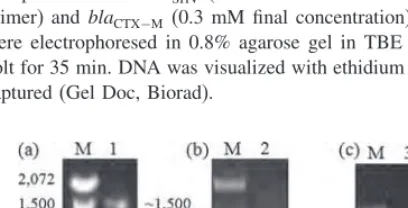
were electrophoresed in 0.8% agarose gel in TBE buffer, at 60 volt for 35 min. DNA was visualized with ethidium bromide and captured (Gel Doc, Biorad).
Fig. 1. 16SrRNA gene (∼1500 bp) detected by using Uniplex-PCR
(a).blaTEMdetected by using Uniplex PCR (lane 2) (b), and blaSHV and
blaCTX−Mgenes simultaneously detected by using Duplex-PCR (lane 3) (c)
fromK.pneumoniaeisolate no. 0177.
Table II. blaTEM,blaSHV, andblaCTX−Mgenes detected by using Uniplex-and Duplex-PCR from 97 ESBL-producingK.pneumoniaeisolates.
No. Identified genes No. (%)
1. blaTEM 7 (7.2)
All 97 clinical isolates were positive tested for the presence of 16s rRNA gene (Fig. 1(a)) and all isolates wereK.pneumoniae.Gene of blaTEM was detected using Uniplex-PCR, while blaSHV and bla CTX-M genes by Duplex-PCR (Figs. 1(b) and 1(c)).
Amplicons were electrophoresed in 0.8% agarose gel and visualized with ethidium bromide. (M=marker, 100 bp DNA
Ladder, Invitrogen). It was found that most of isolates had coexistences of more than one gene. As shown in Table II, 49 (50.5%) ESBL-producing K.pneumoniae were positive harbor-ing blaTEM, blaSHV, and blaCTX−M. Coexistences of two genes
were also observed in this study. Twelve (12.4%) of isolates had
blaTEM andblaSHV, 15 (15.5%) hadblaTEM andblaCTX−Mand 8
(8.2%) hadblaSHVandblaCTX−M
This study showed that K.pneumoniae that produce ESBL mostly possessed multiple genes, which were also reported in previous study conducted by Goyal et al.10This group found that
57.1% of ESBL-producingK.pneumoniaehad multiple genes of ESBL.10Several studies also showed thatK.pneumoniaeisolates
had more than one type of ESBLs, although it was not as high as that of this study.6"11Higher prevalence of ESBLs detected in
this study might be caused by misuse of antibiotics,2 or better
screening method of MDROs in the hospital laboratory. Severin6
reported that most of ESBL type found in Surabaya is CTX-M ESBL.6 Other studies in Singapore, Vietnam and Thailand also
showed prominent spreading of CTX-M ESBL type.4"12"13In this
study, CTX-M type prevalence was also found to be high but still SHV type was predominantly found among the isolates, sug-gesting there might be spreading of both SHV and CTX-M in Denpasar, Bali.
4. CONCLUSION
This study showed ESBL produced by K.pneumonia isolated from clinical specimens in Bali are mostly encoded byblaTEM,
blaSHV, and blaCTX−M. The molecular characterization of these
types ESBL is noteworthy for clinical microbiologist to perform early and prompt laboratory detection, and also to plan infection prevention and control program in preventing spread of multidrug resistance organisms. It can act as an alarm for the clinicians in management of patients with serious K.pneumoniae infections, especially to use antibiotics prudently. At the end, we can save our planet from spreading of multidrug resistant microorganism (MDROs). Further investigation of molecular epidemiology of hospital- or community-ESBLs with bigger number of samples and involving several health care enter would be promising to get good database for ESBLs-producing microorganisms in Bali.
R E S E A R C H
A R T I C L E
Adv. Sci. Lett. 21, 219–221, 2015
Acknowledgments: This work was financially sup-ported by Hibah Penelitian Unggulan Udayana Lembaga Penelitian dan Pengabdian kepada Masyarakat (LPPM) Udayana University, Bali, Indonesia under Grant No. 238-8/UN14.2/PNL.01.03.00/2014. We thank Wahyu Hidayati (Molecular Biology Laboratory staff), Ida Bagus Nyoman Putra Dwija (Clinical Microbiology Laboratory, Faculty of Medicine staff) and Ni Wayan Nilawati (Clinical Microbiology Laboratory Sanglah General Hospital staff) for their technical supports.
References and Notes
1. M. E. Falagas and D. E. Karageorgopoulos,J. Hosp. Infect.73, 345(2003).
2. D. L. Paterson and R. A. Bonomo,Clin. Microbiol. Rev.18, 657(2005).
3. P. A. Bradford,Clin. Microbiol. Rev.14, 933(2001).
4. R. Bonnet,Antimicrob. AgentsCh.48, 1(2004).
5. R. H. P. Dhillon and J. Clark,Crit. Care Research and Practice(2012).
6. J. A. Severin, N. M. Mertaniasih, K. Kuntaman, E. S. Lestari, M. Purwanta, N. L. D. Toom, D. O. Duerink, U. Hadi, A. V. Belkum, H. A. Verbrugh, and W. H. Goessens,J. Antimicrob. Chemoth.65, 465(2010).
7. V. Jarlier, M. H. Nicolas, and G. Fournier,Rev. Infect. Dis.10, 867(1988).
8. S. Tofteland, B. Haldorsen, K. H. Dahl, G. S. Simonsen, M. Steinbakk, T. R. Walsh, and A. Sundsfjord,J. Clin. Microbiol.45, 199(2007).
9. W. G. Weisburg, S. M. Barns, D. A. Pelletier, and D. J. Lane,J. Bacteriol.
73, 697(1991).
10. A. Goyal, K. N. Prasad, A. Prasad, S. Gupta, U. Ghosal, and A. Ayyagari,
Indian J. Med. Res.129, 695(2009).
11. C. F. Lin, S. K. Hsu, C. H. Chen, J. R. Huang, and H. H. Lo,J. Med. Microbiol.
58, 665(2010).
12. P. M. Hawkey,Clin. Microbiol. Infec.14, 159(2008).
13. N. H. T. Trang, T. V. T. Nga, J. I. Campbell, N. T. Hiep, J. Farrar, S. Baker, and P. Duy,J. Infection in Developing Countries7, 922(2013).
Received: 19 September 2014. Accepted: 15 October 2014.