Acute Detection of ST-Elevation Myocardial Infarction Missed on
Standard 12-Lead ECG With a Novel 80-Lead Real-Time Digital
Body Surface Map: Primary Results From the Multicenter
OCCULT MI Trial
James W. Hoekstra, MD Brian J. O’Neill, MD Yuri B. Pride, MD Cedric Lefebvre, MD Deborah B. Diercks, MD W. Frank Peacock, MD Gregory J. Fermann, MD C. Michael Gibson, MD Duane Pinto, MD Jim Giglio, MD Abhinav Chandra, MD Charles B. Cairns, MD Marvin A. Konstam, MD Joe Massaro, PhD Mitchell Krucoff, MD
From Wake Forest University, Winston-Salem, NC (Hoekstra, Lefebvre); Wayne State University, Detroit, MI (O’Neill); Beth Israel Deaconess Medical Center, Boston, MA (Pride, Gibson, Gibson, Pinto); University of California Davis, Sacramento, CA (Diercks); Cleveland Clinic Foundation, Cleveland, OH (Peacock); University of Cincinnati, Cincinnati, OH (Fermann); New York Presbyterian Hospital, New York, NY (Giglio); Duke University Medical Center, Durham, NC (Chandra, Krucoff); University of North Carolina, Chapel Hill, NC (Cairns); Tufts University Medical Center, Boston, MA (Konstam); and Boston University, Boston, MA (Massaro).
Study objective:Although 80-lead ECG body surface mapping is more sensitive for ST-elevation myocardial infarction (STEMI) than the 12-lead ECG, its clinical utility in chest pain in the emergency department (ED) has not been studied. We sought to determine the prevalence, clinical care patterns, and clinical outcomes of patients with STEMI identified on 80-lead but not on 12-lead (80-lead-only STEMI).
Methods:The Optimal Cardiovascular Diagnostic Evaluation Enabling Faster Treatment of Myocardial Infarction trial was a multicenter prospective observational study of moderate- to high-risk chest pain patients presenting to the ED. Patients received simultaneous 12-lead and 80-lead ECGs as part of their initial evaluation and were treated according to the standard of care, with clinicians blinded to the 80-lead results. The primary outcome of the trial was door-to-sheath time in patients with 80-lead-only STEMI versus patients with STEMI identified by 12-lead alone (12-lead STEMI). Secondary outcomes included angiographic and clinical outcomes at 30 days.
Results:One thousand eight hundred thirty patients were evaluated, 91 had a discharge diagnosis of 12-lead STEMI, and 25 patients met criteria for 80-lead-only STEMI. Eighty-four of the 91 12-lead STEMI patients underwent cardiac catheterization, with a median door-to-sheath time of 54 minutes, versus 14 of the 25 80-lead-only STEMI patients, with a door-to-sheath time of 1,002 minutes (estimated treatment difference in median⫽881; 95% confidence interval 181 to 1,079 minutes). Clinical outcomes and revascularization rates,
however, were similar between 80-lead-only STEMI and 12-lead STEMI patients.
Conclusion:The 80-lead ECG provides an incremental 27.5% increase in STEMI detection versus the 12-lead. Patients with 80-lead-only STEMI have adverse outcomes similar to those of 12-lead STEMI patients but are treated with delayed or conservative invasive strategies. [Ann Emerg Med. 2009;54:779-788.]
Providefeedbackon this article at the journal’s Web site,www.annemergmed.com. 0196-0644/$-see front matter
SEE EDITORIAL, P. 789.
INTRODUCTION
Background
An estimated 700,000 Americans will experience myocardial infarction, and 500,000 will experience a recurrent myocardial infarction each year.1For this reason, myocardial infarction has become the focus of aggressive diagnostic and treatment algorithms. Presently, the diagnosis of myocardial infarction is based on several diagnostic tools, including the 12-lead ECG, cardiac biomarkers (troponin), and clinical judgment.2
The 12-lead ECG is the cornerstone in the initial evaluation of chest pain and is especially crucial in the diagnosis of ST-elevation myocardial infarction (STEMI). Unfortunately, the initial 12-lead ECG is not highly sensitive for detection of myocardial infarction when ST-elevation is not present.3-6It is also severely limited in its detection of right sided, lateral, and posterior myocardial infarction, and it has an especially low sensitivity for myocardial infarction in patients with interventricular conduction delays.7-11In addition, patients with STEMI that is not identified on 12-lead ECG have been shown to have a poor prognosis.12,13Improved risk stratification of patients presenting with nondiagnostic 12-lead ECGs and “missed” ST-elevation may lead to improved outcomes.
Eighty-lead ECG body surface mapping is an extension of the conventional 12-lead ECG concept and may deliver improvements to the current diagnostic paradigm for myocardial infarction. The 80-lead displays as a topographic map over a larger area of the thoracic surface, including the right ventricular, posterior, and high left lateral regions. The
80-lead technique allows collection and analysis of data from a broader thoracic area, allowing for greater spatial sampling. The principles of the 80-lead technique have been well
established.14-19With the emergence of more user-friendly computer hardware, software, and electrode application, the 80-lead mapping system has now become feasible in clinical practice for the evaluation of chest pain patients.17-19
Several clinical trials have shown the efficacy of a new generation of 80-lead technology in the detection of myocardial infarction.20-24In particular, it appears to be well suited for detecting injury patterns in the right ventricular and posterior regions associated with inferior myocardial infarction.23The enhanced sensitivity of the 80-lead for myocardial infarction and its ease and speed of application should allow the clinician to identify STEMI in ECG-silent areas of the heart within minutes of initial patient contact. This has particular relevance to emergency physicians, who routinely evaluate chest pain patients and are charged with initiating early therapy.
Importance
Patients who are identified as having STEMI by 80-lead but not by 12-lead ECG (referred to as 80-lead-only STEMI patients) have not been previously clinically characterized, especially in a broad emergency department (ED)– based chest pain population. We hypothesize that these 80-lead-only STEMI patients are presently treated in a manner consistent with non-ST-elevation myocardial infarction (NSTEMI) treatment guidelines,2with either a delayed invasive or selectively invasive angiography approach. This approach is in contrast with the rapid reperfusion strategies used for STEMI patients identified by 12-lead ECG, where door-to-balloon times of 90 minutes or less are advocated.25We also hypothesize that 80-lead-only STEMI patients have similar angiographic pathology and similar rates of mortality and morbidity (major adverse cardiac events) compared with 12-lead STEMI patients.
Goals of This Investigation
The purpose of this trial is to characterize the prevalence, management patterns, and outcomes of patients with acute coronary syndromes who are identified as having STEMI by 80-lead ECG only with those who are identified as having STEMI by 12-lead ECG to determine whether the potential exists to improve care by early detection of myocardial infarction in these patients.
MATERIALS AND METHODS Study Design
The Optimal Cardiovascular Diagnostic Evaluation Enabling Faster Treatment of Myocardial Infarction trial was a
multicenter prospective cohort-blinded observational clinical trial evaluating the 80-lead ECG body surface mapping
technology (PRIME ECG; Heartscape, Inc., Columbia, MD) in the care of patients presenting to the ED with chest pain and moderate to high risk for adverse clinical outcomes.
Editor’s Capsule Summary
What is already known on this topic
The 12-lead ECG does not identify the majority of patients with acute myocardial infarction.
What question this study addressed
Whether patients with ST elevation present on an 80-lead ECG but not on the standard 12-lead ECG behave like ST-elevation myocardial infarction (STEMI) patients.
What this study adds to our knowledge
This study of 1,830 moderate- to high-risk patients suggests that the patients identified with these additional ECG leads have some similarities with traditional STEMI patients.
How this might change clinical practice
Setting
Patients were enrolled in 12 high-volume tertiary care center EDs in the United States (Appendix E1; available online at
http://www.annemergmed.com). The study was approved by the institutional review boards of each site. The study was initiated in November 2006 and concluded in May 2008.
Selection of Participants
Patients were enrolled if they were older than 39 years and presenting to the ED with chest pain or symptoms suspicious for acute coronary syndrome, beginning less than 24 hours before arrival, with the last symptoms less than 12 hours before arrival. Patients had to be moderate to high risk for adverse cardiovascular outcomes, as manifested by chest pain plus ischemic ECG abnormalities, known coronary artery disease, or 3 or more cardiac risk factors (diabetes, hypertension, current smoking, family history of coronary artery disease, or hypercholesterolemia). All patients gave written, informed consent.
Patients were excluded from the study if they were unable to give consent, if they had symptoms greater than 24 hours before presentation, or if they had recent (⬍48 hours) STEMI by 12-lead ECG. Also excluded were patients with recent trauma, hemodynamic instability, cardiogenic shock, or pulmonary edema (Killip Class 3 heart failure).
Interventions
Patients were evaluated and treated by emergency physicians at each site according to the standard of care. Following their initial ECG, patients were evaluated for eligibility for the study. After informed consent was obtained, the 80-lead ECG body surface mapping system was applied by study personnel or investigators (stipulated to be applied within 20 minutes of the initial ECG). Serial 80-lead ECGs (and simultaneous imbedded 12-lead ECGs) were obtained at the discretion of the treating physician or the study personnel during the following 2 to 4 hours. At least 2 serial 80-lead ECGs per patient were encouraged. The bedside clinical care team was blinded to the 80-lead results but could see the imbedded 12-lead ECG when each serial test was performed.
Patients were followed during their hospital stay to determine outcomes. Patients who proceeded to coronary angiography were assessed to determine door-to-sheath time (see definitions below). Coronary angiography results were analyzed in a blinded fashion by experienced interventional cardiologists in the Percutaneous/Pharmacologic Endoluminal
Revascularization for Unstable Syndromes and its Evaluation (PERFUSE) core lab (TIMI Group, Boston, MA) to determine angiographic outcomes and revascularization rates. Clinical outcomes were recorded by study personnel at the clinical sites and included troponin levels at presentation and
precatheterization, provocative stress testing results, echocardiographic or nuclear perfusion imaging results, and surgical revascularization procedures. Major adverse cardiac events after discharge from the hospital, including death from
any cause, recurrent myocardial infarction, percutaneous coronary intervention, coronary artery bypass grafting surgery and rehospitalization for coronary complications were assessed at 30 days (see definitions below). Follow-up was obtained by multiple telephone calls, letters, or medical records review. Final diagnoses were obtained from the subject’s discharge summary report.
Eighty-lead and simultaneous 12-lead ECGs were analyzed by the ECG core laboratory at Duke Cardiovascular Research Institute, Durham, NC, by cardiologists and emergency physicians who were blinded to clinical results and the 80-lead mapping system– generated algorithm readings. After
elimination of ECGs with bundle branch blocks or technical failures, the 12-lead and 80-lead ECGs were categorized as ischemic (yes/no), and ischemic ECGs were further categorized as ST-elevation, ST-depression, or T-wave changes (Figure 1). ECG location of the ischemic changes was also documented. If serial ECGs were performed on a given patient, only 1 ECG of the series had to be ischemic for that patient to be labeled as “ischemic” by the core laboratory. No historical ECG tracings were available for comparison, unless they were done in serial fashion within the same ED encounter.
Because of the time pressures of reperfusion therapy in patients with STEMI, we found it difficult to enroll STEMI patients in the trial, yet these STEMI patients were crucial to the door-to-sheath time outcome. After enrollment of the first 225 patients, a protocol amendment was implemented to allow patients with 12-lead STEMI to be entered into and proceed within the protocol without undergoing the 80-lead ECG. This amendment yielded a cohort of STEMI patients who were included in the trial but did not undergo 80-lead ECGs. These patients are included in the analysis of the primary and
secondary outcomes, but direct comparisons of the performance of the 12-lead to the 80-lead ECG were not possible in this group.
Methods of Measurement
Twelve-lead STEMI was defined by discharge diagnosis, as noted on the discharge summary. NSTEMI was defined by
discharge diagnosis, as noted on the discharge summary. Patients were defined as 80-lead-only STEMI if they were (1) not 12-lead STEMI by discharge diagnosis; (2) had increased troponin levels; (3) had core laboratory–adjudicated 80-lead ECG of STEMI; and (4) had core laboratory–adjudicated 12-lead ECG reading of not STEMI. Twelve-12-lead NSTEMI patients were defined as NSTEMI by discharge diagnosis after elimination of patients who met criteria for 80-lead-only STEMI. Door-to-sheath time was defined as the time from ED registration to arterial sheath placement in the catheterization laboratory, in minutes. Troponin was reported for blood draws before angiography. Increased troponin level was defined as any increase over the local range of normal at enrolling sites. Troponin levels are reported as eitherx-fold increases or absolute values in nanograms per milliliter. Death was defined as all-cause mortality within 30 days after discharge from index hospitalization. Recurrent myocardial infarction was defined as rehospitalization within 30 days, with a discharge diagnosis of STEMI or NSTEMI. Rehospitalization was defined as any hospital admission for cardiovascular diagnoses within 30 days from index hospitalization, including discharge diagnosis of myocardial infarction or treatment including percutaneous coronary intervention or coronary artery bypass grafting. Major adverse cardiac events or major adverse clinical events were defined as the combination of death, recurrent myocardial infarction, and rehospitalization. Revascularization was defined as percutaneous coronary intervention or coronary artery bypass grafting after coronary angiography.
Data Collection and Processing
All data were collated and analyzed by Cardiovascular Clinical Trials, Inc., Boston, MA.
Outcome Measures and Primary Data Analysis
The primary outcome of the trial was door-to-sheath time; the primary analysis was the comparison of door-to-sheath time distribution in 80-lead-only STEMI patients versus door-to-sheath time in traditional 12-lead STEMI patients, in only those patients who underwent coronary angiography during their index hospitalization. The comparison was performed with rank analysis of covariance, adjusting for the covariates age, sex, prevalence of diabetes, current smoking status, and histories of hypertension and hypercholesteremia. A 2-sided significance level of 0.05 was used to declare treatments significantly different.
Angiographic outcomes, including revascularization rates, culprit artery locations, and percentage of stenosis in the culprit artery, were also presented for each group. Revascularization rates were statistically compared between 80-lead-only STEMI and 12-lead STEMI by using odds ratios (ORs) and their 2-sided 95% confidence intervals (CIs).
Secondary outcomes of incidence of death, myocardial infarction, and rehospitalization at 30 days were presented for 12-lead STEMI, 80-lead-only STEMI, and 12-lead NSTEMI patients, with 2-sided 95% CIs of the difference between ORs
for only STEMI versus 12-lead STEMI and 80-lead-only STEMI versus 12-lead NSTEMI. For dichotomous outcomes in which event rates were large enough, Cls were calculated, adjusting for the same covariates as above; otherwise, CIs were unadjusted. All patients in each group were included, whether or not they underwent coronary angiography. Mean troponin increases and culprit artery stenosis percentages for each group were presented but not compared statistically.
In clinical care, outside of study protocols, the 80-lead ECG is typically not used for patients who have already received a diagnosis of STEMI by 12-lead ECG. The 80-lead ECG offers little clinical value in these patients, and its use may delay reperfusion. On the other hand, it is very useful in patients when the initial 12-lead ECG is nondiagnostic but STEMI is suspected. To assess the incremental value of the 80-lead ECG reading of ST elevation in these patients who were not 12-lead STEMI, the secondary outcomes of incidence of death, myocardial infarction, and death/myocardial infarction at 30 days were analyzed, stratified according to core
laboratory–adjudicated 12-lead and 80-lead ECG readings of ST elevation. The strata were as follows: ST-elevation and not ST-elevation by 80-lead ECG, and ST-elevation and not ST-elevation by 12-lead ECG. Unadjusted pairwise ORs between strata and their 2-sided 95% CIs were calculated (multivariate-adjusted ORs were not calculated because of the relatively low event rates in the strata).
All analyses were conducted with SAS, version 9.1 (SAS Institute, Inc., Cary, NC).
The following was the planned sample size for the study, with the primary comparison between the 80-lead-only STEMI and the 12-lead STEMI group.26The sample size was estimated
according to a 10% increase in the detection of 80-lead-only STEMI within the NSTEMI group, a 20% incidence of myocardial infarction in the total study population, a 10% dropout rate, and at least a 2.25 times higher median door-to-sheath time in the 80-lead-only STEMI group compared with the 12-lead STEMI group. A sample size of 1,400 patients or 280 myocardial infarctions was chosen to provide at least 80% power to detect a significant difference in the door-to-sheath time distribution between 80-lead-only STEMI and 12-lead STEMI at a 2-sided alpha of 0.05, under these assumptions. The final sample size was increased to 1,800 patients according to a blinded interim analysis by the study data safety and monitoring board. Specifically, the event rate (myocardial infarction) was lower than initially estimated, necessitating the addition of 400 patients to the sample size to attain the appropriate number of 80-lead-only STEMI patients.
RESULTS
Characteristics of Study Subjects
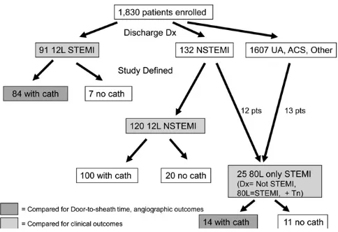
diagnosis of STEMI by the treating physicians using the 12-lead ECG only (designated 12-lead STEMI,Figure 2), whereas 132 received a diagnosis of NSTEMI and 1,607 as unstable angina, acute coronary syndrome, or other chest pain diagnoses.
Twenty-five patients met criteria for 80-lead-only STEMI. Twelve of these patients came from the NSTEMI diagnosis group, and 13 had received a diagnosis of unstable angina or acute coronary syndrome. We found that the discharge diagnoses of unstable angina or acute coronary syndrome were quite variable, and that many of the patients receiving a diagnosis by clinicians of unstable angina or acute coronary syndrome had an increased troponin level. Thirteen of the patients had increased troponin levels and ST elevation results on their 80-lead ECG and thus met criteria for 80-lead-only STEMI.
One hundred twenty patients who received a discharge diagnosis of NSTEMI did not meet core laboratory–adjudicated criteria for 80-lead-only STEMI (designated as 12-lead
NSTEMI inFigure 2). Baseline characteristics between all three groups (STEMI, 12-lead NSTEMI, and 80-lead-only STEMI) are shown inTable 1.
Main Results
Of the 91 patients with 12-lead STEMI, 84 (92.3%) underwent cardiac catheterization; of the 25 patients with 80-lead-only STEMI, 14 (56.0%) underwent cardiac catheterization (80-lead-only versus 12-lead OR 0.1; 95% CI 0.04 to 0.4). Among patients who underwent cardiac catheterization, the median door-to-sheath time was
significantly shorter among 12-lead STEMI (54.0 minutes) than 80-lead-only STEMI patients (1,002.5 minutes) (Hodges-Lehman estimate of treatment difference in median⫽881; 95% CI 181 to 1,079 minutes) (Table 2).
Eighty-lead-only STEMI patients undergoing cardiac catheterization had rates of revascularization (percutaneous coronary intervention or coronary artery bypass grafting) similar to those of 12-lead STEMI patients undergoing cardiac
catheterization (12/14 [85.7%] versus 79/83 [95.2%]; OR 0.6; 95% CI 0.3 to 1.8). One 12-lead STEMI patient had missing revascularization information. Angiographic outcomes in the 80-lead-only STEMI patients are shown inTable 3.
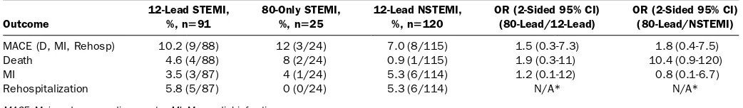
Clinical outcomes were evaluated among all patients with 12-lead STEMI, 80-lead-only STEMI, and 12-lead
NSTEMI, regardless of whether they underwent cardiac catheterization. Eighty-lead-only STEMI patients had 30-day rates of death, recurrent myocardial infarction, and
rehospitalization for cardiovascular reasons similar to those of patients with 12-lead STEMI (Table 4). In addition, 80-lead-only STEMI patients had 30-day rates of death, myocardial infarction, and rehospitalization that were descriptively higher than but statistically similar to those of patients with 12-lead NSTEMI (Table 4). Mean peak troponin levels and culprit lesion angiographic stenosis percentages for 12-lead STEMI, 80-lead-only STEMI, and 12-lead NSTEMI groups are shown inTable 5.
The 25 patients identified as 80-lead-only STEMI constituted a 27.5% increase in the number of patients
identified with STEMI over the 91 patients identified as 12-lead STEMI by the treating physician (4.9% of the cohort had 12-lead STEMI versus 6.3% with 12-12-lead STEMI plus 80-12-lead- 80-lead-only STEMI). They also represented 1.4% of the overall study population and 10.4% of those patients (n⫽241) with increased cardiac biomarker levels but without a diagnosis of 12-lead STEMI.
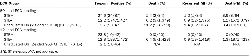
To further explore the incremental value of the 80-lead ECG, we compared the rates of troponin positivity, death, and recurrent myocardial infarction for patients with and without a reading of ST-elevation by 80-lead ECG, excluding those patients receiving a diagnosis of 12-lead STEMI by the treating physician. Outcomes among patients not receiving a diagnosis of 12-lead STEMI by the treating physician (n⫽1,739) were assessed according to the ECG core laboratory definitions of ST elevation or no ST elevation. Two hundred twenty-five patients were eliminated because of insufficient data or inevaluable ECGs (left bunde branch block or technical failures). In the remaining 1,514 patients with available outcome data, an 80-lead reading of ST elevation was associated with a statistically higher rate of death (OR 11.2; 95% CI 1.8 to 67) and a strong trend toward a higher rate of death and recurrent myocardial infarction (OR 3.4; 95% CI 1.0 to 11) than those of patients without a reading of ST-elevation on 80-lead (Table 6). ST elevation on the 12-lead ECG, however, was not predictive of adverse outcomes in this population.
LIMITATIONS
There are limitations to our study that deserve mention. The sample size of 80-lead-only STEMI patients was relatively small. Our study was sufficiently powered to detect a difference in door-to-sheath time, but it was not sufficiently powered to detect differences in clinical outcomes. Conclusions about clinical and angiographic outcomes must take this into account. We chose to analyze relatively gross process differences (such as Figure 2.Flow diagram of the Optimal Cardiovascular
door-to-sheath time) and catheterization outcomes (such as revascularization rates) in our study. Other variables in treatment, such as medications, patient disposition, and physician practice variables, may have affected the patient outcomes but were not analyzed in our study.
Second, the inability to perform 80-lead ECGs in 68 of 91 12-lead STEMI patients because of logistic issues and consent issues with rapid reperfusion makes it difficult to do formal sensitivity/specificity analysis of the 80-lead versus the 12-lead
ECG in STEMI, which is regrettable but could not be avoided. We chose to substitute an incremental value analysis rather than sensitivity/specificity analysis for this reason. Formal sensitivity/ specificity analysis of the 80-lead ECG for myocardial infarction has been reported elsewhere.19-24
Third, all patients with left bundle branch block or right bundle branch block were classified as having technical failure by the core laboratory. Of the 1,830 subjects, 135 were eliminated because of inadequate ECG tracings or bundle
Table 1.Demographics of 12-lead STEMI, 80-lead-only STEMI, and 12-lead NSTEMI groups.
Demographic
12-Lead STEMI Nⴝ91
80-Lead STEMI Nⴝ25
12-Lead NSTEMI Nⴝ120
Age, y, mean (SD) 63.8 (13.0) 66.4 (13.2) 63.6 (12.3)
Male, No. (%) 55 (60) 17 (68) 84 (70)
Diabetes, No. (%) 26 (29) 10 (40) 46 (38)
Hypertension, No. (%) 52 (57) 22 (88) 100 (83)
Hypercholesterol, No. (%) 53 (58) 18 (72) 80 (67)
Smoker, No. (%) 28 (31) 8 (32) 37 (31)
Table 2.Angiographic outcomes, 12-lead STEMI versus 80-lead-only STEMI.
Outcome 12-Lead STEMI 80-Lead Only STEMI Effect Size* (2-Sided 95% CI)
Door-to-sheath time, median (IQR), min 54.0 (30-112) 1,002.5 (229-1626) 881 (181-1079)
With angiogram, % 92.3 (84/91) 56 (14/25) 0.1 (0.04-0.4)
Revascularization, % 95.2 (79/83) 85.7 (12/14) 0.3 (0.1-1.8)
*Effect size is Hodges-Lehmann treatment difference in median for door-to-sheath time (calculated as 80L minus 12L) and OR for remaining characteristics (calculated as 80L/12L).
Table 3.Angiographic outcomes: 80-lead-only STEMI patients.
Patient # DTST, min
Peak TnI, x-fold
elev Culprit Artery TIMI Flow
%
Occlusion Revasc
80-Lead ECG Ischemia Location
Final Dx
1 110 59 LCX 0 100 Yes Posterior NSTEMI
2 132 215 RCA 3 80 Yes Ant/Right NSTEMI
3 161 97 SVG to RCA 3 90 Yes Right NSTEMI
4 229 28 RCA 0 100 Yes Right NSTEMI
5 252 1095 NA 0 NA Yes Right/Post NSTEMI
6 462 186 NA NA NA Yes Right/Post NSTEMI
7 972 3.8 NA NA NA No Ant/lateral ACS
8 1,087 2.7 SVG to RCA 3 95 Yes Right ACS
9 1,626 18 Multiple 0 NA Yes Posterior NSTEMI
10 3,209 2.1 SVG To RCA 3 95 Yes Right ACS
11 4,165 34 Multiple 3 NA Yes Ant/Lat NSTEMI
12 1,033 31 LAD 3 85 Yes Inf/Ant/Lat NSTEMI
13 1,588 5.9 RCA 0 100 Yes Right NSTEMI
14 2,753 1.0 LAD 3 50 No Ant/Lat Other
DTST,Door-to-sheath time;TnI,troponin I;TIMI Flow,thrombolysis in myocardial infarction flow rate.
Table 4.Clinical outcomes in 12-lead STEMI versus 80-lead-only STEMI versus 12-lead NSTEMI patients.
Outcome
12-Lead STEMI, %, nⴝ91
80-Only STEMI, %, nⴝ25
12-Lead NSTEMI, %, nⴝ120
OR (2-Sided 95% CI) (80-Lead/12-Lead)
OR (2-Sided 95% CI) (80-Lead/NSTEMI)
MACE (D, MI, Rehosp) 10.2 (9/88) 12 (3/24) 7.0 (8/115) 1.5 (0.3-7.3) 1.8 (0.4-7.5)
Death 4.6 (4/88) 8 (2/24) 0.9 (1/115) 1.9 (0.3-11) 10.4 (0.9-120)
MI 3.5 (3/87) 4 (1/24) 5.3 (6/114) 1.2 (0.1-12) 0.8 (0.1-6.7)
Rehospitalization 5.8 (5/87) 0 (0/24) 5.3 (6/114) N/A* N/A*
branch blocks. As such, there may be patients who were eliminated from the 80-lead-only STEMI group and the incremental value analysis despite being true STEMI patients. The bundle branch block group will be the subject of a future analysis, which should add significantly to our knowledge of the clinical utility of the 80-lead ECG.
Fourth, patients with ST-depression or T-wave inversion characterized as ischemic by the core laboratory were not included in this analysis. Ischemic changes outside of
ST-elevation were not considered in this analysis but may have influenced treating clinicians in their decisions. Ischemic changes other than ST-elevation will be the subject of a future analysis. Similarly, patients with ST elevation by 80-lead ECG but negative troponin-level results may have represented true unstable angina or coronary artery spasm patients. These patients, although important, did not meet our study definition for 80-lead-only STEMI.
Fifth, the troponin values presented inTable 5represent peak troponin values before cardiac catheterization and may not represent peak troponin values for the patient’s overall
hospitalization. Troponin levels post-percutaneous coronary intervention are often not tested and difficult to mandate in observational studies. In addition, our primary outcome was door-to-sheath time, which is a process outcome. Troponin levels precatheterization may be a driver of invasive
management, and is important to assess them as part of that decisionmaking process. Clinical outcomes, which may correlate with peak troponin levels, were a secondary outcome in this study. For these reasons, the mean troponin increases between groups were presented but not statistically compared.
The patients in the Optimal Cardiovascular Diagnostic Evaluation Enabling Faster Treatment of Myocardial Infarction
clinical trial may not have perfectly mirrored the optimum patient characteristics for clinical utilization patterns of the 80-lead ECG in “real practice.” The treating clinician was blinded to the results of the 80-lead ECG. As such, the treating clinician may not have ordered the 80-lead in real life or intended to use the 80-lead in his or her clinical decisionmaking.
This study did not analyze the cost-effectiveness of the 80-lead ECG. Cost-effectiveness will be analyzed in a separate article. We analyzed only the positive predictive ability of the 80-lead ECG to detect 80-lead-only STEMI. Each 80-lead vest costs approximately $160, and using the 80-lead ECG in 1,830 patients to detect 25 80-lead-only STEMI patients would not be cost-effective. This article does not analyze the predictive value of the 80-lead ECG for NSTEMI or acute coronary syndrome, nor does it analyze the negative predictive value of a normal 80-lead ECG. To assess the cost-effectiveness of the 80-lead ECG, all these effects need to be analyzed together, which will be reported elsewhere.
DISCUSSION
The purpose of this study was to characterize the prevalence, management patterns, and outcomes of patients with acute coronary syndromes who are identified as having STEMI by 80-lead ECG only versus those who are identified as having STEMI by 12-lead ECG, to determine whether the potential exists to improve care by speeding the diagnosis of STEMI in these patients. We were able to detect 25 patients in our study with study-defined 80-lead-only STEMI versus 91 patients receiving a diagnosis by clinicians of 12-lead STEMI and 120 receiving a diagnosis by clinicians of 12-lead NSTEMI. The 25 patients with 80-lead-only STEMI had significantly longer Table 5.Peak troponin values and culprit lesion stenosis percentages at cardiac catheterization in patients with 12-lead STEMI, 80-lead-only STEMI, and 12-lead NSTEMI.
Diagnosis N (Measured Total) Variable Mean 25th Percentile 75th Percentile 95% CI
12-Lead STEMI 87/91 Peak troponin value 19.7 0.45 28 12.4 to 27
74/91 Culprit stenosis 88.9 80 100 85.4 to 92
80-Lead-Only STEMI 25/25 Peak troponin value 10.3 0.10 3.5 ⫺1.9 to 23
9/25 Culprit stenosis 86.7 81 100 74.5 to 99
12-Lead NSTEMI 120/120 Peak troponin value 4.8 0.20 3.2 3 to 6.6
62/120 Culprit stenosis 76.3 60 90 72 to 80.7
Table 6.Association of 80-lead and 12-lead ECG readings of ST elevation with death and myocardial infarction outcomes after elimination of data for patients with 12-lead STEMI.
ECG Group Troponin Positive (%) Death (%) Recurrent MI (%) Death/MI (%)
80-Lead ECG reading
STE⫹ 27.6 (24/87) 2.4 (2/84) 1.2 (1/84) 3.6 (3/84)
STE– 12.2 (174/1,427) 0.2 (3/1,379) 0.9 (12/1,375) 1.1 (15/1,379)
Unadjusted OR (2-sided 95% CI) (STE⫹/STE–) 2.7 (1.7-4.5) 11.2 (1.8-67.9) 1.4 (0.2-10.7) 3.4 (1.0-11.9) 12-Lead ECG reading
STE⫹ 23.8 (10/42) 0 (0/40) 0 (0/40) 0 (0/40)
STE– 12.8 (188/1,472) 0.4 (5/1,423) 0.9 (13/1,419) 1.3 (18/1,423)
Unadjusted OR (2-sided 95% CI) (STE⫹/STE–) 2.1 (1.0-4.4) N/A N/A N/A
door-to-sheath time than 12-lead STEMI patients and were treated with a significantly delayed and conservative cardiac catheterization strategy. Their angiographic and clinical outcomes, however, were very similar to those of 12-lead STEMI patients.
Our study results are consistent with past clinical studies of the 80-lead ECG body surface mapping technology, but with the largest and most risk-diverse population studied to date, to our knowledge. The 80-lead ECG has never been studied in an undiagnosed ED chest pain population before this study. Several clinical trials have shown the efficacy of 80-lead
technology in the detection of myocardial infarction in high-risk patients only.20-24Menown et al20prospectively evaluated validation-set chest pain patients whose initial 12-lead ECG showed ST depression only. They demonstrated sensitivity for myocardial infarction in a 12-lead multivariate ECG model of 38% and specificity 81% versus sensitivity by the 80-lead ECG of 88% and specificity 75%. The 80-lead ECG has also displayed the potential for early detection of STEMI in specific regions of the myocardium. In particular, it appears to be well suited for detecting injury patterns in the right ventricular and posterior regions associated with inferior myocardial
infarction.23This was also demonstrated by Ornato et al,21 whose multicenter trial compared the 80-lead to 12-lead ECG in detecting STEMI among patients with biomarker-confirmed myocardial infarction and a discharge diagnosis of myocardial infarction. The 80-lead ECG showed a 26% greater sensitivity than the 12-lead ECG for identifying ST elevation, whereas specificity between the 2 diagnostic modalities was
comparable.21Our study, consistent with previous studies, showed a 27.5% relative increase in STEMI detection with the 80-lead ECG over the 12 lead.
The standard 12-lead ECG is not optimally effective in detecting STEMI in patients with infarcts in the posterior, right, inferior, and high lateral areas of the heart.7-11 Conversely, the 80-lead ECG is more sensitive for detecting STEMI in these areas.18-24As would be expected, the 80-lead-only STEMI patients in our study demonstrated ST elevation most commonly in the right and posterior regions. At angiogram, they demonstrated involvement most often in the posterior (left circumflex artery) and inferior (right coronary artery) arterial distributions. The culprit arteries at angiogram had high-grade stenoses in the majority of cases, and their arterial distribution also correlated nicely with the areas of ST elevation on the 80-lead ECGs. In addition, as demonstrated in Table 5, the culprit artery stenosis percentage at coronary angiogram in the 80-lead-only STEMI patients also closely resembled that of the 12-lead STEMI patients more than that of the 12-lead NSTEMI patients.
Although our study was not sufficiently powered to demonstrate differences in clinical outcomes, the death, myocardial infarction, and rehospitalization rate in the 80-lead-only STEMI population was very similar to that seen in the 12-lead STEMI population and trended worse than outcomes
in 12-lead NSTEMI patients. This is also consistent with past studies that demonstrated similar or worse outcomes for patients with angiographically defined STEMI.12,13A recent analysis of the NSTEMI patients in the Platelet IIb/IIIa Antagonist for the Reduction of Acute Coronary Syndrome Events in the Global Organization Network trial by Wang et al12showed that 27% of the NSTEMI patients in the trial had angiographic findings consistent with STEMI (single culprit lesion with thrombolysis in myocardial infarction flow rates (TIMI) 0/1 flow). In the study, NSTEMI patients with high-grade stenoses at angiogram had significantly higher 6-month mortality (OR 1.72; 95% CI 1.07 to 2.79) than NSTEMI patients with traditional “open arteries” observed at angiogram. In a retrospective review of the Trial to Assess Improvement in Therapeutic Outcomes by Optimizing Platelet Inhibition with Prasugrel, 26.2% of the 1,198 patients presenting with isolated anterior depression13 had a low TIMI flow grade of 0/1 during angiography and a positive troponin level result and were classified as “STEMI.” The 30-day death or myocardial infarction rate for the STEMI patients was 8.6% compared with 6.3% for NSTEMI and 2.9% for unstable angina (3-way⫽0.006). Our results demonstrated a 12.5% 30-day death, myocardial infarction, and
rehospitalization rate in the 80-lead-only STEMI patients, which was the highest of the 3 diagnostic groups, although not statistically so. In addition, peak preangiogram troponin levels trended higher in 80-lead-only STEMI versus NSTEMI patients as well, although they were not statistically analyzed. These results are consistent with the findings in past studies reporting that patients with missed STEMI are at high risk for adverse outcomes.12,13,27
The treatment patterns demonstrated in this study for 80-lead-only STEMI patients are also consistent with past studies. In the study by Gibson et al13of 1,198 patients with acute coronary syndrome and isolated anterior ST depression, only 5% received a diagnosis by treating physicians of STEMI, and none received coronary angiogram within 6 hours. The median time to catheterization was 29 hours in this study, consistent with a classic NSTEMI management strategy.2,28Clinicians are presumably reluctant to use an aggressive “time is muscle” catheterization approach in patients without STEMI on their initial 12-lead ECG, and at this point the data from past studies do not support such an approach. Our study, in conjunction with the Gibson et al13and Wang et al12studies, indicates that there may be a population of “missed STEMI” patients who are at high risk for adverse outcomes and may benefit from early, more aggressive catheterization if they can be identified by 80-lead ECG.
Because 68 of the 91 STEMI patients in our study did not undergo 80-lead ECG analysis, we were unable to perform traditional sensitivity/specificity analysis of the 80-lead ECG versus the 12-lead ECG for either STEMI or troponin-increased myocardial infarction. We chose instead to analyze the
consistent with clinical use of the 80-lead ECG in the ED. The 80-lead ECG is useful for the detection of ischemic changes in high-risk chest pain patients after an initial 12-lead ECG result is either negative or nondiagnostic for STEMI.26,29As such, we analyzed the predictive value of the 80-lead versus the 12-lead ECG for death, myocardial infarction, and troponin positivity in all patients who were not 12-lead STEMI. In our study, an 80-lead reading of ST elevation in these traditional NSTEMI patients was much more predictive of death and myocardial infarction than a reading of ST-elevation on the 12-lead ECG. Both tests predicted troponin increase well. However, our study was not powered for clinical outcomes, and these results, although consistent with those of past studies, are limited by the small study population. There will always be false-positive 80-lead ECGs for STEMI, and there will always be false-positive 12-lead ECGs for STEMI, but past studies have shown that the specificity of a reading of ST elevation on the 80-lead or 12-lead ECG is similar and high for myocardial infarction. Any ECG reading must be correlated to the clinical picture. Our study indicates that once data for patients with 12-lead STEMI are eliminated, a reading of ST elevation on the 80-lead ECG is much more predictive of adverse outcomes than a similar reading on the 12-lead ECG.
In our study, the 80-lead ECG body surface mapping system provided an incremental 27.5% increase in STEMI detection versus the 12-lead ECG. These patients with 80-lead-only STEMI are presently treated with a significantly delayed and conservative cardiac catheterization strategy, yet they have angiographic and clinical adverse outcomes similar to those of STEMI patients, detected by 12-lead ECG. These patients may have a potential for improved care and early intervention with early STEMI detection using the 80-lead ECG. Finally, in the patient without STEMI by 12-lead ECG, the incremental value of an 80-lead ECG reading of ST-elevation is significantly more predictive of adverse cardiovascular outcomes than a similar reading on the 12-lead ECG alone.
Supervising editor:Judd E. Hollander, MD
Author contributions:JWH and MK were the co–principal investigators. JWH was responsible for primary article preparation. JWH, JG, MAK, and MK were responsible for study design. BJO, CL, DD, GJF, and AC were the site principal investigators. BJO, CL, DD, and JG enrolled patients. BJO, JG, and MAK were members of the steering committee. BJO, YP, CD, DD, WFP, GJF, CMG, DP, MAK, and MK were responsible for article editorial assistance. YP, CMG, and DP conducted angiographic analysis. CL, AC, CBC, and MK conducted ECG core laboratory analysis. JWH, JM, and MK conducted data analysis. JM conducted statistical analysis. JWH takes responsibility for the paper as a whole.
Funding and support:ByAnnalspolicy, all authors are required to disclose any and all commercial, financial, and other relationships in any way related to the subject of this article that might create any potential conflicts of interest. See the Manuscript Submission Agreement in this issue for examples
of specific conflicts covered by this statement. The trial was funded by Heartscape, Inc., the makers of the 80-lead technology. The data were collated and analyzed by an independent contract research organization (CCT, Inc.). The steering committee had full control of the study design, and CCT had complete control of the data, without sponsor influence. The authors do not hold stock in the company, although a few have served as consultants to the company in trial design and product development. The authors have had complete access to and control of the data and complete freedom with data analysis and results presentation in this article, without influence from Heartscape.
Publication dates:Received for publication April 3, 2009. Revision received June 16, 2009. Accepted for publication June 24, 2009. Available online September 19, 2009.
Reprints not available from the authors.
Address for correspondence:James Hoekstra, MD,
Department of Emergency Medicine, Wake Forest University Health Sciences, Medical Center Blvd, Winston-Salem, NC 27023; 336-716-4626, fax 336-716-5438; E-mail
REFERENCES
1. American Heart Association.Heart Disease and Stroke
Statistics—2007 Update. Dallas, TX: American Heart Association; 2007.
2. Anderson JL, Adams CD, Antman EM, et al. ACC/AHA 2007 guidelines for the management of patients with unstable angina/ non-ST elevation myocardial infarction— executive summary.J Am Coll Cardiol. 2007;50:652-726. Available at:http://www.acc.org
orhttp://www.heart.org. Accessed August 26, 2009.
3. Herring N, Paterson DJ. ECG diagnosis of acute ischaemia and infarction: past, present and future.QJM. 2006;99:219-230. 4. Fesmire FM, Percy RF, Bardoner JB, et al. Usefulness of
automated serial 12-lead ECG monitoring during the initial emergency department evaluation of patients with chest pain.Ann Emerg Med. 1998;31:3-11.
5. Welch RD, Zalenski RJ, Frederick PD, et al. Prognostic value of a normal or nonspecific initial electrocardiogram in acute myocardial infarction.JAMA. 2001;286:1977-1984.
6. Menown IB, MacKenzie G, Adgey AA. Optimizing the initial 12-lead electrocardiographic diagnosis of acute myocardial infarction.Eur Heart J.2000;21:275-283.
7. Menown IB, Allen J, Anderson JMC, et al. Early diagnosis of right ventricular or posterior infarction associated with inferior wall left ventricular acute myocardial infarction.Am J Cardiol. 2000;85: 934-938.
8. Brady WJ, Hwang V, Sullivan R, et al. A comparison of 12- and 15-lead ECGS in ED chest pain patients: impact on diagnosis, therapy, and disposition.Am J Emerg Med. 2000;18:239-243. 9. Menown IB, MacKenzie G, Adgey AA. Optimizing the initial 12-lead
electrocardiographic diagnosis of acute myocardial infarction.Eur Heart J.2000;21:275-283.
10. Masoudi FA, Magid DJ, Vinson DR, et al. Implications of the failure to identify high-risk electrocardiogram findings for the quality of care of patients with acute myocardial infarction: results of the Emergency Department Quality in Myocardial Infarction (EDQMI) study.Circulation. 2006;114:1565-1571. 11. Zalenski RJ, Cooke D, Rydman R, et al. Assessing the diagnostic
12. Wang TY, Zhang M, Fu Y, et al. Incidence, distribution, and prognostic impact of occluded culprit arteries among patients with non-ST elevation acute coronary syndrome undergoing diagnostic angiography.Am Heart J. 2009;157:716-723. 13. Gibson CM, Pride YB, Mohanavelu S, et al. Angiographic and
clinical outcomes amongst patients presenting with acute coronary syndromes and anterior ST-segment depression. Circulation. 2008;118(suppl 2):654.
14. Muller JE, Maroko PR, Braunwald E. Evaluation of precordial electrocardiographic mapping as a means of assessing changes in myocardial ischemic injury.Circulation. 1975;52:16-27. 15. Madias JE, Venkataraman K, Hodd WB Jr. Precordial ST-segment
mapping 1. Clinical studies in the coronary care unit.Circulation. 1975;52:799-809.
16. Lux RL. Electrocardiographic mapping. Noninvasive
electrophysiological cardiac imaging.Circulation. 1993;87:1040-1042.
17. Taccardi B. Body surface distribution of equipotential lines during atrial depolarization and ventricular repolarization.Circ Res. 1966;19:865-878.
18. McMechan SR, MacKenzie G, Allen J, et al. Body surface ECG potential maps in acute myocardial infarction.J Electrocardiol. 1995;28:184-190.
19. McClelland AJ, Owens CG, Menown IB, et al. Comparison of the 80-lead body surface map to physician and to 12-lead
electrocardiogram in detection of acute myocardial infarction. Am J Cardiol.2003;92:252-257.
20. Menown IB, Allen J, Anderson JM, et al. ST depression only on the initial 12-lead ECG: early diagnosis of acute myocardial infarction.Eur Heart J. 2001;22:218-227.
21. Ornato JP, Menown IB, Riddell JW, et al. 80-Lead body map detects acute ST elevation myocardial infarction missed by standard 12-lead electrocardiography.J Am Coll Cardiol.2002; 39:332A.
22. Menown IB, Allen J, Anderson JM, et al. Early diagnosis of right ventricular or posterior infarction associated with inferior wall left ventricular acute myocardial infarction.Am J Cardiol.2000;85: 934-938.
23. Kornreich F, Montague TJ, Rautaharju PM. Body surface potential mapping of ST segment changes in acute myocardial infarction. Implications for ECG enrollment criteria for thrombolytic therapy. Circulation. 1993;87:773-782.
24. Maynard SJ, Menown IB, Manoharan G, et al. Body surface mapping improves early diagnosis of acute myocardial infarction in patients with chest pain and left bundle branch block.Heart. 2003;89:998-1002.
25. Antman EM, Hand M, Armstrong PW, et al. 2007 Focused update of the 2004 guidelines for the management of patients with ST-segment elevation myocardial infarction.J Am Coll Cardiol.2008. Available at:http://www.heart.orgorhttp:// www.jacc.org.
26. Lefebvre C, Hoekstra JW. Early detection and diagnosis of acute myocardial infarction: the potential for improved care with next-generation, user-friendly ECG body surface mapping.Am J Emerg Med. 2007;25:1063-1072.
27. Pope JH, Aufderheide TP, Ruthazer R, et al. Missed diagnosis of cardiac ischemia in the emergency department.N Engl J Med. 2000;342:1163-1170.
28. Zia MI, Goodman SG, Peterson ED, et al. Paradoxical use of invasive cardiac procedures for patients with non-ST segment elevation myocardial infarction: an international perspective from the CRUSADE Initiative and the Canadian ACS Registries I and II. Can J Cardiol.2007;23:1073-1079.
29. Self WH, Mattu A, Martin M, et al. Body surface mapping in the ED evaluation of the patient with chest pain: use of the 80-lead electrocardiogram system.Am J Emerg Med. 2006;24:87-112.
CORRECTION
Duke University Medical Center Baystate Medical Center
Wake Forest University Baptist Medical Center William Beaumont Hospital
Columbia University Medical Center Cleveland Clinic
Medical University South Carolina Hospital Tampa General Hospital