How to cite:
Nurtyas Y, Subandiyah K, Fitri LE (2017) The Correlation of Regulatory T (TReg) and Vitamin D3 in Pediatric Nephrotic Syndrome. J. Trop. Life. Science 8 (1): 43 – 47.
*Corresponding author: Yunika Nurtyas
Medical Faculty, Brawijaya University Jalan Veteran, Malang, Indonesia 65145
VOL. 8, NO. 1, pp. 43 – 47, January 2018 Submitted April 2017; Revised May 2017; Accepted December 2017
The Correlation of Regulatory T (TReg) and Vitamin D3 in Pediatric Nephrotic Syndrome
Yunika Nurtyas 1 *, Krisni Subandiyah 2, 3, Loeki Enggar Fitri 4
1 Faculty of Medicine, Univeristas Brawijaya, Malang, Indonesia
2 Department of Pediatrics, Faculty of Medicine, Univeristas Brawijaya, Malang, Indonesia 3 dr. Saiful Anwar General Hospital, Malang, Indonesia
4 Department of Parasitology, Faculty of Medicine, Univeristas Brawijaya, Malang, Indonesia
ABSTRACT
Nephrotic syndrome (NS) is an autoimmune disease that correlates to the imbalance of regulatory T cells (TReg).
This study was aimed to investigate the effect of vitamin D as adjuvant therapy of TReg population in pediatric
nephrotic syndrome. This study was designed randomized clinical trial, double blind, with pre- and post-test control groups involving 15 subjects newly diagnosed with NS. Subjects were divided into 2 groups, namely K1 for group treated with prednisone+vitamin D and K2 group for prednisone treatment only. The population of TReg in
periph-eral blood mononuclear cells (PBMC) was analyzed using flowcytometry. Vitamin D serum level was measured through ELISA method. Results showed that there was a significant elevation of TReg (independent t-test, p = 0.010)
in K1 group, which was higher than in K2 group. The Pearson test in the K1 group showed that vitamin D level was positively correlated with TReg (p = 0.039, r = 0.779).
Keywords: Nephrotic syndrome, vitamin D, TReg
INTRODUCTION
Idiopathic nephrotic syndrome is a glomerular dis-ease characterized by several clinical manifestations such as severe proteinuria, hypoalbuminemia, hyperlipide-mia, and edema [1, 2]. The prevalence of nephrotic syn-drome stands at 12 – 16 cases per 100,000 children, which is mostly found at the age of 2 – 5 years old. In Indonesia, there were 6 cases found in every 100,000 children below 14 years old per year in children, which is dominated by male (male: female ratio = 2 : 1) [3].
Pathogenesis of nephrotic syndrome is based on the immunological aberration, characterized by abundant circulating factor [4] and immuno-regulatory imbalance [5]. Conversely, regulatory T-cells (TReg) is an important
tolerogenic T-cell possessing protective effect on podo-cyte destruction [6, 7]. Vitamin D has been known as immunomodulator that able to induce TReg
differentia-tion as it has pleiotropic effects [8, 9]. There-fore, this study was aimed to investigate the effect of vitamin D as adjuvant therapy to the TReg population in
pediatric nephrotic syndrome.
MATERIALS AND METHODS
This study was conducted at Biomedical Laboratory, Faculty of Medicine, Brawijaya University. The duration of the study ranged between February until July 2015.
Study Design
This study was designed as a randomized clinical trial (RCT) double blind, with pre-and post-test control group. There were 2 groups namely K1 (prednisone and vitamin D3) and K2 (prednisone only). TReg population
and vitamin D levels were measured before and after treatment. Treatment for K1 were prednisone 2 mg/kg body weight/day (maximal dose 80 mg/day) and vitamin D3 oral preparation (D-Vit, PT. Gracia PharmindoTM)
University of Brawijaya, Malang No. 314/EC/KEPK-S2/05/2015.
Subjects
There were 15 subjects included in this study (7 sub-jects in K1; 8 subsub-jects in K2). Subsub-jects were taken from the Pediatric Nephrology Outpatient Care and Pediatric Ward, Dr. Saiful Anwar General Hospital, Malang dur-ing February – July 2015. The inclusion criteria for this study were newly diagnosed nephrotic syndrome patients aged 1 – 14 years old whose parents allowed them to participate in this study (informed consent). Meanwhile, the exclusion criteria for this study were sec-ondary nephrotic syndrome, congenital nephrotic syn-drome, relapse nephrotic synsyn-drome, and steroid depend-ent nephrotic syndrome.
Isolation of Peripheral Blood Mononuclear Cells (PBMC)
Blood samples in EDTA vacutainer were homoge-nized and added with PBS at a ratio of 1 : 1. The blood sample-PBS mixture was transferred slowly into falcon tube walls filled with Ficoll-Hipaque d = 1.077 g/dL (1 : 1). This mixture was centrifuged at 1,500 rpm at room temperature for 30 minutes which resulted in the for-mation of 4 layers namely plasma, PBMC, Ficoll-Hipaque, and erythrocyte. The PBMC ring was slowly transferred into 15mL centrifuge bottle. It was washed with 10mL PBS and centrifuged at 1200 rpm, at room temperature for 10 minutes. The supernatant was re-moved, washed using PBS, and centrifuged again at 1,200 rpm at room temperature for 10 minutes. After the second process of washing and centrifugation, PBMC will be formed as pellet at the bottom of centri-fuge bottle.
Measurement of regulatory T-cell population
The population of TReg was measured trough
flowcytometry method. Antibodies that were used phy-coerythrin (PE) anti-human FOXP3, FITC anti-human CD4, and PE/Cy5 anti-human CD25 (eBioscience, San Diego, CA). PBMC was suspended at certain density (2
× 106 cells/mL) in culture medium (RPMI equipped with
penicillin 100 U/mL, streptomy-cin 100 µg/mL, gluta-mine 2 mM, 10% calf fetal serum). The cell suspension was transferred into 24 wells then was stimulated with phorbol myristate acetate (PMA) 50 ng/mL and
iono-mycin 1 µM for 4 hours in monensin 500 ng/mL (Alexis
Biochemical, San Diego, CA). The incubator was set at temperature of 37°C and air pressure of 5% CO2. After
4 hours, the cell culture was transferred into sterile tubes and centrifuged at 1,500 rpm for 15 minutes.
T-cell lymphocyte was transferred into new tubes, washed with phosphate-buffered saline (PBS), and then incubated with fluorescein isothiocyanate (FITC)
anti-human CD4 and PE CD25 at 4°C for 30 minutes. After
incubation, speciments were stained with PE anti-hu-man Foxp3. Specimens were transferred into cuvette ready for flowcytometry analysis. The population of TReg
was analyzed with BD Cell Quest Pro.
Measurements of vitamin D level
Vitamin D level was measured through ELISA
method as previously described. Briefly 200 µL pre -di-luted serum samples were added into each well then it incubated for 2 hours at 25°C. After being washed, 100
µL enzyme conjugate was added into the tubes and in-cubated for 30 minutes at room temperature. Following this, 100 µL chromogen/substrates solution was added and incubated for 15 minutes at room temperature in
dark room. Finally, 100 µL stop solution was added to
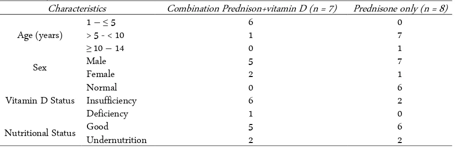
Table 1. Subject Characteristics
Characteristics Combination Prednison+vitamin D (n = 7) Prednisone only (n = 8)
Age (years)
1 –≤ 5 6 0
> 5 - < 10 1 7
≥10 – 14 0 1
Sex Male 5 7
Female 2 1
Vitamin D Status
Normal 0 6
Insufficiency 6 2
Deficiency 1 0
Nutritional Status Good 5 6
Undernutrition 2 2
each well. After 30 minutes, specimens were ready for analysis using ELISA reader at 650 nm.
Statistical analysis
Data distribution and homogeneity were statistically analyzed. Moreover, statistical differences of TReg and
vi-tamin D levels between groups were analyzed by independent t-test. The differences of TReg, Th17, and
vitamin D before and after treatment were analyzed by paired t-test. The correlation of TReg and vitamin D level
was analyzed with the Pearson correlation test. Data was analyzed at 95% confidence interval (α = 0.05) using SPSSversion 17.0. for Windows.
RESULTS AND DISCUSSION
Subject and baseline characteristics
Subject characteristics such as age, sex, vitamin D status, outcomes (steroid sensitive or resistant), and nu-tritional status were shown in Table 1. Morover the clinical outcome of subjects were shown in Table 2. Remission before 4 weeks and remission after 4 weeks were found in both groups that earlier was also found in K1. Most of the subjects were diagnosed with steroid sensitive nephrotic syndrome (SSNS), there was only one patient did not get remission and is classified as ster-oid resistant nephrotic syndrome (SRNS).
Based on the age factor, subjects were mostly from kids aged under 10 years old. The subjects were domina-ted by male or 12 boys from 15 subjects. This finding was also in accordance with previous studies and has been considered to be correlated with abnormal T cell clones in male thymus gland [10]. Based on nutritional status, it is revealed that most subjects had good tional status. However, it is important to evaluate nutri-tional status of children with nephrotic from syndrome because they are at high risk of suffering from malnour-ishment.
Vitamin D level status in nephrotic syndrome pa-tients (9 of 15) was low. This result was in accordance with previous study conducted in the General Hospital Dr. Cipto Mangunkusumo that 22 of 26 nephrotic pa-tients had low vitamin D levels (10 insufficiency, 16 de-ficiency) [11]. Loss of vitamin D-bounded protein through urine had been considered as etiologic factor for low plasma concentration in nephrotic patients [12]. Low vitamin D levels cause hyper-reactivity o dendritic cell, T cell, B cell, TReg suppression, and
pro-inflamma-tory cytokines elevation [13] that would lead to ne-phrotic syndrome. Several factors affect 25(OH)D level such as age, race, season, and milk consumption [14].
Table 2. Clinical outcome Outcome
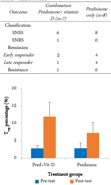
Figure 1. Treg percentage before treatment (pre-test) and after treatment (post-test)
Regulatory T cell population
The results demonstrated that there was no signi-ficant difference in the population of TReg in K1 and K2
(independent t-test,p = 0.97 pretest) before treatment. However, the differences were found significantly after treatment (independent t-test, p = 0.03 post test). Fur-thermore, the elevation of the TReg population in K1 and
K2 (before and after treatment, independent t-test, p = 0.01) were significantly different. Figure 1 shows the TReg percentage before treatment, after treatment, and its
enhancement after treatment. Furthermore, the enhan-cement of TReg percentage was significantly different in
both groups (paired t-test, K1 p = 0.00, K2 p = 0.00).
Vitamin D level and Treg population
The vitamin D level was found higher in K1 than K2 (before and after treatment, independent t-test, p = 0.00). Figure 2 shows vitamin D levels before treatment, after treatment, and its elevation after treatment. Fur-thermore, the elevation of vitamin D level was signifi-cantly different in both groups (paired t-test, K1 p = 0.00, K2 p = 0.00).
The elevation of TReg population in prednisone and
vitamin D treated group was higher than in prednisone only treated group. Reduction of TReg population and its
dysfunction in nephrotic syndrome would lead to dis-ability to suppress effector T cells [15]that is associated to proteinuria [16, 17]. TReg also acts as
tory T cells through secretion of several anti-inflamma-tory cytokines such as IL-10 and TGF-β[18].
Vitamin D administration could induce and stimu-late TReg directly through antigen presenting cells or
dendritic cells also indirectly through endocrine or in-tracrinal conversion of 25(OH)D becoming 1,25(OH)2 D3 [18]. Furthermore, vitamin D administration was correlated with elevation of TReg Foxp3+ population [9,
19]. Several mechanisms focused on how vitamin D af-fects TReg have been studied. Administration of 1,25(O
H)2D3 could enhance STAT5 phosphorylation in Foxp3+ cells via TGF-β and IL-2 that lead to T
Reg
differ-entiation [20, 21]. Conversely, low vitamin D levels would lead to IL-6 upregulation instead of TGF-β down-regulation causing Th17 differentiation [21].
Glucocorticoid had been known as one of anti-in-flamation drugs that induces T cell apoptosis, T cell en-ergy, and suppress T cell function [22]. Furthermore, glucocorticoid could induce IL-10 upregulation resulted in immature dendritic cells or macrophage thus induc-ing differentiation of TReg/suppressor T cells [22].
Adju-vant therapy with vitamin D3 could induce immuno-supressive effects of TReg through upregulation of Foxp3
and IL-10 [23, 24, 25].
CONCLUSION
In conclusion, there was significant elevation of TReg
population in the prednisone and vitamin D treated group than in prednisone only treated group. However, the vitamin D level was positively correlated with TReg
population.
ACKNOWLEDGMENT
Authors thank to Brawijaya University for facilitat-ing this research.
REFERENCES
1. Bagga A (2008) Management of steroid sensitive nephrotic: revised guidelines. Indian Journal of Nephrology 18 (1): 31 – 39. doi: 10.4103/0971-4065.41289.
2. Zhang S, Audard V, Fan Q et al. (2011) Immunopathogenesis of idiopathic nephrotic syndrome. In: Hererra GA (ed) Ex-perimental models for renal diseases: Pathogenesis and diag-nosis. Basel, Karger. pp 94 – 106. doi: 10.1159/000313 947. 3. UKK Nefrologi Ikatan Dokter Anak Indonesia (2008)
Tatalaksana sindrom nefrotik idiopatik pada anak. Jakarta, Badan Penerbit Ikatan Dokter Anak Indonesia.
4. Hafez MA, Shimada M, Lee PY et al. (2009) Idiopathic ne-phrotic syndrome and atopy: Is there a common link?. Amer-ican Journal of Kidney Disease 54 (5): 945 – 953. doi: 10.1053/j.ajkd.2009.03.019.
5. Wang L, Li Q, Wang L et al. (2013) The role of Th17/IL-17 in the pathogenesis of primary nephrotic syndrome in chil-dren. Kidney and Blood Pressure Research 37 (1): 332 – 345. doi: 10.1159/000350161.
6. Wang (2008) Regulatory T cells in renal disease. International Journal of Clinical and Experimental Medicine 1 (4): 294 – 304.
7. Pereira WF, Brito-Melo GEA, Guimaraes FTL et al. (2014) The role of the immune system in idiopathic nephrotic syn-drome: A review of clinical and experimental studies. Inflam-mation Research 63 (1): 1 – 12. doi: 10.1007/s00011-013-0672-6.
8. Terrier B, Derian N, Schoindre Y et al. (2012) Restoration of regulatory and effector T cell balance and B cell homeostasis in systemic lupus erythematosus patients through vitamin D supplementation. Arthritis Research and Therapy 14 (1): 1 – 10. doi: 10.1186/ar4060.
of Immunology 42 (10): 2697 – 2708. doi: 10.1002/eji.2012 42370.
10. van den Berg JG, Weening JJ (2004) Role of the immune sys-tem in pathogenesis of idiopathic nephrotic syndrome. Clin-ical Science 107 (2): 125 – 136. doi: 10.1042/CS20040095. 11. Septarini AD, Tambunan T, Amalia P (2012) Calcium and
vitamin D supplementation in children with frequently re-lapsing and steroid-dependent nephrotic syndrome. Paediat-rica Indonesiana 52 (1): 16 – 21. doi: 10.14238/pi52. 1.2012.16-21.
12. Esmaeeili M, Azarfar A, Hoseinalizadeh S (2015) Calcium and vitamin D metabolism in pediatric nephrotic syndrome: An update on the existing literature. International Journal of Pediatrics 15 (3): 103 – 109. doi: 10.22038/IJP.2015.3932. 13. Ginanjar E, Sumariyono, Setiati S, Setiyohadi B (2007)
Vita-min D and autoimmune disease. Acta Medica Indonesiana 39 (3): 133 – 141.
14. Weng FL, Schults J, Heskovitz RM et al. (2005) Vitamin D insufficiency in steroid-sensitive nephrotic syndrome in re-mission. Pediatric Nephrology 20 (1): 56 – 63. doi: 10.1007/ s00467-004-1694-7.
15. Araya C, Diaz L, Wasserfall C et al. (2009) T regulatory cell function in idiopathic minimal lesion nephrotic syndrome. Pediatric Nephrology 24 (9): 1691 – 1698. doi: 10.1007/s004 67-009-1214-x.
16. Liu LL, Qin Y, Cai JF et al. (2011) Th17/ Treg imbalance in adult patients with minimal change nephrotic syndrome. Clinical Immunology 139 (3): 314 – 320. doi: 10.1016/ j.clim.2011.02.018.
17. Shao XS, Yang XQ, Zhao XD et al. (2009) The prevalence of Th17 cells and FOXP3 regulatory T cells (Treg) in children with primary nephrotic syndrome. Pediatric Nephrology 24 (1): 1683 – 1690. doi: 10.1007/s00467-009-1194-x.
18. Lang CL, Wang MH, Chiang CK, Lu KC (2014) Vitamin D
and the Immune system from the nephrologist’s viewpoint.
ISRN Endocrinology 14 (1): 1 – 11. doi: 10.1155/2014/ 105456.
19. Smolders J, Thewissen M, Peelen E et al. (2009) Vitamin D status is positively correlated with regulatory T cell function in patients with multiple sclerosis. PLoS ONE 4 (8): 63 – 45. doi: 10.1371/journal.pone.0006635.
20. Chambers ES, Suwannasaen D, Mann EH et al. (2014) 1,25-dihydroxyvitamin D3 in combination with transforming growth factor-β increases the frequency of Foxp3+ regulatory T cells through preferential expansion and usage of interleu-kin-2. Immunology 143 (1): 52 – 60. doi: 10.1371/journal. pone.0006635.
21. Gordillo R, Spitzer A (2009) The nephrotic syndrome. Pedi-atric in Review 30 (3): 94 – 104. doi: 10.1371/journal.pone. 0006635.
22. Franchimont D (2004) Overview of the actions of glucocorti-coids on the immune response: a good model to characterize new pathways of immunosuppression for new treatment strategies. Annals of the New York Academy of Sciences 1024 (1): 124 – 137. doi: 10.1196/annals.1321.009.
23. Heine G, Niesner U, Chang HD et al. (2008) 1,25-dihy-droxyvitamin D3 promotes IL-10 production in human B cells. European Journal of Immunology 38 (8): 2210 – 2218. doi: 10.1002/eji.200838216.
24. Barrat FJ, Cua DJ, Boonstra A et al. (2002) In vitro generation of interleukin-10-producing regulatory CD4+ cells is induced by immunosuppressive drugs and inhibited by T helper type 1 (Th1)- and Th2-inducing cytokines. Journal of Experi-mental Medicine 195 (5): 603 – 616. doi: 10.1084/jem.20011 629.