VOL. 6, NO. 3, pp. 176 - 183, September, 2016 Submitted January 2016; Revised May 2016; Accepted September 2016
Advanced Glycation End Products (AGEs) Antibody Protects Against AGEs-Induced Apoptosis and
NF-ĸB p65 Subunit Overexpression in Rat Glomerular Culture
Oktavia Rahayu Adianingsih 1*, Diana Lyrawati 1, Nur Samsu 2
1Laboratory of Pharmacy, Faculty of Medicine, Brawijaya University, Malang, Indonesia
2Division of Nephrology and Hypertension, Department of Internal Medicine, dr. Saiful Anwar Public Hospital, Malang, Indonesia
ABSTRACT
Advanced glycation end products (AGEs) have been thought to be a major cause of diabetic nephropathy (DN). The mechanisms underlying the involvement of AGEs antibody in diabetic nephropathy are not fully understood. The present study was designed to investigate the protective effect of AGEs antibody on AGEs-induced glomerular damage. Isolated glomeruli were pre-incubated either with 10 µg/mL polyclonal anti-AGEs antibody (AGE-pAb) or monoclonal anti-Nɜ
-carboxymethyl-lysine antibody (CML-mAb) as a model of AGEs antibody to block interaction of AGEs with receptor for AGEs (RAGE) and incubated afterwards either with 100 µg/mL bovine serum albumin (BSA) or AGE-modified bovine serum albumin (AGE-BSA) for 48 h. Annexin V/nephrin double-staining was performed to determine apoptosis. Using immunofluorescence, we found that administration of AGE-BSA not only significantly increased glomerular cells apoptosis and nuclear factor kappa B (NF-ĸB) p65 expression, but also reduced expression of nephrin, an important structural and signal molecule of podocytes slit diaphragm. Blocking the interaction of AGE-RAGE with AGEs antibody significantly protected glomerular cells from AGEs-induced apoptosis and NF-ĸB p65 overexpression. We found that AGE-pAb conferred superior protective effect compared with CmL-mAb for the same reduction in apoptosis and NF-ĸB p65 expression. In sharp contrast, CmL-mAb led to preserve expression of podocytes nephrin better than AGE-pAb. These results demonstrate that the antibody against AGEs may be beneficial for preventing the glomerular damage in DN.
Keywords: Advanced glycation end products; antibody, apoptosis, diabetic nephropathy, Ne-(carboxymethyl)lysine
Diabetes mellitus (DM) is a significant health prob-lem with a worldwide mortality around 382 million people in 2013, and this number is expected to rise to 592 million by 2035 [1]. AGEs are a causative factor in diabetic vascular complication such as diabetic nephropathy (DN) which further lead to end-stage re-nal disease [2,3]. Accumulation of AGEs is present in all renal compartments in diabetic patient with three mechanisms of toxicity include interaction AGEs with receptor for AGE (RAGE), in situ glycation and tissue deposition [4]. Inhibition of AGEs is widely regarded as an implicit goal in clinical medicine for the treat-ment of DN [5]. So far, most studies have been fo-cused on the potential of therapies that not only target
various pathways upregulated by AGEs, but also target AGEs itself by preventing AGE formation, breaking AGE-protein cross-linking, or neutralizing AGE [6– 11]. In addition to the development of anti-AGE activ-ity from natural product or synthetic compound, im-munization of AGEs in diabetic mice has recently at-tached attention [12].
AGEs have antigenic properties that may exert an autoimmune response. Autoantibodies against AGEs are detected in serum of patients with DM and non-DM which may play a role in the macrophage uptake of AGEs-modified protein via AGEs-immune com-plexes (AGE-IC) formation [13,14]. Disruption of the balance between AGEs formation and AGEs elimina-tion will lead to accumulaelimina-tion of AGEs [14,15].
How-JTLS | J. Trop. Life. Science 176 Volume 6 | Number 3 | September | 2016
INTRODUCTION
*Corresponding author: Oktavia Rahayu Adianingsih
Laboratory of Pharmacy, Faculty of Medicine, Brawijaya University Jalan Veteran, Malang, East Java 65145, Indonesia
E-mail: [email protected]
How to cite:
Adianingsih OR, Lyrawati D, Samsu N (2016) Advanced Glycation End Products (AGEs) Antibody Protects Against
ever, our knowledge of the molecular mechanism gov-erning role of AGE-Ab and AGE-IC in diabetic com-plication is still very limited. Thus, in this research, we aimed to demonstrate the role of AGE-Ab in prevent AGEs-induced glomerular damage.
Ethics
This research was approved by the Health Research Ethics Commission from Faculty of Medicine, Brawi-jaya University, Malang, Indonesia, with registration number 356B/EC/KEPK-S2/06/2015.
Chemicals
All chemicals were purchased from Sigma (St. Louis, MO) unless otherwise indicated. AGE-mAb was purchased from Circulex (Nagano, Japan). AGE-pAb was purchased from Abcam (Cambridge, England). BSA (fraction V) was purchased from Roche Diagnos-tic GmbH (Mannheim, Germany). AGE-BSA was pur-chased from BioVision (California, USA). For im-munofluorescence assay, the following primary anti-bodies were used: rabbit polyclonal anti-NF-ĸB p65 (Bioss, Woburn, USA), goat monoclonal anti-nephrin (Santa Cruz Biotechnology, California, USA), rabbit polyclonal anti-nephrin (Bioss, Woburn, USA) and An-nexin V conjugated fluorescein-isothiocyanate (FITC) (BioLegend, Fell, Germany). Secondary antibodies were used: goat anti-rabbit FITC, rabbit anti-goat IgG-rhodamine and goat anti-rabbit IgG-IgG-rhodamine (Santa Cruz Biotechnology, California, USA). Fetal bovine serum (FBS) was purchased from Gibco (Grand Island, NY, USA).
Rat glomerular isolation and culture
Isolation and culture of glomeruli were done as de-scribed previously with some modifications [16–19]. Briefly, male Wistar rats at age 7 - 9 weeks were sacri-ficed by cervical dislocation and perfused with ice-cold Hank’s balanced salt solution (HBSS) through the heart. The kidneys were perfused with ice-cold HBSS and dissected into small pieces (1 - 2 mm3 cubes) with a surgical blade in ice-cold HBSS. Next, the tissues were digested in collagenase solution containing 1 mg/mL collagenase A in HBSS at 37oC for 1 hour with gentle agitation. The collagenase-digested tissues were gently pressed through a 100 µm cell strainer (BD Bio-science, Bedford, USA), followed by flushing with 5 mL of ice-cold sterile HBSS. The cell suspension was then centrifuged at 200 × g for 5 minutes. The super-natant was discarded and the cell pellet was
resus-pended in 2 mL of ice-cold HBSS. Glomeruli were iso-lated manually with micro-hematocrit tube and trans-ferred at least two times into another dish to remove any remaining debris which included when isolation process. All procedure were performed on ice, except for the collagenase digestion. Isolated glomeruli were cultured on type I collagen-coated glass-bottom culture dishes (MatTek Corporation, Ashland, MA) in RPMI 1640 medium containing 10% FBS supplemented with 10% penicillin-streptomycin and 1% insulin-transfer-rin-selenium A liquid media supplement. Glomeruli were cultured at 37oC in a moist 95% air/5% CO2 at-mosphere.
Incubation of glomeruli with AGEs and blocking with AGEs antibody
Treatment was performed after 3 days of culture of isolated glomeruli. To block the binding of AGEs to RAGE, 10 µg/mL AGE-pAb or AGE-mAb pre-incu-bated for 1 hour before 100 µg/mL BSA or AGE-BSA were added for 48 hours. All experiments were re-peated two times in each indicated condition.
Double labelling immunofluorescence staining
The indirect immunofluorescence technique was applied to fixated glomeruli. Double labelling of cul-tured glomeruli was performed to evaluate the effect of AGE-Ab on AGEs-induced apoptosis and NF-ĸB p65 expression. In brief, after being incubated with AGE-BSA in the absence or pre-treatment of AGE-Ab with a 1 hour interval, glomeruli on glass bottom dish were fixed with 4% paraformaldehyde for 15 minutes, washed with PBS three times, permeabilized with 2% Triton X-100 for 5 minutes, blocked with blocking so-lution (2% BSA and 2% FBS in PBS) for 30 minutes, and incubated with primary antibody for 1 hour. For double-label immunostaining of NF-ĸB p65 expression, anti-NF-ĸB p65 and anti-nephrin primary antibody were premixed as follows and applied simultaneously. After washing with PBS three times, glomeruli were in-cubated with fluorescein isothiocyanate (FITC)- and rhodamine-conjugated secondary antibody for 1 hour, then washed again with PBS three times. Sequential double staining was performed to evaluate apoptosis using FITC-conjugated annexin V for 1 hour, blocked with blocking solution for 30 minutes, incubated with anti-nephrin primary antibody for 1 hour, and then in-cubated with rhodamine-conjugated secondary anti-body for 1 hour. The negative control was performed using 2% BSA in PBS instead of the primary antibody.
Quantitative imaging
Fluorescence images were acquired using confocal laser scanning microscope (Fluoview FV1000, Olym-pus, Tokyo, Japan) and recorded on a computer using the Olympus Fluoview ver 1.7a viewer. The expression of protein was determined as described previously [19]. Digital pictures of the green and red channels were quantitated using the ImageJ 1.50 software (NIH, MD, USA). Three fields were selected for analysis of each stain. The images were converted to 8-bit grayscale. Then, an outline was drawn around each glomeruli and selected as the region of interest (ROI) to measure area (A) and integrated density (ID) of fluorescence. Next, the mean gray value of background readings (MGV) was measured by selecting five distinct areas in the background with no fluorescence. The corrected optical density (COD) = ID – (A Ö × MGV), was cal-culated. Data were plotted using Prism 6.0 software (GraphPad, San Diego, CA, USA).
Statistical analysis
All data are presented as mean ± SD of two inde-pendent experiments. The statistical analysis was per-formed using the IBM SPSS Statistics 20 software for Windows (IBM Corp., Armonk, NY, USA). Differ-ences between groups were determined by Kruskal-Wallis tests, followed by Mann-Whitney post-tests to calculate statistical significance. Correlation analyses between variables were evaluated using the non-para-metric Spearman rank coefficient. Group differences at the level of p < 0.05 were considered statistically signif-icant.
The previous study demonstrated that AGEs accu-mulation resulted in glomerular damage by activating RAGE in podocyte, endothelial cells, mesangial cells, and tubular cells [4]. In this study, we provided in vitro evidences that blocking AGE-RAGE interaction with AGEs antibody protects glomerular cells from apoptosis and NF-ĸB overexpression induced by AGEs. To the best of our knowledge, this is the first study demonstrating that AGEs antibody protects glomerular damage induced by AGEs. Previously study demon-strated that AGE-RAGE interaction can also be inhib-ited by pre-incubation of RAGE-Ab. This antibody protects functional and morphological damages of hu-man podocytes induced by peritoneal dialysis fluid-and glucose degradation products [20].
AGEs antibody down-regulated AGEs-induced glomerular NF-ĸB p65 overexpression
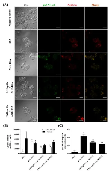
To examine the effect of AGEs on NF-ĸB p65 ex-pression in glomerular cells, we pre-incubated glomeruli with pAb or mAb before AGE-BSA exposure. As shown in Figure 1A-B, AGEs signi-ficantly increased NF-ĸB p65 expression in cultured glomeruli. However, AGEs-induced over-expression of NF-ĸB p65 was significantly inhibited by preincubation of AGE-Ab. AGE-pAb significantly decreased NF-ĸB p65 expression better than AGE-mAb. Since nephrin expression has been associated with podocyte survival, we studied the effect of AGE-Ab on nephrin expres-sion. The presence of AGE-BSA significantly reduced nephrin expression, thus increased NF-ĸB/nephrin ex-pression ratio (Figure 1C). Both AGE-pAb and CmL-mAb significantly reduced NF-ĸB/nephrin expression ratio compared with AGE-BSA-treated group. These findings indicated that AGE-Ab down-regulated AGEs-induced NF-ĸB p65 over-expression.
AGEs are believed to play a major role in the de-velopment of DN. Interaction of AGE-RAGE acti-vates a series of intracellular signaling pathway, inclu-ding NF-ĸB transcription factor which initiates and stimu-lates the production of pro-inflammatory mole-cules that contribute to the pathogenesis of DN [3]. Our study showed that AGE-BSA increased glome-rular NF-ĸB p65 expression. This finding supported by Bier-haus et al. [21] that binding AGEs to RAGE results in sustained activation of NF-ĸB as the result of increased levels of de novo synthesized NF-ĸB p65. Sustained ac-tivation NF-ĸB is mediated by initial degradation of IĸB protein followed by new synthesis of NF-ĸB p65 mRNA and protein in the presence of newly synthe-sized IĸBα and IĸBb [21]. Research conducted by Peng
et al. [22] showed that AGEs signal via RAGE gener-ates a signal-specific post-translational modification, or a “barcode” to NF-κB that mediates a specific gene
ex-pression pattern. AGE-RAGE signaling results in NF-ĸB activation via phosphorylation of NF-NF-ĸB p65 sub-unit at T254, S311, S536 residues [22]. AGEs also in-duces NF-ĸB activation mediated by suppression of sir-tuin 1 (SIRT1) expression in podo-cytes, which leads acetylation of NF-ĸB p65 subunit [23].
Unlike many other receptors, the activation of RAGE by AGEs positively upregulates RAGE expres-sion, resulting in sustained RAGE signaling through a NF-ĸB-dependent mechanism [24,25]. Moreover, acti-vation of NF-ĸB also upregulates AGEs formation via
JTLS | J. Trop. Life. Science 177 Volume 6 | Number 3 | September | 2016
RESULTS AND DISCUSSION
Figure 1. AGEs antibody downregulated glomerular NF-ĸB p65 expression induced by AGEs. A. Representative confocal microscopic images of NF-ĸB p65 (green) and nephrin (red) fluorescence in the glomeruli. BSA was chosen as normal control while AGE-BSA as positive control. The primary antibody was replaced by 2% BSA in PBS for negative control. Magnification 400 ×. Scale bars = 50 µm. B. Bar graphs show the average optical density of NF-ĸB p65 and nephrin per
JTLS | J. Trop. Life. Science 177 Volume 6 | Number 3 | September | 2016
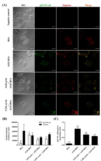
Figure 2. AGEs antibody ameliorated glomerular cells apoptosis induced by AGEs. A. Images show glomeruli in either normal condition or treated with AGE-BSA for 48 hours in the absence or presence of AGE-Ab. Apoptotic cells were stained with FITC-conjugated Annexin V (shown in green) due to phosphatidylserine externalization and nephrin (shown in red). Magnification 400 ×. Scale bars = 50 µm. B. Optical density of annexin V and nephrin was calculated using ImageJ. C. Expression ratio of annexin V/nephrin. Data were expressed as mean ± SD. AGE = advanced glycation end product; AGE-pAb = polyclonal anti-AGEs antibody; BSA = bovine serum albumin; CmL-mAb = monoclonal anti-N-carboxymethyl-lysine antibody. *p < 0.05, vs BSA; **p < 0.05, vs AGE-BSA.
suppressing the expression of glyoxalase which inacti-vates the AGE precursor methylglyoxal [25]. RAGE ac-tivation in mesangial cells increases angiotensin-II (Ang-II) production then activates trans-forming growth factor-b–Smad signaling that induces mesan-gial cell hypertrophy [26]. In addition, Ang-II induces podocyte injury and nephrin inactivation (dephospho-rylation) through caveolin-1-dependent mechanism and C-terminal-Src kinase- C-terminal-binding protein-Fyn axis [27,28]. Reduction of nephrin surface expres-sion is also induced by activation of protein kinase C-mediated nephrin endocytosis [29]. Nephrin deficiency activates NF-ĸB and disrupts clustering of membrane raft micro-domains to prevent nephrin-mediated sig-naling, which affects the podo-cytes or other glomeru-lar cells that interact with podo-cytes [30,31].
AGEs antibody ameliorated AGEs-induced apoptosis of glomerular cells
Figure 2A-B illustrate the effect of AGEs and AGE-Ab on apoptosis. AGE-BSA treatment resulted in a sig-nificant increase of glomerular cells apoptosis and de-crease of podocyte nephrin which significantly inhib-ited by pre-incubation of AGE-Ab. Previously study showed that AGE-BSA induces apoptosis by enhancing FOXO4 binding to a forkhead binding ele-ment in the promoter of Bcl2lll and increasing the acetylation of FOXO4 at lysine residues mediated by downregulation of sirtuin 1 (SIRT1) in cultured podocytes and in glomeruli of diabetic patients [32]. Likewise, AGE-Ab significantly reduced apoptosis/ nephrin expression ra-tio compared with AGE-BSA-treated group (Figure 2C). Interestingly, we found that nephrin expression significantly preserved in the group of glomeruli under AGE-mAb-treated group better than AGE-pAb-treated group. These results suggested that AGE-Ab had a pro-tective activity on AGEs-induced glomerular cells apop-tosis and nephrin deple-tion.
The correlation between NF-ĸB p65 expression and apoptosis
A positive correlation was observed between NF-ĸB p65 expression and apoptosis (r = 0.818) as well as be-tween NF-ĸB/nephrin and apoptosis/nephrin ex-pres-sion ratio (r = 0.828). Taken together, these data pro-vided indirect evidence that AGE-Ab-mediated down-regulation of NF-ĸB p65 expression may contri-bute to inhibition of glomerular cells apoptosis. The results of the present study support the protective role of AGE-Ab against glomerular cells apoptosis, NF-ĸB p65 over-expression and nephrin depletion induced by AGEs. In
this study, we found that polyclonal AGEs anti-body protects apoptosis and NF-ĸB p65 overexpression better than monoclonal anti-CmL antibody. We specu-late that AGE-IC formation contributes to these effects. According to Mera et al. [13], we hypothesize that polyclonal AGEs antibody recognizes multiple epitopes on AGE-BSA, thus increases the formation of AGE-IC which induces macrophages-mediated AGE-BSA phagocytosis [13]. However, activated macrophages can downregulate nephrin expression [33]. In this study, monoclonal CmL-specific antibody preserves nephrin expression. CmL is one of the major immuno-gen of AGEs that accumulates in all renal compart-ment of diabetic patient. CmL increases expression of ZEB2 by NF-ĸB activation and results in epithelial-mesenchymal trans-formation of podocyte which is be-lieved to play a vital role in podocyte depletion and the pathogenesis of albuminuria during DN [34]. Our re-sults showing only minor reduction of nephrin expres-sion on pre-incubation of polyclonal AGEs anti-body. The monoclonal anti-CmL antibody obviously exerts pro-tective effect to podocyte with an aim to pre-serve nephrin expression. Previously study reported that both polyclonal and monoclonal CmL anti-body have a significant reactivity to CmL-proteins. Moover, this monoclonal antibody significantly re-acted with AGE-BSA as well as BSA modified by sev-eral aldehydes such as glyoxal [35].
In conclusion, the present data confirm that pre-in-cubation of AGEs antibody inhibits apoptosis and NFĸB p65 overexpression in glomeruli exposed to AGEs. Our study demonstrates for the first time that the antibody against AGEs may prevent AGEs-induced glomerular damage.
The authors gratefully acknowledge the Central Laboratory of Life Sciences and the Laboratory of Bioscience, Brawijaya University, Malang, for providing the laboratory facilities.
1. Guariguata L, Whiting DR, Hambleton I et al (2014) Global estimates of diabetes prevalence for 2013 and projections for 2035. Diabetes Res Clin Pract. 103 (2): 137–149.
2. Ott C, Jacobs K, Haucke E et al (2014) Role of advanced glycation end products in cellular signaling. Redox Biol. 2:411–429.
ACKNOWLEDGMENT
3. Yamagishi SI, Matsui T (2010) Advanced glycation end products, oxidative stress and diabetic nephropathy. Oxid Med Cell Longev. 3 (2): 101–108.
4. Daroux M, Prévost G, Maillard-lefebvre H, Gaxatte C (2010) Advanced glycation end-products : Implications for
diabetic and non-diabetic nephropathies. Diabetes Metab. 36: 1–10.
5. Miyata T, Dan T (2008) Inhibition of advanced glycation end products (AGEs): An implicit goal in clinical medicine for the treatment of diabetic nephropathy ?
Diabetes Res Clin Pract. 825: 25–29.
6. Pashikanti S, Alba DR De, Boissonneault GA, Cervantes-laurean D (2010) Free Radical Biology & Medicine Rutin metabolites : Novel inhibitors of nonoxidative advanced
glycation end products. Free Radic Biol Med. 48 (5): 656– 663.
7. Joglekar MM, Panaskar SN, Arvindekar AU (2013) Inhibition of advanced glycation end product formation by cymene – A common food constituent. J Funct Foods. 6: 107–115.
8. Losso JN, Bawadi HA, Chintalapati M (2011) Inhibition of the formation of advanced glycation end products by thymoquinone. Food Chem. 128 (1): 55–61.
9. Sang H, Gu J, Yuan J, Zhang M (2014) The protective effect of smilax glabra extract on advanced glycation end products-induced endothelial dysfunction in HUVECs via RAGE-ERK1/2-NF- B pathway. J Ethnopharmacol.κ
155(1):785–795.
10. Feng L, Zhu M, Zhang M et al (2013) Protection of glycyrrhizic acid against AGEs-induced endothelial dysfunction through inhibiting RAGE/NF-κB pathway
activation in human umbilical vein endothelial cells. J Ethnopharmacol. 148 (1): 27–36.
11. Reddy VP, Beyaz A (2006) Inhibitors of the Maillard reaction and AGE breakers as therapeutics for multiple diseases. Drug Discov Today. 11: 646–654.
12. Mashitah MW, Azizah N, Samsu N et al (2015) Immunization of AGE-modified albumin inhibits diabetic nephropathy progression in diabetic mice. Diabetes, Metab Syndr Obes Targets Ther. 8: 347–355.
13. Mera K, Nagai R, Takeo K et al (2011) An autoantibody against N-(carboxyethyl) lysine (CEL): Possible involvement in the removal of CEL-modified proteins by macrophages. Biochem Biophys Res Commun. 407 (2): 420–425.
14. Turk Z, Ljubic S, Turk N, Bojan B (2001) Detection of autoantibodies against advanced glycation endproducts and AGE-immune complexes in serum of patients with diabetes mellitus. Clin Chim Acta. 303: 105–115. 15. Baydanoff S, Konova E, Ivanova (1996) Determination of
anti-AGE antibodies in human serum. Glycoconj J.
13:335–339.
16. Katsuya K, Yaoita E, Yoshida Y et al (2006) An improved method for primary culture of rat podocytes. Int Soc Nephrol. 69:2101–2106.
17. Takemoto M, Asker N, Gerhardt H et al (2002) Technical Advance A New Method for Large Scale Isolation of Kidney Glomeruli from Mice. Am J Pathol. 161 (3): 799– 805.
18. Liu X, Fan Q, Yang G et al (2013) Isolating glomeruli from mice : A practical approach for beginners. Exp Ther
Med. 5: 1322–1326.
19. Mallipattu SK, Liu R, Zheng F et al (2012) Krüppel-like factor 15 (KLF15) is a key regulator of podocyte differentiation. J Biol Chem. 287(23):19122–19135. 20. Müller-Krebs S, Kihm LP, Madhusudhan T et al (2012)
Human RAGE antibody protects against AGE-mediated podocyte dysfunction. Nephrol Dial Transplant. 27 (8): 3129–3136.
21. Bierhaus A, Schiekofer S, Schwaninger M et al (2001) Diabetes-associated sustained activation of the transcription factor nuclear factor-kappaB. Diabetes. 50(12):2792–2808.
22. Peng Y, Kim J, Park H et al (2016) AGE-RAGE signal generates a specific NF- κB RelA “ barcode ” that directs
collagen I expression. Nat Publ Gr. 1–10.
23. Liu R, Zhong Y, Li X et al (2014) Role of transcription factor acetylation in diabetic kidney disease. Diabetes. 63 (7): 2440–2453.
24. Fritz G (2011) RAGE: a single receptor fits multiple ligands. Trends Biochem Sci. 36 (12): 625–632.
25. Yao D, Brownlee M (2010) Hyperglycemia-induced reactive oxygen species increase expression of the receptor for advanced glycation end products (RAGE) and RAGE ligands. Diabetes. 59: 249–255.
26. Fukami K, Ueda S, Yamagishi S et al (2004) AGEs activate mesangial TGF-beta-Smad signaling via an angiotensin II type I receptor interaction. Kidney Int. 66 (6): 2137–2147.
27. Ren Z, Liang W, Chen C et al (2012) Angiotensin II induces nephrin dephosphorylation and podocyte injury: role of caveolin-1. Cell Signal. 24 (2): 443–450.
28. Yu L, Lin Q, Feng et al (2013) Inhibition of nephrin activation by c-mip through Csk–Cbp–Fyn axis plays a critical role in Angiotensin II-induced podocyte damage. Cell Signal. 25 (3): 581-588.
29. Tossidou I, Teng B, Menne J et al (2010) Podocytic PKC-alpha is regulated in murine and human diabetes and mediates nephrin endocytosis. PLoS One. 5 (4): e10185. 30. Li X, Chuang PY, D’Agati VD et al (2015) Nephrin
Preserves Podocyte Viability and Glomerular Structure and Function in Adult Kidneys. J Am Soc Nephrol. 1–17.
31. Hussain S, Romio L, Saleem M et al (2009) Nephrin Deficiency Activates NF- B and Promotes Glomerularκ
Injury. J Am Soc Nephrol. 20 (8): 1733–43.
32. Chuang PY, Dai Y, Liu R et al (2010) Alteration of forkhead box O (foxo4) acetylation mediates apoptosis of podocytes in diabetes mellitus. PLoS One. 6 (8): e23566. 33. Ikezumi Y, Suzuki T, Karasawa T et al (2008) Activated
macrophages down-regulate podocyte nephrin and podocin expression via stress-activated protein kinases. Biochem Biophys Res Commun. 376 (4): 706-711. 34. umar PA, Welsh GI, Raghu G et al (2016) Carboxymethyl
lysine induces EMT in podocytes through transcription factor ZEB2: Implications for podocyte depletion and proteinuria in diabetes mellitus. Arch Biochem Biophys. 590: 10–19.
35. Koito W, Araki T, Horiuchi S, Nagai R (2004) Conventional Antibody against N -(Carboxymethyl)ε
Lysine (CmL) Shows Cross-Reaction to N -ε