The Roles of Polyvinyl Alcohol (PVA) as the Capping Agent
on the Polyol Method for Synthesizing Silver Nanowires
Junaidi
1,a, Kuwat Triyana
2,3,b, Edi Suharyadi
2,3, Harsojo
2,3and Linda Yongling Wu
41
Departement of Physics, Universitas Lampung, Bandar Lampung, Indonesia
2
Departement of Physics, Universitas Gadjah Mada, Yogyakarta, 55281 Indonesia
3
Nanomaterials Research Group, Universitas Gadjah Mada, Yogyakarta, 55281 Indonesia
4
Singapore Institute of Manufacturing Technology, 71 Nanyang Drive, Singapore
Email: [email protected]; [email protected] (corresponding author)
Keywords: silver nanowires, silver nitrate, polyvinyl alcohol, capping agent, transparent electrode
Abstract. We report our investigation of roles of polyvinyl alcohol (PVA) as a high-performance capping agent in synthesizing silver nanowires (AgNWs) using polyol method. For this purpose, we varied the concentration of silver nitrate (AgNO3), from 0.3 M to 1.0 M, and molar ratios of
[PVA:AgNO3] from 2 to 6. The UV-vis spectra show the AgNWs growth optimally at a molar ratio
of 4.5 with the absorbance peaks of 378 nm and 380 nm. Meanwhile, from XRD patterns, it was found that the crystal structure of the AgNWs can be identified as a face-centered cubic (fcc) with a lattice constant according to the spacing distance between the {111} planes of 4.087 Å. Finally, scanning electron microscopy (SEM) and transmission electron microscopy TEM images show the
diameter and length of the AgNRs are 150 to 230 nm and 50 to 120 µm, respectively. These results show that the AgNWs synthesized using PVA having a long size.
Introduction
Metal nanowires and nanorods have attracted much attention due to a high potential as a transparent conductive material for producing solar cells, touch screens, and electronic components [1,2]. Indium tin oxide (ITO) has long been used as a transparent conductive electrode for the optoelectronic applications because of its high conductivity and transparency. However, the intrinsic brittleness and low abundance are among the weak points of ITO [3–5]. Other alternative promising materials for substitution of ITO have been synthesized including copper nanowires (CuNWs), silver nanorods (AgNRs), and silver nanowires (AgNWs).
Some methods have been widely used to synthesize AgNWs and AgNRs including solvothermal method [6,7,8], microwave irradiation [9], and polyol method [10–13] including its modification [14]. Up to present, most AgNWs were synthesized using polyol method with a capping agent of polyvinyl pyrrolidone (PVP) [15,16]. Compared to the other methods, the polyol is a simple in process, low temperature, low cost, and high yield. For synthesizing AgNWs, typically the silver nitrate (AgNO3) is reduced by ethylene glycol (EG) to form neutral nanosilver and capped by a
water-soluble polymer of polyvinyl pyrrolidone (PVP) as a capping agent and a stabilizer. By using this method, it has been reported that the multifaceted silver nanoparticle in {111} plane direction grew due to lower free energy compared to {200}. For controlling this condition, the growing of the diameter of nanowires can be made in tens nanometer [11,14,17].
For mass production, it is necessary to synthesize AgNWs by using an inexpensive capping agent materials. For this purpose, polyvinyl alcohol (PVA) may be the best alternative materials as a capping agent for synthesizing AgNWs. PVA is much cheaper and higher mechanical properties compared to PVP [18–20]. PVA is a polymer composed of monomer N-vinyl alcohol (CH2
-CH-OH) that easily soluble in water and alcohol. Many advantages of PVA, such as non-toxicity, a biocompatible, high mechanical strength, low membrane permeability, high dielectric constant, and
the ability to form a good film. The electrical conductivity of PVA reaches of 9.73 x 10⁻⁹ S.cm-1 at a temperature of 303 K. Non-toxic properties of PVA make these polymers are safe to use on a larger scale [21].
This paper reports our investigation on employing the cheaper water-soluble polymer of PVA for synthesizing AgNWs using polyol method with the focus on the effect of concentration and molar ratio of [PVA:AgNO3]. In this paper, we use the lightweight molecular of PVA instead of PVP to
improve the performance of the resulted nanowires. The temperature optimum when using PVA for synthesizing AgNWs by a polyol method is about 140 C [22]. By varying the molar ratio of [PVA:AgNO3] on the synthesis of AgNWs, it may be beneficial to understand the physical
mechanism of AgNWs formation and furthermore for large-scale production in the future.
Materials and Methods
The materials used for synthesizing of AgNWs through polyol method included silver nitrate (AgNO3, 99%, Merck), polyvinyl alcohol (PVA, Sigma-Aldrich, Mw. 31.000-50.000 g/mol),
ethylene glycol (EG, 99%, Merck), and ethanol (EtOH, 98%, Merck). Using the same method reported previously [22], two steps of the polyol method must be considered as follows. Firstly, a 20 mL of EG was heated in an Erlenmeyer flask at 150 °C and stirred at 350 rpm for 20 minutes. Secondly, a 10 mL of AgNO3/EG solution and 10 ml of PVA/EG solution were injected into EG
solution drop by drop at 0.5 cc/min. The solution was kept for 2 hours at a constant temperature of 150 C by the stirring process. The solution containing produced of AgNWs then cooled naturally to room temperature. It was followed by being separated and washed with ethanol through several times of centrifugation at a speed of 6000 rpm. Finally, the AgNWs were stored in an ethanol solution for further characterization. For the investigation, the concentration of AgNO3 was varied
as 0.3 M, 0.5 M, and 1.0 M. Furthermore, the molar ratio of [PVA:AgNO3] was varied as 2, 3, 4,
4.5, 5, and 6, respectively.
UV-vis spectrometer (Shimadzu, UV-1700) was used to measure the absorption spectrum of AgNWs solution in the wavelength range of 300 to 600 nm. Meanwhile, clusters identification of pure PVA, AgNWs, and AgNPs were observed using Fourier transform infrared spectroscopy (Shimadzu, FTIR-8201 PC). Furthermore, the crystal structure of AgNWs was analyzed using XRD
(Shimadzu R6000) by CuKα (λ = 1.54184 Å) with a scanning 2θ in the range of 30 to 90. The morphology and size of AgNWs were observed using scanning electron microscopy (JEOL, JSM-6510) by accelerating voltage of 10 kV. Furthermore, the structure and electron diffraction patterns were analyzed using transmission electron microscopy (JEOL, JEM-2010) by accelerating voltage of 120 kV.
Results and Discussion
Temperature effect on the formation of AgNWs and AgNRs using polyol method was reported [22]. The process of nanowires formation started as neutral silver nanoparticles (AgNPs) growing into multi-twinned particles (MTPs) seeds and followed by AgNWs [25,26]. In this process, the addition of PVA and EG was to modify and stabilize the MTPs prior to form nanowires. Therefore, the different molar ratio of [PVA:AgNO3] might influence nanoparticles and nanowires structures.
We found that the strongest interaction was detected in AgNWs using FTIR which is due to the hydroxide chain in PVA as shown in Fig. 1. Therefore, we suggest this chain might play a significant role in capping the MTPs seeds before growing into AgNWs. Moreover, we also found all wires have multi-twined edges suggesting that in the addition of PVA with correct concentration and molar ratio. A different molar ratio of [PVA:AgNO3] resulted in changing nanostructures, such
as nanoparticles, nanorods, or nanowires. At a high molar ratio of [PVA:AgNO3], the PVA capped
found that the optimum for the molar ratio of [PVA:AgNO3] was between 4 to 5. Fortunately, this
optimum molar ratio is similar to by using PVP as the capping agent. The AgNWs in this process was formed at a low concentration of Ag.
UV-vis Spectroscopy Analysis. The UV-vis spectra of AgNWs in ethanol solution for various concentrations of AgNO3 were shown in Fig. 1(a). For low concentration, the absorbance peaks due
to the present of AgNWs were observed at the wavelength of 378 nm and 380 nm. From the literature, the absorbance peaks of AgNWs occurred at 350 to 380 nm [23,28]. Next, for a high concentration of AgNO3 (1 M), the absorbance peaks of AgNWs occurred at 410 nm and 450 nm.
When the vefry high concentration of AgNO3, It can be concluded that the process of transforming
MTPs to become nanorods or nanowires. At a high concentration of AgNO3, Ag atoms become
supersaturation. In such circumstances, the Ag atoms are experiencing agglomeration into MTPs seeds by fluctuations [27].
Figure 1. UV-vis spectra of AgNWs or AgNPs in ethanol solution synthesized with variation of (a) silver nitrate concentration, (b) molar ratio of [PVA:AgNO3]; and (c) FTIR spectra of pure PVA,
AgNWs, and AgNPs.
Figure 1(b) shows the UV-vis spectra for variation molar ratio of PVA and AgNO3. The AgNWs
can’t grow for the molar ratio of [PVA:AgNO3] less than 4 and more than 5. The AgNWs only grow
for the molar ratio of [PVA:AgNO3] of 4 to 5. In these conditions, the related the absorbance peaks
occurred at 380 nm for the molar ratio of 4 and at 378 nm for the molar ratio of 4.5. For high concentrations of PVA, AgNPs may grow become AgNWs. Therefore, the molar ratio of [PVA:AgNO3] as a capping agent and as a precursor shows a very significant impact on the shape
and size of Ag nanostructures.
FTIR Spectroscopy Analysis. Figure 1(c) shows the FTIR spectra of three samples, i.e. pure PVA; AgNPs for the molar ratio of [PVA:AgNO3] of 5; and AgNWs for the molar ratio of
[PVA:AgNO3] of 4.5. From pure PVA, the absorption peak occurred at around 1273 cm-1 indicates
a -CO- stretching vibration. Meanwhile, the absorption peaks occurred at around 1635 cm-1 for the carbonyl group (-C=O) stretching vibration and 2924 cm-1 for -CH stretching vibration, respectively. Additionally, there are broad peaks occurred at around 3448, 3749, and 3873 cm-1 indicates the hydroxyl group (OH) stretching vibration. The weak peaks at 2924 and 2862 cm-1 are the peak of CH3 and -H2- stretching vibration, respectively.
FTIR spectra of AgNWs or AgNPs in ethanol solution show the sharp absorption peaks at around 1010 cm-1, which indicates -CO- stretching vibration. Meanwhile, the absorption peaks at around
1651 cm-1 and 2947 cm-1 indicate the existence of stretching vibration of the carbonyl group (-C=O) and –CH, respectively. Moreover, a broad peak at around 3317 cm-1 indicates the hydroxyl group (OH) stretching vibration as the strongest peak. The weak peaks, on the other hand, at 2947 and 2831 cm-1 show the occurrence of CH3 and -H2- stretching vibration, respectively.
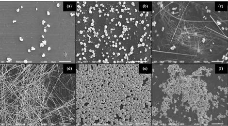
SEM Analysis. The morphology and size of the AgNWs with various concentrations were observed using SEM. Figure 2(a) shows the SEM image of product synthesized using AgNO3 with a
concentration of 0.3 M in which resulted in both nanowires and nanoparticles. The dominated AgNWs product was obtained optimally when the concentration of AgNO3 was 0.5 M (Fig 2(b)).
The average of diameter and length of AgNWs for this condition were ~200 nm and ~100 µm, respectively. Finally, when the concentration of AgNO3 was 0.5 M, the final products were
dominated by agglomerated AgNPs (see Fig 2(c).
Figure 2. SEM images of product synthesized using different concentrations of silver nitrate i.e. (a) 0.3 M, (b) 0.5 M, and (c) 1.0 M.
Figure 3. SEM images of product synthesized using for variation molar ratios of [PVA:AgNO3] at
(a) 2, (b) 3, (c) 4, (d) 4.5, (e) 5, and (f) 6.
Figure 3(a) shows the SEM images of the product synthesized using a low concentration of PVA (1 M) or when the molar ratio of [PVA:AgNO3] was 2. In this experiment, a small amount of MTPs
was produced with the diameter range of 1 to 3 µm. In this condition, AgNO3 caused MTP seeds
broken before being capped by PVA. When the molar ratio of [PVA:AgNO3] was 3 (see Fig. 3(b)),
it was obtained MTPs in larger quantities. The mixed product of AgNWs and AgNPs started to form in large amounts when the molar ratio of [PVA:AgNO3] was 4 (see Fig. 3(c)) with the estimated
diameters and lengths of AgNWs were about 100 to 200 nm and 30 to 60 µm, respectively. The AgNWs were optimally formed when the molar ratio of [PVA:AgNO3] of 4.5 as shown in Fig. 3(d).
The diameter and length of AgNWs are obtained to be 150 to 230 nm and 50 to 120 µm, respectively.
(a) (b) (c)
(d) (e) (f)
SEM images show that the AgNWs synthesized using PVA having a longer size than using PVP. It was because both the bonds between Ag-O and the cap with hydroxyl of PVA became stronger. Therefore, the amount of Ag particles formed the higher concentration of MTPs that subsequently resulted in a higher concentration of nanorods. When the molar ratio of [PVA:AgNO3] was too
high, the AgNWs were very difficult to growth because the concentration of H+ ions from the hydroxyl group of alcohol was also too high. Fig. 3(e) and 3(f) show that MTPs seeds were homogeneously formed with the diameter of 1 to 3 µm. In this condition, only a small amount of AgNWs were formed with the diameter of 300 to 800 nm and length of 5 to 10 µm. When the concentration of capping agent was high, the size of MTPs beceme bigger. It might cause the diameter of AgNWs also became bigger [21].
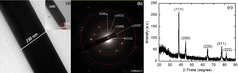
TEM Analysis. Figure 4(a-b) shows the typical TEM image and the pattern of selected area electron diffraction (SAED) recorded individually from AgNWs when the molar ratio [PVA:AgNO3] was 4.5. We found that the diameters of AgNWs were in the range of 220 to
230 nm.
Figure 4. (a-b) TEM images of silver nanowires and (c) XRD pattern of silver nanowires.
From the SAED pattern, all diffractions can be indexed with the face-centered-cubic (fcc). The TEM images show the PVA as capping agent served as a soft template for the formation of anisotropic AgNWs. Meanwhile, the PVA adsorbed on the {100} facets of the Ag crystal, and subsequently, the anisotropic AgNWs grew in the {111} facets direction. The anisotropic growth of AgNWs occurred selectively in {100} and {111} facets. The growth from PVA in {100} facets was passive while that of {111} faces was more active. Therefore, the silver atoms were reduced to be heading towards {111} facets. The interaction between PVA and silver atoms at {100} facets was stronger than that of {111} facets in which during the nucleation of silver nanoparticles and a capping agent (PVA), the {100} facets suppressed the growth orientation of AgNWs [29]. Furthermore, due to higher chemical stability and reactively, the silver atoms always chose to spread to the ends of nanowires. Also, the Ag+ coordinated immediately with the lone pair of the oxygen atom of the carbonyl group of PVA and subsequently growth the AgNWs.
XRD Analysis. The XRD pattern of AgNWs is depicted in Fig. 4(c). The crystal structure of AgNWs produced four diffraction peaks from XRD pattern analysis. All peaks can be indexed to the cubic phase of Ag, which the angle of diffraction 2θ was at 38.14 (111), 44.89 (200), 65.12 (220), 77.07 (311) and 81.11o
(222). The number in the parenthesis indicates the corresponding crystal plane. According to the standard of JCPDS card of 04-0783 from ASTM, the XRD patterns indicate that the AgNWs was crystallized. The crystalline of the AgNWs can be identified as a face-centered cubic (fcc). The AgNWs synthesized in this study also showed a high aspect ratio where the diffraction signal peak of XRD at (111) was larger about 2-fold than those at (200). The calculated lattice constant according to the spacing distance (dhkl) of the {111} planes was 4.087 Å. It is very close to the literature value of 4.086 Å [22,29].
Mechanical strength and low membrane permeability of the PVA resulted in AgNWs with a longer size. It means that AgNWs were not be easily broken when synthesized at high temperature. Furthermore, this condition makes AgNWs with a capping agent of PVA have a very large ratio of
length and diameter (l/d) AgNWs. The l/d from AgNWs affects the haze and conductivity of AgNWs based thin films. The length of AgNWs is still crucial for improving the performance of high transparent materials, and, therefore, some effort is still worth for such an improvement [20,31,32].
Summary
In the polyol process using PVA and EG, the molar ratio of [PVA:AgNO3] is a very important
factor that determines whether the product to become nanoparticles, nanorods, and nanowires of silver. The low molar ratio of [PVA:AgNO3] will prevent the nanoparticles to form AgNWs. The
optimum results may be obtained by managing this ratio. At present, the optimum homogeneous AgNWs of 200 nm in diameter and 100 µm in length with fcc structures.
Acknowledgment
This work was supported by a research grant of “International Research Collaboration and Scientific Publication, Contract No. 1021/UN1-P.III/LT/DIT-LIT/2016” by Ministry of Research, Technology and Higher Education of the Republic of Indonesia.
References
[1] W. Zhang, P. Chen, Q. Gao, Y. Zhang, and Y. Tang, High-concentration preparation of silver nanowires: Restraining in Situ nitric acidic etching by steel-assisted polyol method, Chem. Mater. 20(5) (2008) 1699–1704.
[2] M. R. Johan, N. Azri, K. Aznan, S. T. Yee, I. H. Ho, S. W. Ooi, N. D. Singho, and F. Aplop, Synthesis and growth mechanism of silver nanowires through different mediated agents (CuCl2
and NaCl) polyol process, J. Nanomat. Article ID 105454 (2014).
[3] Y. S. Kim, M. H. Chang, E. J. Lee, D. W. Ihm, and J. Y. Kim, Improved electrical conductivity of PEDOT-based electrode films hybridized with silver nanowires, Synth. Met.195 (2014) 69– 74.
[4] D. S. Hecht, C. Ladous, P. Drzaic, and S. I. D. Fellow, Carbon-nanotube film on plastic as transparent electrode for resistive touch screens, J. SID 17(11) (2009) 941–946.
[5] D. Kim, L. Zhu, D. Jeong, K. Chun, Y. Bang, S. Kim, J. Kim, and S. Oh, Transparent flexible heater based on hybrid of carbon nanotubes and silver nanowires, Carbon 63(2013) 530–536. [6] M. Song, J. Feng, W. Li, Q. Hu, Z. Zhang, B. Liu, and X. Zhao, Synthesis and Characterization
of Silver Nanowires by Solvothermal, Advanced Mater. Res. 66 (2009) 159–162.
[7] T. Tetsumoto, Y. Gotoh, and T. Ishiwatari, Mechanistic studies on the formation of silver nanowires by a hydrothermal method, J. Colloid Interface Sci. 362(2) (2011) 267–273.
[8] C. Xu, Y. Wang, H. Chen, D. Nie, and Y. Liu, Hydrothermal synthesis of silver crystals via a sodium chloride assisted route, Mater. Lett.136 (2014) 175–178.
[9] Y. Tang, W. He, S. Wang, Z. Tao, and L. Cheng, One step synthesis of silver nanowires used in preparation of conductive silver paste, J. Mater. Sci. Mater. Electron. 25(7) (2014) 2929–2933. [10] M. Kang, E. Chung, S. Kim, and S. W. Rhee, “Ag nanowires prepared by a modified polyol
method with 1,4-benzoquinone additives, Bull. Korean Chem. Soc. 35(11) (2014) 3209–3212. [11] A. B. V Kiran Kumar, C. Wan Bae, L. Piao, and S. H. Kim, Silver nanowire based flexible
electrodes with improved properties: High conductivity, transparency, adhesion and low haze, Mater. Res. Bull. 48(8) (2013) 2944–2949.
[12] W. M. Schuette and W. E. Buhro, Polyol synthesis of silver nanowires by heterogeneous nucleation; mechanistic aspects influencing nanowire diameter and length, Chem. Mater. 26 (2014) 6410-6417.
[14] Y. Sun, B. Mayers, T. Herricks, and Y. Xia, Polyol synthesis of uniform silver nanowires: A plausible growth mechanism and the supporting evidence, Nano Lett. 3(7) (2003) 955–960. [15] D. A. Dinh, K. N. Hui, K. S. Hui, P. Kumar, and J. Singh, Silver nanowires: a promising
transparent conducting electrode material for optoelectronic and electronic applications, Rev. Adv. Sci. Eng. 2(4) (2013) 1–22.
[16] K. Azuma, K. Sakajiri, H. Matsumoto, S. Kang, J. Watanabe, and M. Tokita, Facile fabrication of transparent and conductive nanowire networks by wet chemical etching with an electrospun nanofiber mask template, Mater. Lett.115 (2014) 187–189.
[17] T. Araki, J.Jiu, M. Nogi, H. Koga, S. Nagao, T. Sugahara, Low haze transparent electrodes and highly conducting air dried films with ultra-long silver nanowires synthesized by one-step polyol method, Nano Res. 7 (2014) 236–245.
[18] J. Lu, Q. Nguyen, J. Zhou, and Z. Ping, Poly(vinyl alcohol)/Poly(vinyl pyrrolidone)
interpenetrating polymer network : synthesis and pervaporation properties, J. Appl. Polym. Sci. 89 (2003) 2808–2814.
[19] A. Bernal, I. V. O. Kuritka, and P. Saha, Poly (vinyl alcohol)-poly (vinyl pyrrolidone) blends: Preparation and characterization for a prospective medical application, Mathematical Methods and Techniques in Engineering and Environmental Science (2011) 431–434.
[20] N. Rajeswari, S. Selvasekarapandian, S. Karthikeyan, M. Prabu, G. Hirankumar, H. Nithya, and C. Sanjeeviraja, Conductivity and dielectric properties of polyvinyl alcohol-polyvinylpyrrolidone poly blend film using non-aqueous medium, J. Non. Cryst. Solids. 357(22–23) (2011) 3751–3756.
[21] F. F. Hatta, M. Z. A. Yahya, A. M. M. Ali, R. H. Y. Subban, M. K. Harun, and A. A. Mohamad, Electrical conductivity studies on PVA/PVP-KOH alkaline solid polymer blend electrolyte, Ionics 11(5–6) (2005) 418–422.
[22] Junaidi, K. Triyana, H. Sosiati, E. Suharyadi and Harsojo, Effect of temperature on silver nanorods synthesized by polyol method, Advanced Mater. Res.1123 (2015) 256–259.
[23] Q. N. Luu, J. M. Doorn, M. T. Berry, C. Jiang, C. Lin, and P. S. May, Preparation and optical properties of silver nanowires and silver-nanowire thin films, J. Colloid Interface Sci. 356(1) (2011) 151–158.
[24] N. V. Nghia, N. N. K. Truong, N. M. Thong, and N. P. Hung, Synthesis of Nanowire-Shaped Silver by Polyol Process of Sodium Chloride, Int. J. Mater. Chem. 2(2) (2012) 75–78.
[25] A. Taguchi, S. Fujii, T. Ichimura, P. Verma, Y. Inouye, and S. Kawata, Oxygen-assisted shape control in polyol synthesis of silver nanocrystals, Chem. Phys. Lett. 462(1–3) (2008) 92–95. [26] X. Tang, M. Tsuji, P. Jiang, M. Nishio, S.-M. Jang, and S.-H. Yoon, Rapid and high-yield
synthesis of silver nanowires using air-assisted polyol method with chloride ions, Colloids Surfaces A Physicochem. Eng. Asp. 338(1–3) (2009) 33–39.
[27] Y.-H. Chang, Y.-C. Lu, and K.-S. Chou, Diameter Control of Silver Nanowires by Chloride Ions and Its Application as Transparent Conductive Coating, Chem. Lett. 40(12) (2011) 1352– 1353.
[28] J. Y. Lin, Y. L. Hsueh, and J. J. Huang, The concentration effect of capping agent for synthesis of silver nanowire by using the polyol method, J. Solid State Chem. 214 (2014) 2–6.
[29] H. Mao, J. Feng, X. Ma, C. Wu, and X. Zhao, One-dimensional silver nanowires synthesized by self-seeding polyol process, J. Nanoparticle Res. 14(6) (2012).
[30] L. Sun, L. Wang, Y. Song, C. Guo, Y. Sun, C. Peng, Z. Liu, and Z. Li, Aggregation-based growth of silver nanowires at room temperature, Appl. Surf. Sci. 254(9) (2008) 2581–2587. [31] O. W. Guirguis and M. T. H. Moselhey, Thermal and structural studies of poly (vinyl alcohol)
and hydroxypropyl cellulose blends, Nat. Sci. 4(1) (2012) 57–67.
[32] C. V. Subba Reddy, X. Han, Q.-Y. Zhu, L.-Q. Mai, and W. Chen, Dielectric spectroscopy studies on (PVP+PVA) polyblend film, Microelectron. Eng. 83(2) (2006) 281–285.
![Figure 1. UV-vis spectra of AgNWs or AgNPs in ethanol solution synthesized with variation of (a) silver nitrate concentration, (b) molar ratio of [PVA:AgNO3]; and (c) FTIR spectra of pure PVA, AgNWs, and AgNPs](https://thumb-ap.123doks.com/thumbv2/123dok/3993142.1936562/3.595.66.527.247.373/figure-ethanol-solution-synthesized-variation-nitrate-concentration-spectra.webp)