Klotho: An antiaging protein involved in mineral
and vitamin D metabolism
P Uren˜a Torres
1,2,3,4, D Prie´
2,3, V Molina-Ble´try
1, L Beck
3, C Silve
4and G Friedlander
2,31Service de Ne´phrologie et Dialyse, Clinique de l’Orangerie, Aubervilliers, France;2Service de Physiologie-Explorations Fonctionnelles,
Hoˆpital Necker, Paris, France;3INSERM Unit 811, Hoˆpital Necker-Enfants Malades, Paris, France and4INSERM Unit 773, Faculte´ de
Me´decine Xavier Bichat, Paris, France
Klotho gene mutation leads to a syndrome strangely resembling chronic kidney disease patients undergoing dialysis with multiple accelerated age-related disorders, including hypoactivity, sterility, skin thinning, muscle atrophy, osteoporosis, vascular calcifications, soft-tissue calcifications, defective hearing, thymus atrophy, pulmonary emphysema, ataxia, and abnormalities of the pituitary gland, as well as hypoglycemia, hyperphosphatemia, and
paradoxically high-plasma calcitriol levels. Conversely, mice overexpressing klotho show an extended existence and a slow aging process through a mechanism that may involve the induction of a state of insulin and oxidant stress resistance. Two molecules are produced by the klotho gene, a membrane bound form and a circulating form. However, their precise biological roles and molecular functions have been only partly deciphered. Klotho can act as a circulating factor or hormone, which binds to a not yet identified high-affinity receptor and inhibits the intracellular insulin/ insulin-like growth factor-1 (IGF-1) signaling cascade; klotho can function as a novelb-glucuronidase, which deglycosylates steroidb-glucuronides and the calcium channel transient receptor potential vallinoid-5 (TRPV5); as a cofactor essential for the stimulation of fibroblast growth factor (FGF) receptor by FGF23. The two last functions have propelled klotho to the group of key factors regulating mineral and vitamin D metabolism, and have also stimulated the interest of the nephrology community. The purpose of this review is to provide a nephrology-oriented overview of klotho and its potential implications in normal and altered renal function states.
Kidney International(2007)71,730–737. doi:10.1038/sj.ki.5002163; published online 28 February 2007
KEYWORDS: aging; calcium; phosphorus; vascular calcifications; parathyroid hormone; renal osteodystrophy
In Greek mythology, the duration of life is controlled by the three daughters of Zeus and Themis: Klotho (Clotho) who combs and spins the thread of life, Lachesis who determines the length of life by measuring the threads length, and Athropos who cuts the string causing a life to end. In science, a Japanese group, which was exploring aging mechanisms, conferred the name of Klotho to a gene that they fortuitously discovered in 1997.1
Indeed, the klotho gene was disrupted or mutated in its 50-flanking promoter region by the random insertion of an exogenously introduced nonfunctional gene, the rabbit Na/H exchanger under the control of the human elongation factor promoter.1The coding region of this mutated klotho gene was still preserved, but its expression was markedly reduced generating a mouse strain with a strong hypomorphic allele. Homozygous mice, for this hypomorphic allele, showed shortened lifespan and a syndrome strangely resembling chronic kidney disease patients undergoing dialysis with multiple accelerated age-related disorders, including hypoactivity, sterility, skin thinning, muscle atrophy, osteoporosis, vascular calcifications, soft tissue calcifications, defective hearing, thymus atrophy, pulmonary emphysema, ataxia, and abnormalities of the pituitary gland, as well as hypoglycemia, hyperphosphate-mia, and paradoxically high-plasma calcitriol levels.
Conversely, mice overexpressing klotho showed an extended existence and a slow aging process through a mechanism that may involve the induction of a state of insulin and oxidant stress resistance.2 Moreover, several single-nucleotide polymorphisms in the human klotho gene have been found to be associated with lifespan, osteoporosis, stroke, and coronary artery diseases. All these observations support the suggestion that klotho plays an important role in aging and senescence-related disorders.
Nephrologists have recently been extremely interested by the possible physiological functions of klotho because of its predominant renal expression, its colocalization together with the epithelial calcium channel transient receptor potential vallinoid-5 (TRPV5) in kidney distal tubular cells,3 and its interaction with fibroblast growth factor-23 (FGF23).4 The purpose of this review is to provide a nephrology-oriented overview of klotho and its potential implications in normal and altered renal function states.
Received 14 November 2006; revised 26 December 2006; accepted 2 January 2007; published online 28 February 2007
Disclosure statements: P Uren˜a Torres reports receiving consulting and lectures fees from Abbott, Amgen, Astra Zeneca, and Shire
MOLECULAR CHARACTERISTICS OF KLOTHO: GENE, mRNA, AND PROTEIN
The human klotho gene is a 5-exon gene located on chromosome 13q12 within a region longer than 50 kb. Its promoter region lacks a TATA-box consensus sequence and contains four potential binding sites for SP1.5Two transcripts arise from this single gene; one full-length transcript of 5.2 kb encoding a 1012-aminoacid (130 kDa), single-pass, mem-brane protein. This memmem-brane form can be released into the circulation after losing its short transmembrane domain and slightly lowering its molecular weight. Moreover, it is possible that the secreted form is ultimately metabolized to a smaller size protein of 65–70 kDa. The other transcript derived from an alternative mRNA splicing, encodes the N-terminal half of klotho, a protein of 549 amino acids with a molecular weight of approximately 65–70 kDa.5–7
On the basis of their predicted structures, both proteins belong to the b-glycosidase family. The expression of the secreted form predominates over that of the membrane form. The human protein shows 86% of amino-acid identity with the mouse klotho protein. The extracellular domain of klotho is composed of two internal repeats (KL1, KL2), each one of approximately 450 amino acids long with a similarity of 21% to each other. These two domains form a butterfly-shaped molecule on the surface of the cellular membrane. They share 20–40% sequence identity with the b-glucosidase of both bacteria and plants and with mammalian lactase glycosyl-ceramidase.6,7 Another speciality of klotho proteins is that the secreted form and the membrane form develop oligomeric complexes, suggesting a post-translation klotho processing and possible regulatory mechanisms for klotho secretionin vivo.
The tissue distribution of klotho mRNAs expression reveals that it is expressed, in descending order, in kidney, brain, reproductive organs, pituitary gland, parathyroid glands, urinary bladder, skeletal muscle, placenta, thyroid gland, and colon.1 In the kidney, klotho mRNAs and proteins are localized in the distal tubular cells. In these cells, klotho is diffusely expressed in the cytoplasm and not at the apical side.8 It is colocalized with other proteins involved in tubular calcium reabsorption such as the epithelial calcium channel TRPV5 and calbindin 28 K,3,8 suggesting that klotho is implicated in renal calcium homeostasis (Figures 1 and 2).
In brain, klotho is expressed at the apical plasma mem-brane of ependymal cells in the choroids plexus of both the lateral ventricles and the third ventricle. Klotho protein is also expressed in the stria vascularis and spiral ligament of the inner ear probably serving as a modulator of ion transport as in the renal distal tubular cells.9 In the heart, klotho expression is recognized exclusively in the sinoatrial node region, where it plays an essential role in sinoatrial node function as a dependable pacemaker under conditions of stress.10 In reproductive organs, in the testis, klotho is expressed in the inner layers of seminiferous tubules containing elongating spermatids or mature germ cells. It is absent in spermatogonia, primary spermatocytes, rounds
spermatids, and Sertoli cells. In the ovary, klotho is expressed exclusively in the most mature follicles; it is absent or weakly expressed in primary and secondary follicles.8
MODE OF ACTION OF KLOTHO
Nine years after its identification, the exact biological role and molecular function of klotho have been only partly deciphered. For instance, first, klotho can act as a glyco-sidase because of its high similarity with other members of the glycosidase family. However, this has been questioned because klotho lacks glutamic acid residues that are responsible for the catalytic activity of this enzyme family. Nonetheless, recent results obtained in in vitro experiments
support the glycosidase activity of klotho; when a purified chimeric klotho-human IgG1 Fc protein is incubated in the presence of a series of 4-methylumbelliferylb-glycosides serving as putative substrates, an enzymatic activity of klotho is demonstrated only with the 4-methylumbelliferyl b-D -glucuronide.6 This enzymatic activity of klotho-human IgG1 Fc protein is reduced by the addition of specific inhibitors ofb-glucuronidase. Furthermore, naturally occur-ring b-glucuronides such as b-estradiol 3-b-D-glucuronide, strone 3-b-D-glucuronide, and estriol 3-b-D-glucuronide are also hydrolyzed by klotho-human IgG1 Fc protein.6 In addition, klotho hydrolyses sugar residues on TRPV5, avoiding its retrieval from the cell surface. Interestingly, this stimulatory effect of klotho can be entirely mimicked by a purified bovine b-glucuronidase and blocked by the D-saccharic acid 1,4-lactone, a klotho inhibitor.3Collectively, these data strongly suggest that klotho functions as a novel b-glucuronidase, and steroid b-glucuronides and calcium channels TRPV5 are potential candidates for klotho actions. Second, klotho can act as a circulating factor and this is supported by the fact that klotho protein, probably resul-ting from the secretion of the membrane form, is detectable in urine, serum, and cerebrospinal fluid.3This protein binds to a high-affinity but yet not identified cell-surface klotho receptor and activates the protein kinase C (PKC) pathway in kidney and testicular cells; klotho also stimulates cAMP pathway in several cell types.11The activation of this receptor by klotho leads to the suppression of tyrosine phosphory-lation of insulin/insulin-like growth factor (IGF-1) receptors and insulin receptor substrates, association of insulin recep-tor substrates with phosphatidylinositol 3-kinase, and serine phosphorylation of Akt/PKB.2 Therefore, klotho protein is a circulating factor that inhibits the intracellular insulin/IGF-1 signaling cascade. This activity probably contributes to the antiaging effects of klotho, because inhibition of insulin-like signaling is an evolutionarily conserved mechanism for extending lifespan.2,12
phosphorylation of FGF receptor substrate and extracellular signal-regulated kinase in a variety of cell types.4 The interaction between klotho, FGFR, and FGF23 is a new type of receptor modulation, which has been further illustrated in a recent report.13Indeed, klotho binds to FGFR1(IIIc) and its concerted action constitutes the FGF23-specific receptor; without klotho the function of FGF23 is literally abolished.13
REGULATION OF KLOTHO
The regulation of Klotho by several factors and in different organs is depicted in Table 1. In the kidney, klotho protein level is markedly increased in estrogen deficiency as in the aromatase-deficient mice model and is decreased after estra-diol therapy (Oz et al., abstract No. 1013, ASBMR, 2006).
Calcium, phosphate, and 1.25(OH)2D3 alone have minimal effect on klotho expression; however, combined calcium/ phosphate augmentation causes a seven-fold increase in klotho mRNA expression in distal tubular cells (Yu et al.,
abstract No. SU 423, ASBMR, 2006). This increase in klotho expression is associated with increased FGF23 activity, suggesting that klotho exerts a critical function in the modu-lation of FGF23 activity in situations of high-serum calcium/ phosphate. On the other hand, mice overexpressing FGF23, under the control of b1-type I collagen gene promoter, exhibit an increased renal klotho expression (Marsellet al.,
abstract No. SU 421, ASBMR, 2006). Klotho mRNA expres-sion in the kidney is reduced in several animal models of human diseases characterized by a sustained circulatory and/or oxidant stress, including spontaneously hypertensive rat, deoxycorticosterone acetate–salt hypertensive rat, 5/6 nephrectomized rat,14,15 noninsulin-dependent diabetes mellitus rat (the Otsuka long-Evans Tokushima fatty rat), ischemia–reperfusion injury models,16 and rat with acute myocardial infarction.17,18Klotho protein is reduced in renal cell carcinoma tissues compared with those in nontumor regions.19 Angiotensin II downregulates renal klotho gene
Bone Intestine
Parathyroid glands
2
PTH
Serum phosphate Low phosphate diet
NPT2b Klotho
NPT2c NPT2a
NPT2c NPT2a
FGF23
1,25OH2D3 1
Kidney FGFR1(IIIc)/
FGF23 3
TRPV5 Demonstrated mechanisms
Possible mechanisms
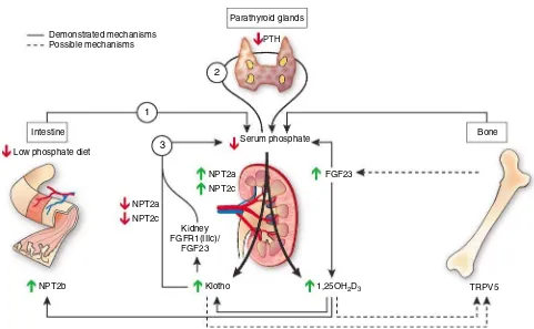
Figure 1|Phosphate regulation by klotho: Hypotheses. The first arrow starts from the intestine, where a reduced dietary phosphate intake diminishes serum phosphate concentration and leads to a decrease in PTH secretion, which physiologically reduces urinary phosphate excretion. In addition, to save phosphate, the renal action of FGF23 will decrease facilitating tubular phosphate reabsorption by the stimulation of sodium-dependent phosphate cotransporters (NPT2a and NPT2c). It will also facilitate the synthesis of 1,25(OH)2D3in spite of low PTH levels.
The increase in calcitriol levels stimulates sodium-dependent phosphate cotransporter type IIb expression and intestinal phosphate absorption. Then, to counteract the activation of these three phosphate-saving mechanisms and to avoid hyperphosphatemia, the renal synthesis of klotho is increased. This increase in renal klotho will facilitate the phosphaturic action of FGF23. Klotho binds to FGFR1(IIIc) and forms the specific FGF23 receptor. Furthermore, klotho negatively regulates the synthesis of 1,25(OH)2D3by enabling FGF23 binding to its receptor
and thereby its inhibitory effect on 1-b-hydroxylase activity. At the bone level, klotho could stimulate bone resorption and phosphate release by acting on TRPV5, which is a recently identified osteoclast function modulator. The increased levels of 1,25(OH)2D3could also stimulate
expression by an AT1 receptor-dependent pathway, but a pressor-independent mechanism.20,21 Klotho expression is also reduced in the kidney by oxidant stress injury by H2O2
22
and in chronic renal diseases.14With aging, klotho expression decreases in heart and liver.23,24
Klotho expression is modified during adipocyte differen-tiation. Klotho is expressed in 3T3-L1 preadipocyte cell line, and adipose differentiation is accompanied with a gradual increase in the expression of klotho. In the same cells, triiodothyronine increased significantly the expression of klotho. Peroxisome proliferator-activated receptor-b(PPRAb) agonists also increase klotho expression in adipocytes.25
ANTIAGING EFFECTS OF KLOTHO
Klotho-deficient mice exhibit a syndrome resembling human premature aging, with multiple pathological phenotypes in tissues including reproductive organs. This phenotype can be rescued by exogenous expression of klotho cDNA,1 and interestingly, klotho gene overexpression extends lifespan by 20–30%.2Likewise, several wild-type mouse strains in which the klotho protein have suffered four amino-acid
substitu-tions, which results in higher levels of klotho, exhibit longer lifespan, reduced atherosclerosis risk factors, and better hearing than other mouse strains.26
Klotho extends lifespan by inhibiting the aging process through a surprising mechanism, that is the induction of insulin resistance.2 Indeed, by inducing the inhibition of insulin/IGF-1 signaling klotho also increases the resistance to oxidative stress at the cellular and subcellular level in mammals. Furthermore, klotho protein activates the forkhead transcription factors (FoxO) that are negatively regulated by insulin/IGF-1 signaling, thereby inducing expression of manganese superoxide dismutase. This in turn facilitates removal of reactive oxygen species and confers oxidative stress resistance.12,27
Klotho could also hence extend lifespan by protecting the cardiovascular system through endothelium-derived NO production. Many experimental data support this hypothesis: klotho reduces H2O2-induced apoptosis and cellular senes-cence in vascular cells,28 the impaired endothelium-dependent vasodilation of the aorta and arterioles of hetero-zygous klotho-deficient mice are restored by parabiosis with
Bone Intestine
Parathyroid glands
2
PTH
Serum calcium Low calcium diet
TRPV6 Klotho 1,25OH2D3
1
TRPV5 3
TRPV5 Demonstrated mechanisms
Possible mechanisms
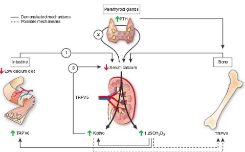
Figure 2|Calcium regulation by klotho: Hypotheses. The first arrow starts from the intestine, where a reduced dietary calcium intake diminishes serum calcium concentration and leads to an increase in PTH secretion. PTH will activate three mechanisms to normalize serum calcium: it will stimulate urinary calcium reabsorption, renal 1,25(OH)2D3synthesis, and bone remodeling. The renal production of klotho would
be stimulated to increase the expression and function of the epithelial calcium channel TRPV5 and therefore tubular calcium reabsorption. Indeed, klotho stimulates calcium reabsorption in the distal convoluted tubule by deglycosylating and stabilizing the epithelial calcium channel TRPV5 on the surface of cellular membrane. Klotho could also favor intestinal calcium absorption by facilitating the expression and function of TRPV6. At the bone level, klotho could stimulate bone resorption and calcium release by acting on TRPV5, which is a recently identified osteoclast function modulator. Besides stimulating intestinal calcium absorption, the increased levels of 1,25(OH)2D3could also stimulate
wild-type mice18 or by in in vivo klotho gene delivery.29,30
The klotho-induced insulin resistance could prevent cellular lipid overload by reducing insulin-stimulated availability of the lipogenic substrate glucose and thereby could decrease cellular apoptosis.31
To determine whether the klotho gene was involved in human aging, a population-based association study, using two microsatellite markers flanking the klotho gene and DNA sequencing, revealed that a functional variant of klotho (KL-VS) was associated with human survival, defined as postnatal life expectancy (475 years) and longevity.32 In addition, there is a progressive decline with aging of serum klotho levels, as assessed by a recent ELISA using a polyclonal antibody against the C terminus of human secreted klotho protein.33
EFFECTS OF KLOTHO ON BONE, MINERAL, AND VITAMIN D METABOLISM
Klotho-deficient mice show low bone formation and bone resorption activities, which result in a radiographic,
densito-metric, and histomorphometric osteopenia.1Although osteo-blastic cells from these mice proliferate normally in vitro,
their ability to produce alkaline phosphatase and to mineralize extracellular matrix is reduced.34 In contrast, cultured osteoclastic cells have normal bone resorption activity and survival rate, but their differentiation process from osteoclast precursor cells is disturbed. Moreover, osteoprotegerin, a secreted factor that inhibits osteoclasto-genesis, is upregulated in klotho-deficient mice suggesting that there is an independent impairment of osteoblast and osteoclast differentiation, which could be the cause of this low bone turnover osteopathy.34,35
In human, several single-nucleotide polymorphisms in the klotho gene have been found associated with bone mineral density in Asian and Caucasian populations. In the Caucasian population, the single-nucleotide polymorphisms in the promoter region (G395A) and in exon 4 (C1818T), and their haplotypes are significantly associated with low bone density in postmenopausal women (465 years) and in Japanese postmenopausal women, but not in premenopausal
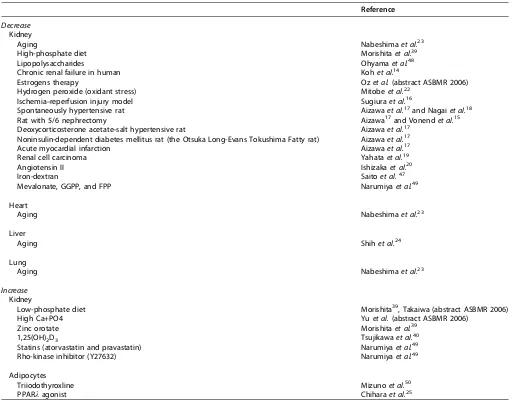
Table 1 | Factors regulating the expression of klotho
Reference
Decrease Kidney
Aging Nabeshimaet al.23
High-phosphate diet Morishitaet al.39
Lipopolysaccharides Ohyamaet al.48
Chronic renal failure in human Kohet al.14
Estrogens therapy Ozet al.(abstract ASBMR 2006)
Hydrogen peroxide (oxidant stress) Mitobeet al.22
Ischemia–reperfusion injury model Sugiuraet al.16
Spontaneously hypertensive rat Aizawaet al.17and Nagaiet al.18
Rat with 5/6 nephrectomy Aizawa17and Vonendet al.15
Deoxycorticosterone acetate–salt hypertensive rat Aizawaet al.17 Noninsulin-dependent diabetes mellitus rat (the Otsuka Long-Evans Tokushima Fatty rat) Aizawaet al.17
Acute myocardial infarction Aizawaet al.17
Renal cell carcinoma Yahataet al.19
Angiotensin II Ishizakaet al.20
Iron-dextran Saitoet al.47
Mevalonate, GGPP, and FPP Narumiyaet al.49
Heart
Aging Nabeshimaet al.23
Liver
Aging Shihet al.24
Lung
Aging Nabeshimaet al.23
Increase Kidney
Low-phosphate diet Morishita39, Takaiwa (abstract ASBMR 2006)
High Ca+PO4 Yuet al.(abstract ASBMR 2006)
Zinc orotate Morishitaet al.39
1,25(OH)2D3 Tsujikawaet al.40
Statins (atorvastatin and pravastatin) Narumiyaet al.49
Rho-kinase inhibitor (Y27632) Narumiyaet al.49
Adipocytes
Triiodothyroxline Mizunoet al.50
or younger postmenopausal women. The polymorphism G395A substitution in the promoter region affects DNA–-protein interaction and may affect the level of expression of klotho.36,37 Another polymorphism in klotho gene (F352V) has been associated with a higher bone mineral density in a Spanish population of postmenopausal women.38
The mouse klotho model shows a disturbed calcium and phosphate homeostasis together with an increase in the serum concentration of active vitamin D (1,25(OH)2D3). Interestingly, most of the aging phenotypes of these mice can be lightened, as well as serum calcium and 1,25(OH)2D3 concentrations reduced, with phosphate restriction in the diet,39,40 suggesting that these phenotypes are downstream events resulting from elevated 1,25(OH)2D3 as shown in the FGF23 knockout mice. Indeed, removal or reduction of 1,25(OH)2D3 in FGF23 and klotho mutant mice, either by dietary restriction or genetic manipulation rescue premature aging-like features and ectopic calcifications.40–42
The increase in serum 1,25(OH)2D3 concentration in klotho-deficient mice is because of the increase in the kidney of the 25-hydroxyvitamin D-1a-hydroxylase (CYP27b1) activity, but the mechanisms of this stimulation are unknown. Of note, in these animals, the normal pathways leading to the upregulation of CYP27b1, such as PTH, calcitonin, and 1,25(OH)2D3, are intact suggesting the existence of other regulatory pathways.40Like 1,25(OH)2D3, dietary phosphate depletion, a recognized stimulus of CYP27b1 expression, also increases the renal expression of klotho, supporting again the hypothesis that klotho could influence renal CYP27b1 expression. Lately, a recent report showed that the stimulatory effects of phosphate depletion on 1,25(OH)2D3 synthesis in renal proximal tubules are modulated by the positive regulatory actions of the secreted form of klotho on CYP27b1 expression (Takaiwa et al.,
abstract No. 1221, ASBMR, 2006).
What could be the explanation for the hypercalcemia observed in klotho mutant mice? The first and more plausible possibility could be the hypervitaminose D and its stimula-tory effects on the intestinal and renal absorption of calcium. The second possibility could be a direct participation of klotho in the regulation of renal calcium reabsorption. However, these animals show, concomitantly with the rise in serum calcium concentration, an increase in the urinary fractional excretion of calcium, and this in the presence of low-serum PTH levels, which suggests that the hypercalcemia is not probably because of renal calcium handling. Moreover, PTH-stimulated tubular calcium reabsorption is markedly diminished43 whereas the basolateral Na/Ca exchanger appears to be preserved, suggesting that other pathways downstream to PTH could be impaired by the disruption of the klotho gene. One of these pathways has been recently elucidated; klotho regulates calcium reabsorption in the distal convoluted tubule via a novel molecular mechanism, by deglycosylating and stabilizing the epithelial calcium channel TRPV5 on the surface of cellular membrane.3 Klotho colocalizes with TRPV5 and calbindin-D28K in the distal
convoluted and connecting tubule of mouse kidney cells, which are nephron segments responsible for active trans-epithelial calcium reabsorption.3 However, the lack of klotho leads to a diminution in the expression of TRPV5 on the cell surface and reduced tubular calcium reabsorption, similarly, mice lacking TRPV5 have reduced klotho expres-sion and diminished renal calcium reabsorption despite enhanced levels of 1,25(OH)2D3. Although the two proteins together with calbindin-D28K are tightly controlled by vitamin D, suggesting a functional link between these proteins in the maintenance of calcium homeostasis, the renal origin of the hypercalcemia appears more unlikely.3,40 The third hypothesis would be that the lack of klotho could favor the instauration of an adynamic bone disease through a reduced osteoclast activity and thereby the hypercalcemia because the skeleton would be unable to play its buffer action. Indeed, it has been demonstrated that TRPV5 is essential for a proper osteoclastic activity; mice lacking TRPV5 have an increase in osteoclast size and number, but calcium resorption is nonfunctional owing to a reduced osteoclast activity.44 Klotho could exert a similar effect on osteoclast TRPV5 expression as in the kidney cells but that remains to be investigated.
phosphaturic effect of FGF23.13 Fifth, the hyperphosphat-emia could be because of the upregulation of stanniocalcins 1 and 2 (STC1 and STC2) in the kidney. These two molecules are implicated in calcium and phosphorus homeostasis and are normally upregulated by vitamin D. Klotho-deficient mice have an increased renal gene expression of STC1 and STC2 compared with wild-type mice46 and feeding these mice with a low-phosphorus diet resulted in a partial reduction in renal expression of STC1 and the normalization of hyperphosphatemia. Finally, it cannot be ruled out that klotho could modulate other sodium phosphate cotranspor-ters, such as Pit1 and Pit2 or modulate a putative phosphate sensor.
OTHER RENAL EFFECTS OF KLOTHO
Klotho gene is predominantly expressed in the kidney, but little is known about its potential role in other physiological processes besides its implication in mineral and vitamin D metabolism. Recent observations suggest that angiotensin II modulates the renal expression of klotho and that abnormal iron metabolism and increased oxidative stress are involved in the mechanism by which angiotensin II modulates klotho expression.21Treatment of normal animal by angiotensin II infusion downregulates renal klotho mRNA and protein expression by a receptor-dependent but pressor-independent mechanism, and leads to abnormal iron deposition in renal cells. Iron-dextran administration downregulates klotho expression, and iron chelation suppresses angiotensin II-induced klotho downregulation in the kidney. In addition, a free radical scavenger (T-0970), which effectively reduce plasma levels of 8-epi-prostaglandin F (2a) (8-epi-PGF(2a)), suppressed angiotensin II-induced renal klotho downregula-tion.47 Interestingly, the angiotensin II-induced tubulo-interstitial damage could be prevented in a transgenic mouse model overproducing klotho protein,21 suggesting that klotho gene induction could have therapeutic possibilities in treating angiotensin II-induced renal damage.
CONCLUSIONS AND PERSPECTIVE
The discovery of klotho has opened an extraordinary field of investigation not only because of its implications in human longevity but also because of its implication in a multitude of other biological processes. Although the exact biological role and molecular function of klotho have been only partly deciphered, several functions seem to be clearly established. Klotho can be an antiaging hormone, which binds to a not yet identified high-affinity receptor and inhibits the intra-cellular insulin/IGF-1 signaling cascade; klotho can function as a novel b-glucuronidase, which deglycosylates steroid b-glucuronides as well as the calcium channels TRPV5; klotho is a cofactor essential for the activation of FGFR1(IIIc) signaling by FGF23. The two last functions have propelled klotho to the group of key factors regulating mineral and vitamin D metabolism and have also stimulated the interest in the nephrology community. There is a hope that in a near future endogenous circulating klotho could be measured
and the values compared between healthy individuals and those with high risks of a variety of metabolic syndromes, including patients with chronic kidney disease and hyper-phosphatemia.
REFERENCES
1. Kuro-o M, Matsumura Y, Aizawa Het al.Mutation of the mouse klotho gene leads to a syndrome resembling ageing.Nature1997;390: 45–51. 2. Kurosu H, Yamamoto M, Clark JDet al.Suppression of aging in mice by
the hormone klotho.Science2005;309: 1829–1833.
3. Chang Q, Hoefs S, van der Kemp AWet al.The beta-glucuronidase klotho hydrolyzes and activates the TRPV5 channel.Science2005;310: 490–493. 4. Kurosu H, Ogawa Y, Miyoshi Met al.Regulation of fibroblast growth
factor-23 signaling by klotho.J Biol Chem2006;281: 6120–6123. 5. Matsumura Y, Aizawa H, Shiraki-Iida Tet al.Identification of the human
klotho gene and its two transcripts encoding membrane and secreted klotho protein.Biochem Biophys Res Commun1998;242: 626–630. 6. Tohyama O, Imura A, Iwano Aet al.Klotho is a novel beta-glucuronidase
capable of hydrolyzing steroid beta-glucuronides.J Biol Chem2004;279: 9777–9784.
7. Mian IS. Sequence, structural, functional, and phylogenetic analyses of three glycosidase families.Blood Cells Mol Dis1998;24: 83–100. 8. Li SA, Watanabe M, Yamada Het al.Immunohistochemical localization of
klotho protein in brain kidney and reproductive organs of mice.Cell Struct Funct2004;29: 91–99.
9. Kamemori M, Ohyama Y, Kurabayashi Met al.Expression of klotho protein in the inner ear.Hear Res2002;171: 103–110.
10. Takeshita K, Fujimori T, Kurotaki Yet al.Sinoatrial node dysfunction and early unexpected death of mice with a defect of klotho gene expression. Circulation2004;109: 1776–1782.
11. Imai M, Ishikawa K, Matsukawa Net al.Klotho protein activates the PKC pathway in the kidney and testis and suppresses 25-hydroxyvitamin D3 1alpha-hydroxylase gene expression.Endocrine2004;25: 229–234. 12. Yamamoto M, Clark JD, Pastor JVet al.Regulation of oxidative stress by
the anti-aging hormone klotho.J Biol Chem2005;280: 38029–38034. 13. Urakawa I, Yamazaki Y, Shimada Tet al.Klotho converts canonical FGF
receptor into a specific receptor for FGF23.Nature2006;444: 770–774. 14. Koh N, Fujimori T, Nishiguchi Set al.Severely reduced production of
klotho in human chronic renal failure kidney.Biochem Biophys Res Commun2001;280: 1015–1020.
15. Vonend O, Apel T, Amann Ket al.Modulation of gene expression by moxonidine in rats with chronic renal failure.Nephrol Dial Transplant 2004;19: 2217–2222.
16. Sugiura H, Yoshida T, Tsuchiya Ket al.Klotho reduces apoptosis in experimental ischaemic acute renal failure.Nephrol Dial Transplant2005; 20: 2636–2645.
17. Aizawa H, Saito Y, Nakamura Tet al.Downregulation of the Klotho gene in the kidney under sustained circulatory stress in rats.Biochem Biophys Res Commun1998;249: 865–871.
18. Nagai R, Saito Y, Ohyama Yet al.Endothelial dysfunction in the klotho mouse and downregulation of klotho gene expression in various animal models of vascular and metabolic diseases.Cell Mol Life Sci2000;57: 738–746.
19. Yahata K, Mori K, Arai Het al.Molecular cloning and expression of a novel klotho-related protein.J Mol Med2000;78: 389–394.
20. Ishizaka N, Mitani H, Nagai R. Angiotensin II regulates klotho gene expression.Nippon Rinsho2002;60: 1935–1939.
21. Mitani H, Ishizaka N, Aizawa Tet al. In vivoklotho gene transfer ameliorates angiotensin II-induced renal damage.Hypertension2002;39: 838–843.
22. Mitobe M, Yoshida T, Sugiura Het al.Oxidative stress decreases klotho expression in a mouse kidney cell line.Nephron Exp Nephrol2005;101: e67–e74.
23. Nabeshima Y. Ectopic calcification in Klotho mice.Clin Calcium2002;12: 1114–1117.
24. Shih PH, Yen GC. Differential expressions of antioxidant status in aging rats: the role of transcriptional factor Nrf2 and MAPK signaling pathway. Biogerontology2006 (July 19, online).
25. Chihara Y, Rakugi H, Ishikawa Ket al.Klotho protein promotes adipocyte differentiation.Endocrinology2006;147: 3835–3842.
26. Bektas A, Schurman SH, Sharov AAet al.Klotho gene variation and expression in 20 inbred mouse strains.Mamm Genome2004;15: 759–767. 27. Kappeler L, De Magalhaes Filho C, Le Bouc Y, Holzenberger M. Ageing,
28. Ikushima M, Rakugi H, Ishikawa Ket al.Anti-apoptotic and anti-senescence effects of klotho on vascular endothelial cells. Biochem Biophys Res Commun2006;339: 827–832.
29. Saito Y, Nakamura T, Ohyama Yet al. In vivoklotho gene delivery protects against endothelial dysfunction in multiple risk factor syndrome.Biochem Biophys Res Commun2000;276: 767–772.
30. Saito Y, Yamagishi T, Nakamura Tet al.Klotho protein protects against endothelial dysfunction.Biochem Biophys Res Commun1998;248: 324–329.
31. Unger RH. Klotho-induced insulin resistance: a blessing in disguise? Nat Med2006;12: 56–57.
32. Arking DE, Krebsova A, Macek Sr Met al.Association of human aging with a functional variant of klotho.Proc Natl Acad Sci USA2002;99: 856–861.
33. Xiao NM, Zhang YM, Zheng Q, Gu J. Klotho is a serum factor related to human aging.Chin Med J (Engl)2004;117: 742–747.
34. Kawaguchi H, Manabe N, Miyaura Cet al.Independent impairment of osteoblast and osteoclast differentiation in klotho mouse exhibiting low-turnover osteopenia.J Clin Invest1999;104: 229–237.
35. Kawaguchi H, Manabe N, Chikuda Het al.Cellular and molecular mechanism of low-turnover osteopenia in the klotho-deficient mouse. Cell Mol Life Sci2000;57: 731–737.
36. Kawano K, Ogata N, Chiano Met al.Klotho gene polymorphisms associated with bone density of aged postmenopausal women. J Bone Miner Res2002;17: 1744–1751.
37. Ogata N, Matsumura Y, Shiraki Met al.Association of klotho gene polymorphism with bone density and spondylosis of the lumbar spine in postmenopausal women.Bone2002;31: 37–42.
38. Riancho JA, Valero C, Hernandez JLet al.Association of the F352V variant of the Klotho gene with bone mineral density.Biogerontology2006 (July 19, online).
39. Morishita K, Shirai A, Kubota Met al.The progression of aging in klotho mutant mice can be modified by dietary phosphorus and zinc. J Nutr2001;131: 3182–3188.
40. Tsujikawa H, Kurotaki Y, Fujimori Tet al.Klotho, a gene related to a syndrome resembling human premature aging, functions in a negative regulatory circuit of vitamin D endocrine system.Mol Endocrinol2003;17: 2393–2403.
41. Razzaque MS, Lanske B. Hypervitaminosis D and premature aging: lessons learned from Fgf23 and Klotho mutant mice.Trends Mol Med2006;12: 298–305.
42. Razzaque MS, Sitara D, Taguchi Tet al.Premature aging-like phenotype in fibroblast growth factor 23 null mice is a vitamin D-mediated process. FASEB J2006;20: 720–722.
43. Tsuruoka S, Nishiki K, Ioka Tet al.Defect in parathyroid-hormone-induced luminal calcium absorption in connecting tubules of Klotho mice.Nephrol Dial Transplant2006;21: 2762–2767.
44. van der Eerden BC, Hoenderop JG, de Vries TJet al.The epithelial Ca2+ channel TRPV5 is essential for proper osteoclastic bone resorption.Proc Natl Acad Sci USA2005;102: 17507–17512.
45. Segawa H, Yamanaka S, Ohno Yet al.Correlation between hyperphosphatemia and type II Na/Pi cotransporter activity in klotho mice.Am J Physiol Renal Physiol2006;292: F769–F779.
46. Yahata K, Mori K, Mukoyama Met al.Regulation of stanniocalcin 1 and 2 expression in the kidney by klotho gene.Biochem Biophys Res Commun 2003;310: 128–134.
47. Saito K, Ishizaka N, Mitani Het al.Iron chelation and a free radical scavenger suppress angiotensin II-induced downregulation of klotho, an anti-aging gene, in rat.FEBS Lett2003;551: 58–62.
48. Ohyama Y, Kurabayashi M, Masuda Het al.Molecular cloning of rat klotho cDNA: markedly decreased expression of klotho by acute inflammatory stress.Biochem Biophys Res Commun1998;251: 920–925.
49. Narumiya H, Sasaki S, Kuwahara Net al.HMG-CoA reductase inhibitors up-regulate anti-aging klotho mRNA via RhoA inactivation in IMCD3 cells. Cardiovasc Res2004;64: 331–336.