MEDIA MEDIKA
INDONESIANA
Hak Cipta©2008 oleh Fakultas Kedokteran Universitas Diponegoro dan Ikatan Dokter Indonesia Wilayah Jawa Tengah
Vaginal Acidity and Whiff Test
for Screening Bacterial Vaginosis in Pregnant Women
Tri Nur Kristina*
ABSTRACT
Keasaman vagina dan tes whiff untuk penapisan bacterial vaginosis pada ibu hamil.
Background: It has been acknowledged that bacterial vaginosis (BV) in pregnant women is associated with miscarriage and premature delivery. Microscopic examination of vaginal secretion should be used in the diagnosis of BV. Nevertheless, usually microscope does not exist in antenatal care facility in the community based setting. Therefore, screening tool of BV among pregnant women attending antenatal care that is simple, cheap, and fast is needed.
Methods: Diagnostic study was conducted among pregnant women attending antenatal care in the Community Health Centre. Vaginal acidity combines with whiff test were used as a screening tool, and compared blindly with Gram stain of vaginal smear as a gold standard to diagnose BV.
Results: This study showed that the sensitivity and specificity of the vaginal acidity combine with whiff test to diagnose BV were 93.1% and 69% respectively. It means that false negative and false positive were 6.9% and 31% respectively. In the screening method, high sensitivity or less false negative is needed. Therefore, only few people who really have the disease (the false negative) can’t be reached by the gold standard of diagnostic test.
Conclusions: It could be concluded that the combination of vaginal acidity and whiff test is a useful tool for screening BV in pregnant women especially in the community-based health facility.
Keywords: Bacterial Vaginosis, sensitivity, specificity
ABSTRAK
Latar belakang: Vaginosis Bakterial (VB) pada ibu hamil diketahui secara luas sebagai faktor risiko terjadinya abortus dan kelahiran prematur. Diagnosis VB harus ditegakkan dengan pemeriksaan sekret vagina menggunakan pemeriksaan mikroskopik, yang pada umumnya tidak tersedia di tempat pemeriksaan kehamilan primer. Oleh karena itu dibutuhkan alat pemeriksaan (penapisan) VB pada ibu hamil yang mudah, murah dan cepat.
Metode: Penelitian ini merupakan studi diagnostik pada ibu hamil yang memeriksakan kehamilannya di Puskesmas. Keasaman vagina dikombinasikan dengan tes whiff digunakan sebagai alat penapisan dan dibandingkan secara membuta dengan pengecatan Gram pada sekret vagina yang digunakan sebagai standar baku emas untuk mendiagnosis VB.
Hasil: Hasil penelitian menunjukkan keasaman vagina dikombinasikan tes whiff memiliki sensitifitas 93,1% dan spesifisitas 69%. Hal ini berarti kemungkinan adanya negatif palsu hanya 6,9% sedangkan kemungkinan positif palsu 31%. Dalam metode penapisan dibutuhkan alat diagnostik yang memiliki sensitifitas tinggi atau hanya sedikit yang negatif palsu, sehingga hanya sedikit pula yang akan luput dari pemeriksaan Gram.
Kesimpulan: Kombinasi keasaman vagina dengan tes whiff dapat dipergunakan untuk penapisan VB pada ibu hamil terutama di fasilitas kesehatan primer.
INTRODUCTION
Normal vaginal flora is dominated by Lactobacilli. Bacterial vaginosis (BV) is associated with an imba-lance of the bacteria that are normally found in the vagina. This imbalance, occurs when different types of bacteria outnumber the normal ones. Instead of Lacto-bacillus bacteria being the most numerous, increased numbers of anaerobic organisms such as G. vaginalis, Bacteroides, Mobiluncus, and Mycoplasma hominis are found in the vagina of women with BV.1 Lactobacillus produces lactic acid from the metabolism of glycogen, which resulted in a normal vaginal pH of 3.8 to 4.2, and this is suboptimal for the growth of G. vaginalis and anaerobes.2 Furthermore, certain species of lactobacilli produce H2O2, which inhibit the growth of G. vaginalis and anaerobes.3 Women with BV may have an abnor-mal vaginal discharge with an unpleasant odor. Some women report a strong fish-like odor, especially after intercourse. However, nearly half of the patients with BV report no noticeable symptoms.4
It has been acknowledged that BV in pregnant women is associated with
miscarriage, preterm delivery, and low birth weight.5-6 Depends on the type of antenatal clinics, gestation age, and geographic characteristic, the pre-valence of BV in pregnant women varies from 16– 30%.5-7 Since nearly half of pregnant women with BV have no symptoms, screening BV in pregnant women would be worthwhile to reduce the complication of this disease.Amsel criteria defines BV as being present if three of the four following criterion are found: 1)
homogeneous vaginal discharge; 2) vaginal pH greater than 4.5; 3) po
-sitive whiff test; and 4) the presence of clue cells on wet microscopy of the vaginal fluid.8 As a refinement to the above definition, some authorities have recommended that at least 20% of the epithelial cells present be defined as clue cells.9 It was also reported that vaginal smear Gram stain examination is a reproducible test for the diagnosis of BV.10 Thus, microscopic examination should be used to diagnose of BV. Nevertheless, usually microscope do not exist in antenatal care facility especially in the community based setting. Therefore, simple, cheap and quick test is needed to screen BV in pregnant women before it can be considered for microscope examination. Besides, most antenatal care in the community settings are done by midwives, therefore the screening tool should also be easily done by these health providers.This study aimed to measure the validity of vaginal acidity combine with w
hiff test as a screening tool of BV among pregnant women attending antenatal care in the community-based setting.METHODS
This is a diagnostic test study, which 84 pregnant women undergoing antenatal care in the Community Health Centre were screened of BV. Pregnant women in the first trimester and/or sexual intercourse in the past 24 hours were excluded from this study.
Trained midwife obtained vaginal fluid for screening test. Vaginal acidity and whiff test were
interpreted blindly by the midwife and 2 medical students. Screening test was considered (+) if 2
out of 3 examiners confirmed as (+). An additional vaginal swab was used to prepare a fixative
smear and sent to a laboratory of Microbiology in the Faculty of Medicine Diponegoro
University, Semarang, Indonesia for Gram staining and measured blindly by the author using
standardized criteria of Spiegel
11 and Thomason.12The vaginal acidity was determined using ColorpHast Indicator Strips (EM Science, Gibbstown, NJ), which has appropriate range. Vaginal acidity greater than 4.5 is considered as positive. The whiff test was performed by adding a drop of 10% potassium hydroxide to the vaginal fluid and sniffing the mixture and considered as positive if a fishy aroma was noted.
Data were analyzed using standard calculations for sen-sitivity, specificity, and positive and negative predictive values. Reliability analysis was used to measure the agreement between the 3 examiners.
RESULTS
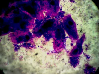
Figure 1. Normal vaginal flora, which dominated by Lactobacilli.
Figure 1 shows the normal vaginal flora, whereas Figure 2 shows Bacterial Vaginosis as defined by Thomason criteria, in which clue cells exceed the normal epithelial cells of vagina. Reliability analysis of vaginal acidity and whiff test between midwife and 2 medical students gave satisfactory result (Table 1 and 2).
Table 1. The reliability of vaginal acidity measurement.
Table 3 presents the utility of single test vaginal acidity for screening of BV. The sensitivity and specificity of vaginal acidity >4.5 compared to Gram stain were 79.3% and 47.3% respectively; the positive predictive value was 44.2%, with the negative predictive value of 81.3%.
Table 3. Vaginal acidity >4.5 compared to Gram stain as the gold standard to diagnose BV.
| |Gram Stain |Total |
Table 4 presents the utility of single test whiff test for screening of BV. The sensitivity and specificity of whiff test compared to Gram stain were 75.9% and 52.7% respectively; the positive predictive value was 54.2%, with the negative predictive value of 80.6%.
Table 4. Whiff test compared to Gram stain as the gold standard to diagnose BV.
| |Gram Stain |Total | | |BV (+) |BV (-) | | |Whiff test |(+) |22 |26 |48 | | |(-) |7 |29 |36 | |Total |29 |55 |84 |
The sensitivity, specificity, and predictive values of the vaginal acidity combined with whiff test for screening of BV compared to Gram stain as the gold standard for the diagnosis of BV is shown in Table 5. The sensitivity and specificity of this combination screening test was 93.1% and 69% respectively; the positive predictive value was 58.5% with negative predictive value of 89.5%.
Table 5. Vaginal acidity >4.5 combined with whiff test as compared to Gram stain.
respectively.10
Although whiff test as suggested by Amsel is con-sidered as a subjective criterion,8 this study showed that vaginal acidity more sensitive but less specific com-pared to whiff test. However, when vaginal acidity combined with whiff test, the sensitivity and specificity increased to 93.1% and 69% respectively. Using the combination of vaginal pH and whiff test, the possibility of false positive and false negative were 6.9% and 31% respectively. In the screening test, sensitivity is more important than specificity. Therefore, only few people who really have the disease can not be reached by the gold standard of diagnostic test.
To increase the sensitivity, it is suggested that pregnant women undergoing screening test using
vaginal pH and w
hiff test with either or both (+) result should be followed by Gram stain vaginal smear. Thus, treatment can be decided as soon as possible.CONCLUSIONS
The combination of vaginal acidity and whiff test can be used as a screening method for BV in pregnant women.
ACKNOWLEDGMENT
The author was grateful to Dr. Siti Zubaedah, the head of Community Health Centre Poncol, Semarang for her support; Erlinda Surya Anis, midwife in the same Community Health Centre; and FMDU’s medical students for their participation in this study.
REFERENCES
1. Rosenstein IJ, Margan DJ, Sheehan M, Lamont RF, Taylor RD. Bacterial vaginosis in pregnancy: distribution of bacterial species in different gram-stain categories of the vaginal flora. J Med Microbiol. 1996; 45:120-26.
2. Wang, J. Bacterial Vaginosis. Prim Care Update Ob/Gyns. 2000; 7:181-85.
3. Hillier SL, Krohn ME, Klebanoff SJ, Eschenbach DA. The relationship of hydrogen peroxide-producing lactobacilli to bacterial vaginosis and genital microflora in pregnant women. Obstet Gynecol. 1992; 79:369–73.
4. Kira EF. The clinical picture and diagnosis of bacterial vaginosis. Akush-Gynecol-Mosk.
1994; 2: 32-5.
5. McGregor JA, French JI, Seo K. Premature rupture of membranes and bacterial vaginosis.
Am J Obstet Gynecol. 1993; 169: 463-
66.6. Riduan JM, Hillier SL, Utomo B, Wiknjosastro G, Linnan M, Kandun N. Bacterial
vaginosis and prematurity in Indonesia; association in early and late pregnancy. Am J
Obstet Gynecol. 1993; 169: 175-
78.7. Aggarwal A, Devi P, Jain R. Anaerobes in bacterial vaginosis. Indian J Med Microbiol.
2003; 21:124-
26.8. Amsel R, Totten PA, Spiegel CA, Chen KCS, Eschenbach DA, Holmes KK. Nonspecific vaginitis: diagnostic criteria and microbial epidemiologic associations. Am J Med. 1983; 74:14– 22.
9. Eschenbach DA, Hillier S, Critchlow C, Stevens C, DeRouen T, Holmes KK. Diagnosis
and clinical manifestations of bacterial vaginosis. Am J Obstet Gynecol. 1988;
158:819-28.
10. Schwebke JR, Hillier SL, Sobel JD, McGregor JA, Sweet CM, Sweet RL. Validity of the
vaginal gram stain for the diagnosis of bacterial vaginosis. Obstetrics & gynecology. 1996;
88:573-
76.11. Spiegel CA, Amsel R, Holmes KK. Diagnosis of bacterial vaginosis by direct Gram stain of vaginal fluid. J Clin Microbiol. 1983;18: 170–77.
12. Thomason JL, Anderson RJ, Gelbart SM. Simplified Gram stain interpretative method for diagnosis of bacterial vaginosis. Am J Obstet Gynecol. 1992;167:16–9.
* Microbiology Department, Faculty of Medicine Diponegoro University, Semarang, Indonesia Email: [email protected]