Hirose and Katsunori Tanaka
Venny Santosa, Sabrina Martha, Noriaki
Formation in DNA Replication
Function during Prereplicative Complex
(MCM-BP), Mcb1, Regulates MCM
Maintenance (MCM)-binding Protein
The Fission Yeast Minichromosome
doi: 10.1074/jbc.M112.432393 originally published online January 15, 2013 2013, 288:6864-6880.
J. Biol. Chem.
10.1074/jbc.M112.432393 Access the most updated version of this article at doi:
. JBC Affinity Sites Find articles, minireviews, Reflections and Classics on similar topics on the
Alerts:
When a correction for this article is posted •
When this article is cited •
to choose from all of JBC's e-mail alerts Click here
Supplemental material:
http://www.jbc.org/content/suppl/2013/01/15/M112.432393.DC1.html
http://www.jbc.org/content/288/10/6864.full.html#ref-list-1
This article cites 65 references, 35 of which can be accessed free at
by guest on October 30, 2014
http://www.jbc.org/
Downloaded from
by guest on October 30, 2014
http://www.jbc.org/
The Fission Yeast Minichromosome Maintenance
(MCM)-binding Protein (MCM-BP), Mcb1, Regulates MCM
Function during Prereplicative Complex Formation in DNA
Replication
*
□SReceived for publication, October 31, 2012, and in revised form, January 5, 2013Published, JBC Papers in Press, January 15, 2013, DOI 10.1074/jbc.M112.432393
Venny Santosa‡§, Sabrina Martha‡, Noriaki Hirose‡, and Katsunori Tanaka‡§1
From the‡Department of Bioscience and the§Research Center for Environmental Bioscience, School of Science and Technology, Kwansei Gakuin University, Sanda, Hyogo 669-1337, Japan
Background:MCM-BP is a novel binding partner of the MCM complex; the mechanisms by which MCM-BP functions and associates with MCM complexes are not well understood.
Results:Genetic analysis showed thatmcb1tsmutants exercise defective regulation of prereplicative MCM complex formation
during DNA replication.
Conclusion:Mcb1 regulates MCM function during prereplicative complex formation in DNA replication.
Significance:This study presents the first evidence of MCM-BP function during prereplicative complex formation.
The minichromosome maintenance (MCM) complex is a rep-licative helicase, which is essential for chromosome DNA repli-cation. In recent years, the identification of a novel MCM-bind-ing protein (MCM-BP) in most eukaryotes has led to numerous studies investigating its function and its relationship to the MCM complex. However, the mechanisms by which MCM-BP functions and associates with MCM complexes are not well understood; in addition, the functional role of MCM-BP remains controversial and may vary between model organisms. The present study aims to elucidate the nature and biological function of the MCM-BP ortholog, Mcb1, in fission yeast. The Mcb1 protein continuously interacts with MCM proteins during the cell cyclein vivoand can interact with any individual MCM subunitin vitro. To understand the detailed characteristics of mcb1ⴙ, two temperature-sensitivemcb1gene mutants (mcb1ts)
were isolated. Extensive genetic analysis showed that themcb1ts
mutants were suppressed by amcm5ⴙmulticopy plasmid and
displayed synthetic defects with many S-phase-related gene mutants. Moreover, cyclin-dependent kinase modulation by Cig2 repression or Rum1 overproduction suppressed the
mcb1ts mutants, suggesting the involvement of Mcb1 in pre-RC formation during DNA replication. These data are consistent with the observation that Mcm7 loading onto rep-lication origins is reduced and S-phase progression is delayed inmcb1tsmutants. Furthermore, themcb1tsmutation led to
the redistribution of MCM subunits to the cytoplasm, and this redistribution was dependent on an active nuclear export system. These results strongly suggest that Mcb1 promotes
efficient pre-RC formation during DNA replication by regu-lating the MCM complex.
Genome integrity depends on successful and faithful DNA replication, which relies on the concerted activity of multiple replication proteins. The temporal and spatial regulation of DNA replication and the cell cycle control system ensure a sin-gle round of replication of chromosome DNA during every cell cycle. The initiation of DNA replication in all eukaryotes involves the assembly of a prereplicative complex (pre-RC)2at
the replication origins in G1phase and the subsequent activa-tion of the pre-RC to the preinitiaactiva-tion complex (pre-IC) at the onset of S-phase. The replication origins are recognized by the origin recognition complex and become a platform for the recruitment of Cdc6 (called Cdc18 in fission yeast) and Cdt1-bound double hexamers of the minichromosome maintenance (MCM) complex to form the pre-RC (reviewed in Ref. 1). Upon entry into S-phase, this complex is activated by the S-phase-specific kinase, Dbf4/Drf1-dependent kinase. The Mcm2–7 complex serves as a platform to recruit Cdc45 and GINS, thereby converting the pre-RC into the pre-IC. Dbf4/Drf1-de-pendent kinase phosphorylates several of the Mcm2–7 proteins and triggers the recruitment of Cdc45 and GINS to form the Cdc45䡠MCM䡠GINS complex. Cdc45䡠MCM䡠GINS complex for-mation then induces the helicase activity of the Mcm2–7 com-plex, promoting the unwinding of the double-stranded DNA at the origin of DNA replication (reviewed in Ref. 2).
The Mcm2–7 hexamer complex is an evolutionarily con-served DNA helicase, which is essential for both the initiation of chromosome DNA replication and elongation (3, 4). Mcm2–7
*This work was supported by a grant-in-aid for scientific research from the Japan Society for the Promotion of Science and the Support Project to Assist Private Universities in Developing Bases for Research by the Ministry of Education, Culture, Sports, Science, and Technology (to K. T.). □S This article containssupplemental Tables S1 and S2 and Figs. S1–S7.
1To whom correspondence should be addressed: Dept. of Bioscience, School
of Science and Technology, Kwansei Gakuin University, 2-1 Gakuen, Sanda, Hyogo 669-1337, Japan. Tel./Fax: 81-79-565-7769; E-mail: katsunori@ kwansei.ac.jp.
2The abbreviations used are: pre-RC, prereplicative complex; pre-IC,
preini-tiation complex; MCM, minichromosome maintenance; BP, MCM-binding protein; HU, hydroxyurea; MMS, methylmethane sulfonate; CPT, camptothecin; DBD, DNA-binding domain; AD, activation domain; DSB, double-stranded DNA break; CDK, cyclin-dependent kinase; GINS, Tetra-meric complex composed of Sld5, Psf1, Psf2, Psf3 (Go-Ichi-Ni-San).
by guest on October 30, 2014
http://www.jbc.org/
proteins are members of the AAA⫹ATPase family of proteins and share a region of homology that encompasses the ATPase motif, referred to as the MCM box (1, 5). The MCM box har-bors distinct motifs, including Walker A, Walker B, and an argi-nine finger. The canonical MCM complex consists of the six subunits of Mcm2–7 and is assembled by the binding of one MCM arginine finger to the P-loop within the Walker A motif of another MCM subunit to form the ATP binding site. This hexameric ring is formed by two subcomplexes; the het-erodimer is formed by Mcm3 and Mcm5 (Mcm3-5), and the trimeric MCM core complex is formed by Mcm4, Mcm6 and Mcm7 (Mcm4-6-7). Mcm2 connects these two subcomplexes to form the Mcm2–7 hexamer complex (6 – 8). Mcm2–5 is thought to act as the gate of the ring structure, the site at which the ring structure opens to encircle DNA (9).
Although a great deal of evidence indicates the importance of the Mcm2–7 complex in DNA replication, there are still unan-swered questions concerning the functional role(s) of the MCM proteins. For example, the relationship between the Mcm4-6-7 core helicase and the larger Mcm2–7 hexamer is not under-stood. It is also unclear why MCM proteins are abundant and exceed the number of replication origins. In fission yeast, a reduction in MCM protein levels causes genome instability due to replication fork collapse and DNA damage (10, 11). In human cells, excess chromatin-loaded MCM complexes are important under conditions of replicative stress, where they activate dormant origins to ensure that DNA replication continues when the replication forks stall (12, 13). Mice expressing hypomorphic Mcm2 (Mcm2IRESCreERT2) or Mcm4 (Mcm4Chaos3) show lower levels of Mcm2–7 loading onto DNA, exhibit replicative stress even under unchallenged con-ditions, and have a high incidence of cancer (14, 15). In addi-tion, some MCM subunits appear to play additional roles that are independent of DNA replication (16 –18).
Although the primary focus of the study of MCM proteins has been to identify the function of the canonical MCM com-plex, several MCM-related complexes have also been charac-terized (19). Two additional MCM family members, Mcm8 and Mcm9, which contain an MCM box, were identified in higher eukaryotes (20 –23). Mcm8 and Mcm9 work downstream of the Fanconi anemia and BRCA2/Rad51 pathways and are required for homologous recombination, which promotes sister chro-matid exchange (24, 25). MCM-binding protein (MCM-BP) was first identified as a protein that strongly associates with human MCM proteins. Tandem affinity purification with tagged human MCM core subunits recovers both MCM-BP and MCM proteins (26). MCM-BP is conserved in most eukaryotes (except budding yeast andCaenorhabditis elegans) and has only limited homology to MCM proteins. Following the first identification in humans, several studies aimed to charac-terize and elucidate the functions of MCM-BP. Co-immuno-precipitation analysis showed that human MCM-BP interacts with all MCM subunits, except Mcm2; conversely, Mcm2 fails to recover MCM-BP (26). A similar pattern of interaction between MCM-BP and MCM subunits was observed in Xeno-pus laevis(27), suggesting that an alternative MCM complex is formed when Mcm2 is replaced by MCM-BP. InArabidopsis thaliana, the MCM-BP ortholog, ETG1, was identified as an
E2F target. The interaction between ETG1 and Mcm2–7 was confirmed by tandem affinity purification with tagged ETG1 (28).
In fission yeast,mcb1⫹(the fission yeast MCM-BP ortholog) is an essential gene, and its deletion results in gradual cell cycle arrest with acdc(cell division cycle) phenotype. Overexpres-sion of the Mcb1 protein or Mcb1 inactivation in temperature-sensitivemcb1mutants induces DNA damage and G2 check-point activation (29, 30). In human cells, the depletion of MCM-BP also leads to centrosome amplification and abnor-mal nuclear morphology, which may be due to G2DNA
dam-age checkpoint activation (31). The loss ofA. thalianaETG1 leads to reduced DNA replication, activation of the G2 checkpoint, and reduced sister chromatid cohesion (28, 32). The depletion of human MCM-BP also leads to reduced sis-ter chromatid cohesion (32). TheXenopusMCM-BP appears to play a role in unloading MCM complexes from chromatin after DNA synthesis (27); however, the depletion of human MCM-BP not only increases the levels of chromatin-associ-ated MCM proteins at the end of S-phase but also leads to a similar increase in soluble levels of MCM proteins through-out S-phase (31), suggesting multiple functions of MCM-BP in DNA replication. The human MCM-BP andA. thaliana ETG1 are largely nuclear throughout the cell cycle (28, 31). The fission yeast Mcb1 is widely distributed in the cytoplasm and nucleoplasm and is bound to chromatin (29); however, XenopusMCM-BP is imported into the nucleus just before the dissociation of Mcm2–7 from chromatin near the end of S-phase. These differences in the localization of MCM-BP may affect other functions associated with the MCM com-plex proteins. Furthermore, human MCM-BP interacts with Dfp4, the regulatory component of the Dbf4/Drf1-depen-dent kinase, as well as with MCM complex components, sug-gesting that MCM-BP may affect DNA replication, at least in part by regulating MCM phosphorylation by Dbf4/Drf1-de-pendent kinase (33). Thus, the functions of MCM-BP in DNA replication have been extensively studied; however, the mechanisms by which MCM-BP functions and associates with MCM complexes are not well understood, and the func-tional role of MCM-BP is controversial and may vary among model organisms.
To address these remaining questions, the functional roles of MCM-BP were examined by genetic analysis of temperature-sensitive mutants in fission yeast. Tight genetic links between mcb1⫹andmcm5⫹were identified. Themcb1tsmutants
dis-played synthetic defects with many S-phase-related gene mutants, and the temperature sensitivity ofmcb1tsmutants was
suppressed by CDK modulation upon Cig2 repression or Rum1 overexpression, suggesting the involvement of Mcb1 in pre-RC formation during DNA replication. In fact, S-phase progression was delayed inmcb1tsmutant cells, and loading of Mcm7 onto
the replication origins during pre-RC formation was reduced. Furthermore,mcb1tsmutations caused most MCM proteins to
exit the nucleus, which was partially rescued bymcm5⫹ over-expression. All of these results strongly indicate the importance of Mcb1 protein in promoting efficient pre-RC formation. This is the first report showing the involvement of MCM-BP in pre-RC formation during DNA replication. Under
by guest on October 30, 2014
http://www.jbc.org/
sion,” we shall discuss the role of MCM-BP in regulating MCM function(s) during DNA replication in the context of these results.
EXPERIMENTAL PROCEDURES
Strains, Growth Media, and Genetic and Molecular Methods— The fission yeast strains and plasmids used in this study are listed insupplemental Tables S1 and S2, respectively. Standard growth media and the general biochemical and genetic meth-ods used for fission yeast were described previously (34). Fission yeast cultures were grown at 30 °C in YES medium (0.5% yeast extract, 3% glucose, and supplements) unless indicated other-wise. Geneticin (G418, 100g/ml; Sigma), hygromycin (150 g/ml; Roche Applied Science), or nourseothricin (clonNAT, 200 g/ml; Werner Bioagents) was added as required. Hydroxyurea (HU; Sigma), methylmethane sulfonate (MMS; Wako), and camptothecin (CPT; Sigma) were used at the indi-cated concentrations. For the induction of expression from the nmt1promoter, cells were grown at 30 °C in the presence of 5 Mthiamine to repress thenmt1promoter until mid-log phase and then washed twice with fresh medium and further incu-bated for 20 h. Tetrad dissection was performed using a Singer Instrument micromanipulator system.
To create themcb1gene mutants, the QuikChange site-di-rected mutagenesis method (Stratagene) was used to mutate the indicated site(s) on a plasmid. All mutations were con-firmed by DNA sequencing. The designed mutations were introduced into the wild-type strain using PCR to insert a 5⫻
FLAG epitope at the C terminus and mark the allele with the kanMX6 gene, as described by Krawchuk and Wahls (35). Introduction of the designed mutations into themcb1gene on the wild-type chromosome was confirmed by colony PCR, fol-lowed by direct sequencing of the PCR product. The expression of the mutated Mcb1 protein tagged with the 5⫻FLAG epitope was confirmed by Western blotting using an anti-FLAG M2 antibody (Sigma). Exchange of thekanMX6marker in existing strains withnatMX6, which gives rise to resistance to the anti-biotic clonNAT, was performed as described by Satoet al.(36). Cell Cycle Synchronization—Yeast strains carrying the cold-sensitive nda3-KM311 mutation in the -tubulin gene (37) were synchronized in M phase by incubation for 4 h at 20 °C before being released at the permissive/restrictive temperature. Cell cycle progression was followed by flow cytometry.
Spotting Assay—To test the response to temperature, HU, CPT, or MMS, the fission yeast strains were grown on YES plates at 25 °C for 2–3 days. The cells were serially diluted (5-fold) and then spotted onto YES plates containing 5–10M CPT, 5–10 mMHU, or 0.005% MMS. The plates were then incubated at the indicated temperatures for 3– 6 days.
Flow Cytometry—Cells were fixed in 70% ice-cold ethanol overnight and then rehydrated in 50 mMsodium citrate. RNA was removed with 100g/ml RNase at 37 °C for 2 h prior to staining with 20 g/ml propidium iodide as described previ-ously (38, 39). Flow cytometry was performed using a BD Bio-sciences FACSCalibur instrument and Macintosh BD Cell-QuestTMsoftware.
Fluorescence Microscopy—Cells expressing Mcm2-GFP, Mcm3-GFP, Mcm4-GFP, Mcm5-RFP, Mcm6-GFP,
Mcm7-GFP, Rad22-YFP, or Rhp54-GFP (grown in EMM5S medium at the indicated temperatures) were harvested and suspended in Milli-Q water. The cells were then treated with DAPI (Dojindo) at room temperature to visualize DNA. The GFP, RFP, YFP, and DAPI signals were detected using a fluorescent microscope (BX51; Olympus). Images were obtained using a CCD camera (DP71; Olympus) and processed using Photoshop (Adobe) software.
Yeast Two-hybrid Assay—The Matchmaker two-hybrid sys-tem 3 (Clontech) was used for the yeast two-hybrid assay according to the manufacturer’s instructions. The indicated proteins were fused to the Gal4 DNA-binding domain (Gal4-DBD) on the pGBKT7 plasmid or to the Gal4 activation domain (Gal4-AD) on the pGAD-GH or pGAD424 plasmid and expressed in the AH109 reporter strain of Saccharomyces cerevisiae.
In Vitro Pull-down Assay and Semi-in Vitro Pull-down Assay— Full-lengthmcb1⫹cDNA was cloned into pET28a for expres-sion as a T7-His6-tagged protein. pRSFDuet-1-swi6 (a gift from
J. Nakayama) was used for expression of His6-tagged Swi6 as a
negative control. Full-lengthmcm2–7cDNAs were cloned into pGEX-KG vectors for expression as GST-tagged fusion pro-teins. T7-His6-tagged Mcb1 and His6-tagged Swi6 were
expressed inEscherichia coliKRX (Promega) and purified using nickel-nitrilotriacetic acid beads (Qiagen). GST-tagged fusion proteins (GST and GST-Mcm2–7) were expressed in E. coli KRX and purified using glutathione-Sepharose (GE Health-care). For thein vitropull-down assay, GST-tagged fusion pro-teins specifically bound to glutathione-Sepharose were incu-bated with purified T7-His6-tagged Mcb1 or His6-tagged Swi6
for 1 h at 4 °C. To test the interaction, T7-His6-tagged Mcb1 or
His6-tagged Swi6 was pulled down by GST-tagged proteins. For the semi-in vitropull-down assay,E. colisoluble crude lysates expressing the T7-His6-Mcb1, T7-His6-Mcb1L254P, or
T7-His6-Mcb1L363P protein, respectively, were used instead of
purified T7-His6-Mcb1.
Preparation of Cell Lysates and Western Blot Analysis —Fis-sion yeast whole-cell extracts were prepared by the trichloro-acetic acid (TCA) method. Briefly, the cell pellets (2⫻108cells)
were suspended in 100l of 20% TCA and disrupted with glass beads using Micro Smash (MS-100; Tomy Seiko). Cell lysates were collected, and beads were washed twice in 100l of 5% TCA. Cell pellets were obtained by centrifugation at 4,000 rpm for 10 min at 4 °C. Then 2⫻ SDS sample buffer was added, followed by 2MTris until the color of the suspension turned from yellow to blue. Samples were boiled at 95 °C for 5 min and cleared by centrifugation at 4,000 rpm for 10 min at 4 °C. Alter-natively, whole-cell extracts were prepared using the boiling method, as described previously (40). The proteins in the extracts were separated by SDS-PAGE and subsequently sub-jected to Western blot analysis using the chemiluminescence system (LAS4000mini, GE Healthcare) or the Odyssey威 infra-red imaging system (LI-COR Biosciences). The antibodies were as follows: anti-HA monoclonal antibody (HA-probe (F7), Santa Cruz Biotechnology, Inc. (Santa Cruz, CA)), anti-GFP monoclonal antibody (GFP, Roche Applied Science), anti-RFP monoclonal antibody (anti-anti-RFP, Abcam), anti-FLAG monoclonal antibody (anti-FLAG M2, Sigma), anti-Mcm2
by guest on October 30, 2014
http://www.jbc.org/
polyclonal antibody (a gift from H. Masukata) (41), anti-Mcm4 polyclonal antibody (a gift from H. Nishitani) (42), anti-Mcm5 polyclonal antibody (a gift from H. Masukata) (41), anti-Mcm6 polyclonal antibody (a gift from H. Masu-kata) (41), anti-Mcm7 polyclonal antibody (a gift from H. Masukata) (43), anti-tubulin monoclonal antibody (TAT-1) (44), anti-His6monoclonal antibody (anti-His6(Clone
His-2), Roche Applied Science), and GST polyclonal anti-body (GST(Z-5), Santa Cruz Biotechnology, Inc.).
Immunoprecipitation—The spheroplast method was used. Yeast cell extracts were prepared for immunoprecipitation, as described previously (45).
The glass beads method was also used. Cells (1–2⫻108cells)
were suspended in 100l of buffer A (50 mMHEPES-KOH, pH 7.5, 300 mMKCl, 0.05% Tween 20, 0.005% Nonidet P-40, 2M NaF, 0.4MNa3VO4, and 2M-glycerophosphate) supple-mented with the necessary inhibitors (1⫻Complete protease inhibitor mixture (Roche Applied Science), 1 mMPMSF, 0.2 mM 4-amidinobenzylsulfonyl fluoride hydrochloride (Sigma), and 1⫻ protease inhibitor mixture (Sigma)). The cells were dis-rupted with glass beads using the Multi Beads Shocker (Yasui Kikai). Glass beads were washed in 100l of buffer B (buffer A⫹1⫻complete protease inhibitor mixture (Roche Applied Science)). The samples were centrifuged at 13,000 rpm for 15 min at 4 °C, and the cleared supernatants were then used for immunoprecipitation with anti-FLAG M2 affinity gel (Sigma). After rotation for 2 h at 4 °C, the antibody beads were washed with 500l of buffer B and buffer C (buffer B⫹1⫻complete protease inhibitor mixture (Roche Applied Science) and 0.2 mM 4-amidinobenzylsulfonyl fluoride hydrochloride (Sigma)). SDS sample buffer was added, and the samples were boiled at 95 °C for 5 min.
Chromatin Immunoprecipitation (ChIP) and Real-time PCR—The ChIP assay and subsequent real-time PCR analysis were conducted according to the method of Fukuuraet al.(46). Immunoprecipitation for Mcm7-3HA was conducted with Dynabeads M-280 anti-mouse IgG beads (Invitrogen) conju-gated overnight with an HA monoclonal antibody at 4 °C. Immunoprecipitation for Orp4 was conducted with Dynabeads M-280 anti-rabbit IgG beads (Invitrogen) conjugated overnight with an anti-Orp4 antibody (a gift from H. Masukata) (47) at 4 °C. DNA from whole-cell extracts and immunoprecipitation samples was then subjected to real-time PCR using SYBR Green I in a LightCycler (Roche Applied Science). Four sets of primers were used for amplification: ars2004-66-F and ars2004-66-R for ars2004(47); ars3002-F (5⬘ -TCATTAGCAAACAAAAGCAAT-TGAG-3⬘) and ars3002-R (5⬘ -AATTTCCGGGCATTAAAAACG-3⬘) forars3002; AT2080-F (5⬘ -CCAATATTAAATAAGCAAT-TTTGAGAGC-3⬘) and AT2080-R (5⬘ -TGAAATTGTTGTT-GGCGCTG-3⬘) forAT2080; and 70-F and nonARS-70-R fornon-ars1(47). The percentage recovery was calculated as follows: immunoprecipitated DNA/total DNA⫻100.
Isolation of mcb1 Temperature-sensitive Mutants —Temper-ature-sensitive mutants were generated and identified as described by Kato et al. (48) with the following changes. Genomic DNA from amcb1-5FLAG-kanMX6strain was used as the starting template. Themcb1-5FLAG-kanMX6construct was amplified using Ex-Taq polymerase (Takara) in the
pres-ence of the increased amounts (2-fold) of dNTPs to increase the chance of base misincorporation. Yeast cells transformed with the PCR product were selected on YES plates supplemented with 100g/ml G418 (Sigma) and examined for sensitivity to high temperature (36.5 °C). Mutations were identified by sequence analysis.
Isolation of Spontaneous Suppressors of the mcb1L254P mutant—Spontaneous suppressors ofmcb1L254Pwere isolated
as follows. Themcb1L254Pcells were grown on YES plates at
25 °C (permissive temperature) for 1 day and then incubated at 34 °C (restrictive temperature) for several additional days. The colonies that spontaneously appeared were picked for further analysis.
Cloning of ded1⫹—Theded1⫹gene was cloned by comple-mentation for the cold-sensitive defects and temperature-resis-tant effects of the suppressors 1 and 2, using the fission yeast genomic library obtained from the National BioResource Pro-ject (49). Sequencing of the plasmid clones isolated from the genomic library indicated the presence of the same gene, ded1⫹, on each plasmid. Theded1⫹locus of suppressors 1 and 2 was amplified by PCR and sequenced, and the mutations in theded1genes of both suppressors were characterized.
RESULTS
Mcb1 Is Constitutively Expressed and Continuously Interacts with MCM Proteins—C-terminally 5⫻FLAG- or GFP-tagged Mcb1 (mcb1-5FLAG or mcb1-GFP, respectively) was con-structed to replace the wild-type copy in the genome. The mcb1-5FLAGormcb1-GFPcells showed normal growth, indi-cating that the tagged copies were functional. We have con-firmed with these strains that Mcb1 is an abundant protein and that protein abundance of Mcb1 is not cell cycle-regulated, findings that agree well with the previous report (data not shown) (29).
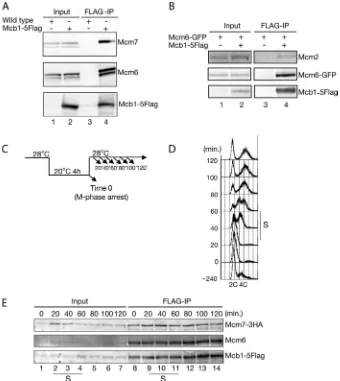
Immunoprecipitation and/or mass spectrometry studies in humans, Xenopus, and fission yeast showed that MCM-BPs (hMCM-BP for human, xMCM-BP forXenopus, and Mcb1 for fission yeast) interact with all MCM complex subunits, except Mcm2 (26, 27, 29, 30). Therefore, MCM-BP was thought to replace Mcm2 within the MCM complex (26); however, in A. thaliana, AtMcm2 was found to co-purify with ETG1, the A. thalianaMCM-BP ortholog, by mass spectrometry analysis (28). To understand this discrepancy, confirmation of the inter-action between Mcb1 and MCMs in fission yeast was sought using co-immunoprecipitation studies. Mcb1-5FLAG was immunoprecipitated, and the immunoprecipitate was blotted for most MCM proteins. As shown in Fig. 1A, Mcb1-5FLAG co-precipitated Mcm6 and Mcm7. In another experiment, Mcb1-5FLAG co-precipitated Mcm6-GFP and Mcm2, although Mcb1-5FLAG co-precipitated Mcm2 at much lower levels than Mcm6-GFP (Fig. 1B). In later experiments, Mcb1-5FLAG co-precipitated all six subunits of the MCM complex (Fig. 7A) (data not shown). To investigate whether the interac-tion between Mcb1 and MCMs is cell cycle-dependent, the interaction between Mcb1 and MCMs was analyzed in syn-chronized cells. Thenda3-KM311 mcb1-5FLAG mcm7-3HA cells were synchronized in M phase and then released, and pro-gression was followed every 20 min (Fig. 1,CandD).
by guest on October 30, 2014
http://www.jbc.org/
precipitation of Mcb1-5FLAG from each sample at the indi-cated time points showed that Mcb1-5FLAG continuously interacts with Mcm7-3HA and Mcm6 independently of the cell cycle phase (Fig. 1E). These results indicate that the Mcb1 pro-tein is constitutively expressed and continuously interacts with MCM proteins throughout the cell cycle.
Mcb1 Can Interact with Any of the Individual MCM Sub-units—To understand the interactions between Mcb1 and each of the MCM complex subunits (Mcm2–7), the interaction between Mcb1 and Mcm2–7 was assessed by a yeast two-hy-brid assay (Fig. 2A). Mcb1 associated with all of the Mcm2–7 subunits, whereas no interaction between Mcb1 and the vector control or between the vector control and Mcm2–7 was detected. The interaction between Mcb1 and Mcm2 and between Mcb1 and Mcm6 was stronger than between Mcb1 and Mcm3/4/5/7 in this two-hybrid assay. The interactions between Mcb1 and Mcm2–7 were further confirmed byin vitro
pull-down analysis using bacterially purified recombinant pro-teins (Fig. 2B). The tagged fusion proteins (GST, GST-Mcm2, GST-Mcm3, GST-Mcm4, GST-Mcm5, GST-Mcm6, and GST-Mcm7) bound to glutathione-Sepharose were incu-bated with purified T7-His6-tagged Mcb1 (T7-His6-Mcb1) or
His6-tagged Swi6 (His6-Swi6). Swi6 is one of the fission yeast
HP1 (heterochromatin protein 1) homologs and is used as a negative control in this assay. T7-His6-Mcb1 interacted with all
GST-Mcm2–7, whereas no interaction between T7-His6-Mcb1 and GST alone or between His6-Swi6 and GST-Mcm5 was
detected. These results show that the Mcb1 protein has the ability to interact with any individual MCM subunit.
Isolation of mcb1 Temperature-sensitive Mutants—Because it appeared unlikely that disrupting Mcb1 function by temper-ature-sensitivemcb1alleles would have an indirect effect on the normal MCM complex, we tried to isolate temperature-sensi-tivemcb1mutants in fission yeast. Themcb1-5FLAG-kanr
con-FIGURE 1.Mcb1 interacts with the MCM complexin vivo.A, wild-type andmcb1-5FLAGcells were grown in YES medium at 30 °C until mid-log phase and harvested. Soluble cell lysates were prepared using the spheroplast method and immunoprecipitated (IP) with anti-FLAG antibody followed by immunoblot-ting for Mcm7 with an anti-Mcm7 antibody, for Mcm6 with an anti-Mcm6 antibody, or for Mcb1-5FLAG with an anti-FLAG antibody.B, soluble lysates of
mcm6-GFPandmcm6-GFP mcb1-5FLAGcells were prepared and immunoprecipitated as described inA, followed by immunoblotting for Mcm2 with an anti-Mcm2 antibody, for Mcm6-GFP with an anti-GFP antibody, or for Mcb1-5FLAG with an anti-FLAG antibody.C, experimental model. Thenda3-KM311 mcb1-5FLAG mcm7-3HAcells grown in YES medium at 28 °C were incubated at 20 °C for 4 h to induce arrest in M phase and released at 28 °C (time 0). Aliquots of cultures taken at the indicated time points were analyzed by flow cytometry and immunoprecipitation.D, the DNA content of thenda3-KM311 mcb1-5FLAG mcm7-3HAcells released from M phase block was analyzed by flow cytometry.S, S-phase.E, soluble lysates ofnda3-KM311 mcb1-5FLAG mcm7-3HAcells were prepared at the indicated time points and immunoprecipitated as described inA, followed by immunoblotting for Mcm7-3HA with anti-HA antibody, for Mcm6 with an anti-Mcm6 antibody, or for Mcb1-5FLAG with an anti-FLAG antibody.
by guest on October 30, 2014
http://www.jbc.org/
struct was amplified by error-prone PCR and introduced into wild-type cells. From the ⬃1,000 G418-resistant transfor-mants, twomcb1mutants that showed sensitivity to high tem-perature (mcb1ts) were isolated. To identify the mutated sites,
the mutant genes in the twomcb1tsmutants were isolated by
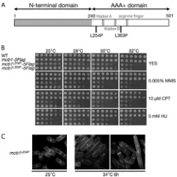
PCR amplification, and the sequences were determined. A sin-gle nucleotide change was identified at the 254th or 363th codon, both of which cause substitutions from the conserved hydrophobic leucine residue to proline. These two amino acids (Leu-254 and Leu-363) are highly conserved among MCM-BP orthologs and are located in the C-terminal domain of Mcb1 (Fig. 3Aandsupplemental Fig. S1). To confirm that the L254P or L363P substitution resulted in the phenotype observed in the twomcb1tsmutants, the L254P or L363P mutation was
intro-duced into wild-type cells using the PCR-based tagging method to place a 5⫻FLAG epitope at the C terminus and mark the allele with the kanMX6 gene. The resulting G418-resistant haploid transformants, designatedmcb1L254P-5FLAGormcb1L363P
-5FLAG, produced the temperature-sensitive phenotypes. Therefore, we concluded that a single amino acid substitution (L254P or L363P) resulted in the phenotypes observed in the two mcb1ts mutants. These two strains, mcb1L254P-5FLAG
(mcb1L254P) andmcb1L363P-5FLAG(mcb1L363P), were used for
further analysis.
Themcb1L254Pandmcb1L363Pcells were incapable of colony
formation at 32 °C (Fig. 3B). Themcb1L254Pcells were
temper-ature-sensitive, even at 30 °C, but themcb1L363Pcells were not.
Therefore, further analysis focused on mcb1L254P, although
mcb1L363Pis also partially included in this study. Moreover, the
mcb1L254Pandmcb1L363Pcells were sensitive to S-phase
stress-ing agents, such as MMS, HU, and CPT. HU depletes the dNTP pool and inhibits DNA synthesis. CPT traps topoisomerase I on DNA and interferes with DNA replication (50). MMS alkylates the template DNA. Both MMS and CPT induce the formation of double-stranded DNA breaks (DSBs) when the replication fork passes damage sites, whereas HU causes fork stalling. However, there was no sensitivity to UV-mediated DNA dam-age (data not shown). These results show that Mcb1 is impor-tant for surviving replication stress.
Themcb1L254Pcells showedcdcphenotypes, with an
elon-gated cell shape and 2C DNA content (Fig. 3C) (data not shown). A small percentage of cells (⫾2–5%) showed an abnor-mal nuclear phenotype, such as abnorabnor-mal DNA segregation,cut (cell uniformed tone) phenotypes, and abnormal septation (Fig. 3C). These phenotypes suggest that themcb1L254Pcells most
likely suffer DNA damage. Actually, themcb1L254Pcells
accu-mulated spontaneous DNA damage, as recognized by Rad22 (supplemental Fig. S2, C and D); showed Rad3 and Chk1 dependence for viability at 30 °C (supplemental Fig. S2A); and induced the phosphorylation of Chk1 kinase at the permissive temperature as well as at the restrictive temperature ( supple-mental Fig. S2B), suggesting activation of the Chk1-dependent DNA damage checkpoint. These results are consistent with the result reported by Li et al.(30), and Ding and Forsburg also conclude that overexpression of Mcb1 triggers cell cycle arrest in a manner that depends upon Rad3 and Chk1 (29).
FIGURE 2.Mcb1 interacts with any of the individual MCM subunits.A, two-hybrid interactions between Mcb1 and Mcm2–7. The interaction of Gal4-DBD-Mcb1 in the AH109 strain (when combined with the Gal4-AD-Mcm2–7 plasmids) was tested in the yeast two-hybrid assay. The interactions were monitored on SC⫺Trp⫺Leu (SC⫺WL; non-selective) medium and SC⫺Trp⫺Leu⫺His⫺Ade (SC⫺WLHA; selective) medium. For vector and Mcb1, the parts of Fig. 7Cwere used in this figure.B, interactions between bacterially expressed Mcb1 protein and Mcm2–7. T7-His6-tagged Mcb1 protein (T7-His6-Mcb1), His6-tagged Swi6
protein (His6-Swi6), and GST-tagged Mcm2–7 proteins (GST-McmX) were produced inE. coli. Purified GST or GST-tagged Mcm2–7 proteins, which are
specifi-cally bound to glutathione-Sepharose, were mixed with purified T7-His6-Mcb1 protein or His6-Swi6 protein and pulled down by glutathione-Sepharose. The
GST pull-down samples were separated on 8% SDS-polyacrylamide gels and blotted with an anti-His6antibody to detect T7-His6-Mcb1 or His6-Swi6 and with
an anti-GST antibody to detect GST-tagged proteins.
by guest on October 30, 2014
http://www.jbc.org/
Furthermore, the viability of the mcb1L254P mutant was
examined at the restrictive temperature. Mid-log phase unsyn-chronized cultures of mcb1-5FLAG and mcb1L254P-5FLAG
cells were shifted up to 36 °C for variable periods of time. Then variable numbers of cells were plated and incubated at 25 °C. The viability of themcb1L254Pcells decreased markedly after 4 h
of incubation at 36 °C, and no viable cells remained after 8 h of incubation (supplemental Fig. S3,AandB). The phenotype was also observed in a singlemcb1L254Pcell grown at 36 °C ( supple-mental Fig. S3C). The cell invariably stopped growing after 2– 4 divisions (4 –16 cells) and showed an elongated cell shape.
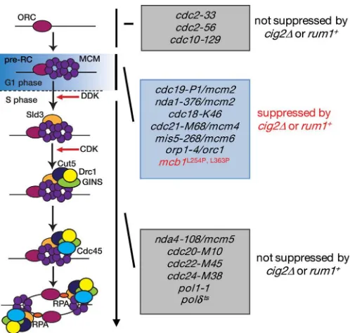
Genetic Interaction between mcb1⫹ and DNA Replication Factors—Combining two mutations in different genes results in synthetic lethality or enhanced temperature sensitivity when the corresponding gene products function in the same pathway. To explore the genetic relationship between Mcb1 and other replication factors, double mutants carryingmcb1L254Pand a
mutation in a component of the pre-RC, the pre-IC, or the DNA polymerases were constructed. Themcb1L254Pcdc19-P1/mcm2
and themcb1L254Pswi7-H4(Pol␣) double mutants exhibited
more severe sensitivity than the parental strains, whereas the sensitivity ofmcb1L254Psld3-10did not differ significantly from
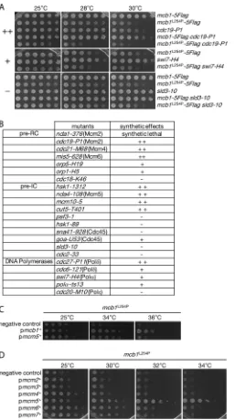
that of single mutants (Fig. 4A). The results of similar experi-ments are summarized in Fig. 4B. A mcb1L254P nda1-376/
mcm2double mutant could not be created, indicating that the combination of the mcb1L254P mutation with thenda1-376/
mcm2mutation results in synthetic lethality or an extremely low viability phenotype (data not shown). The temperature sen-sitivity of themcb1L254Pcells was increased bycdc19-P1/mcm2,
cdc21-M68/mcm4, mis5-628/mcm6, hsk1-1312, nda4 –108/ mcm5,cut5-T401,mcm10-5, andcdc27-P11(Pol␦). The tem-perature sensitivity of themcb1L254Pcells was slightly increased
byorp5-H19, orp1-H5,swi7-H4(Pol␣),goa1-U53/cdc45, pol␣-ts13, andcdc6 –121(Pol␦). By contrast, the temperature sensi-tivity of the mcb1L254Pcells was not affected by cdc18-K46,
hsk1-89,sna41-928/cdc45,sld3-10,cdc2-33,psf3-1, or cdc20-M10(Pol⑀) (Fig. 4B). These results suggest that Mcb1 functions in processes that involve the pre-RC components Nda1/Cdc19/ Mcm2, Cdc21/Mcm4, and Mis5/Mcm6; the pre-IC compo-nents Hsk1, Nda4/Mcm5, and Mcm10; and the DNA polymer-ase component Cdc27/Pol␦.
Overproduction of Mcm5 Suppresses the Temperature Sensi-tivity of mcb1tsMutants—To identify factors that functionally interact with the Mcb1 protein, a screen for fission yeast genes able to suppress the temperature sensitivity ofmcb1L254Pcells
was conducted using a multicopy plasmid. The fission yeast genomic library (pTN-L1 genomic library from the National BioResource Project) (49) was introduced intomcb1L254Pcells,
and transformants that were less sensitive to high temperature were selected. The plasmids recovered from these less sensitive clones contained themcb1⫹or themcm5⫹gene. The product of themcm5⫹gene, the Mcm5 protein, is one of the subunits of the MCM complex. A serial dilution assay showed that overex-pression of Mcm5 suppressed the temperature sensitivity of mcb1L254Pcells at 34 °C but not at 36 °C (Fig. 4C). Complete
suppression was achieved only by the introduction of a multi-copy plasmid expressing themcb1⫹gene. This result prompted us to investigate the specificity of this suppression by introduc-ing multicopy plasmids expressintroduc-ing other members of the MCM complex. As shown in Fig. 4D, overproduction of Mcm2, Mcm3, or Mcm4 did not suppress the temperature sensitivity
ofmcb1L254Pcells at all, but overproduction of Mcm6 or Mcm7
did slightly suppress the temperature sensitivity at 30 °C. Sim-ilar results were obtained with mcb1L363P cells (data not
shown). These results indicate the close genetic relationship betweenmcb1⫹and some MCM genes, especially themcm5⫹ gene. These findings suggest that some MCM functions are impaired by themcb1L254Pmutation.
CDK Modulation Suppresses mcb1ts Mutants—To better understand the defects of themcb1L254Pmutant, a screen for
spontaneous suppressors of themcb1L254Pmutant able to grow
at the restrictive temperature was conducted. Three indepen-dent suppressors that could grow even at 34 °C, here called suppressors 1, 2, and 3, were isolated (Fig. 5A). In addition to their ability to support cell growth at a high temperature, sup-pressors 1 and 2 showed cold-sensitive phenotypes at 20 °C, but suppressor 3 did not (Fig. 5A). To search for the responsible gene, multicopy plasmids were isolated by complementation for these cold-sensitive defects and temperature-resistant effects in suppressors 1 and 2 using the fission yeast genomic library. The sequencing of the clones isolated from the genomic library revealed the presence of the same gene,ded1⫹, on each plasmid (Fig. 5B). Ded1 is a general translation factor that is functionally homologous to RNA helicases from budding yeast FIGURE 3. Isolation and phenotype of mcb1 temperature-sensitive
mutants.A, schematic representation of the Mcb1 protein showing the loca-tion of point mutaloca-tions in the temperature-sensitive mcb1L254P and mcb1L363Palleles. The approximate location of the Walker A, Walker B, and
arginine finger features, which are characteristic of the canonical MCM pro-teins (which are absent in MCM-BP), are shown for reference.B, wild-type,
mcb1-5FLAG, mcb1L254P-5FLAG, and mcb1L363P-5FLAG cells were serially
diluted 5-fold, spotted onto YES medium supplemented with MMS, CPT, or HU at the indicated concentrations, and incubated at 25–32 °C for 3– 6 days.
C, themcb1L254P-5FLAGcells were grown in YES at 25 °C until log phase and
then shifted up to 34 °C for 6 h. Representative DAPI-stained images of the cells are shown.
by guest on October 30, 2014
http://www.jbc.org/
andDrosophila. A ded1⫹ gene was characterized as sum3⫹, moc2⫹, or slh3⫹ in independent studies (51–53). The ded1⫹ locus of suppressors 1 and 2 was amplified by PCR and sequenced, and mutations in theded1genes were detected in both suppres-FIGURE 4.Genetic interaction betweenmcb1ⴙand the DNA replication
components.A, growth of double mutants. 5-Fold serial dilutions of cells with the indicated genotypes were spotted onto YES plates and incubated at the indicated temperatures. The alleles used were mcb1L254P, cdc19-P1/ mcm2,swi7-H4(Pol␣), andsld3–10. Themcb1L254Pshowed strong synthetic
defects withcdc19-P1(⫹⫹),mcb1L254Pshowed weak synthetic defects with swi7-H4(⫹), andmcb1L254Pshowed no or little synthetic defect withsld3-10
(⫺).B, summary of the growth of the double mutants. The growth of the double mutants was examined as in (B).⫹⫹, strong synthetic defects;⫹, weak synthetic defects;⫺, no synthetic defects.C, gene dosage suppression of the temperature sensitivity of mcb1L254Pcells. Aliquots of mcb1L254P leu1⫺32 cells harboring the pTN-L1-rad26⫹ (negative control),
pTN-L1-mcb1⫹, or pTN-L1-mcm5⫹were spotted onto EMM plates after 5-fold serial dilution and incubated at permissive (25 °C) or restrictive (34 and 36 °C) tem-peratures. D, gene dosage suppression of the temperature sensitivity of
mcb1L254Pby other components of the MCM complex. Aliquots ofmcb1L254P
cells harboring pTN-L1-rad26⫹ (negative control) or pTN-L1-mcm2⫹, -mcm3⫹, -mcm4⫹, -mcm5⫹, -mcm6⫹, or -mcm7⫹were spotted onto EMM plates (after 5-fold serial dilution) and incubated at the indicated tempera-tures. In addition to themcm5⫹plasmid, plasmids expressingmcm6⫹or
mcm7⫹ showed weak suppression of the temperature sensitivity of
mcb1L254Pcells.
FIGURE 5.ded1alleles specifically suppress the temperature-sensitive phenotype of themcb1L254Pmutant.A, isolation of spontaneous
suppres-sors of themcb1L254Pmutant. 5-Fold serial dilutions of cells with the indicated
genotypes, including the three spontaneous suppressors ofmcb1L254P
(sup-pressors 1, 2, and 3), were spotted onto YES plates and incubated at the indicated temperatures. Suppressors 1 and 2 suppressed the temperature-sensitive phenotype ofmcb1L254Pcells and showed the additional
cold-sen-sitive phenotypes.B, a multicopy plasmid expressing theded1⫹gene sup-pressed the cold sensitivity of suppressors 1 and 2. Aliquots of wild-type, suppressor 1, and suppressor 2 cells harboring the pTN-L1-rad26⫹(negative control) or pTN-L1-ded1⫹were spotted on EMM plates (after 5-fold serial dilution) and incubated at the indicated temperatures.C, schematic represen-tation of theded1⫹/sum3⫹/moc2⫹/slh3⫹gene. A DEAD-box helicase domain (amino acids 171–393), a DEXDc (DEAD-like helicase superfamily) domain (amino acids 184 – 407), and HELICc (helicase superfamily c-terminal) domain (amino acids 403–533) are indicated bylines. The positions of the mutated sites in sup-pressor 1 (S1; A490S) and 2 (S2; I404T) are indicated.D, the episomal expression of thecig2⫹gene reversed the effect of theded1mutant. Aliquots of wild-type, suppressor 1, and suppressor 2 cells harboring the pREP41 vector or
pREP41-cig2⫹were spotted onto EMM plates (after 5-fold serial dilution) and incubated at the indicated temperatures.E, deletion of thecig2gene rescues the temperature-sensitive phenotype of themcb1L254Pmutant. 5-Fold serial dilutions of wild-type, cig2⌬,mcb1L254P-5FLAG, andmcb1L254P-5FLAG cig2
⌬cells were spotted on YES medium and incubated at 25 and 32 °C for 3– 6 days. Amcb1L254Pandcig2⌬
double mutant grew even at 32 °C.F, a multicopy plasmid expressing therum1⫹ gene can suppress the temperature sensitivity of mcb1L254P. Aliquots of mcb1L254Pcells harboring the pTN-L1-rad26⫹ (negative control) or
pTN-L1-rum1⫹were spotted onto EMM plates (after 5-fold serial dilution) and incubated at the indicated temperatures. Introduction of a multicopy plasmid expressing
rum1⫹partially suppressed the temperature-sensitive phenotype ofmcb1L254P
at both 30 and 32 °C.
by guest on October 30, 2014
http://www.jbc.org/
sors, indicating that mutations in theded1genes are responsible for both suppressors. Both suppressors contain a single amino acid change in theded1gene (Fig. 5C). Suppressor 1 contained an ala-nine to serine mutation at position 490 (A490S), and suppressor 2 contained an isoleucine to threonine mutation at position 404 (I404T). Ded1 is an ATP-dependent RNA helicase, the sequence of which contains DNA motifs such as the DEAD-box helicase, the DEAD-like helicase superfamily (DEXDc), and the helicase super-family C-terminal domain (HELICc). Both mutations are located within the putative HELICc motif.
One of the features of Ded1 is its influence on B-type cyclin Cig2 expression. It is reported thatded1mutants reduce the translation of Cig2 and suppress defects in pre-RC formation during DNA replication. Consequently, increased expression of Cig2 prevents theded1mutants from suppressing such defects, and deletion of thecig2⫹gene (cig2⌬) suppresses mutants that show defects in pre-RC formation (52, 54 –56). Indeed, the introduction of a multicopy plasmid overexpressing cig2⫹ restored the temperature sensitivity of suppressors 1 and 2 (Fig. 5D). Furthermore, double mutants of mcb1L254P and cig2
⌬
grew well at 32 °C (Fig. 5E).
Cig2 promotes the onset of S-phase by activating CDK (55). By contrast, Rum1 inhibits the CDK activity of the Cdc2 kinase, thereby inhibiting the start in the G1phase (57–59). Because Cig2 repression results in the suppression of the temperature-sensitive phenotype of the mcb1L254P mutant, the effect of
Rum1 overexpression was tested. As shown in Fig. 5F, overex-pression of Rum1 resulted in the supoverex-pression of the tempera-ture-sensitive phenotype of the mcb1L254P mutant at 32 °C.
CDK modulation by Cig2 and Rum1 suppresses defects caused by mutations in pre-RC components (Fig. 9). This suppression is specific for pre-RC formation during S-phase because muta-tions in pre-IC components are not suppressed by CDK modula-tion (56, 60, 61). A possible explanamodula-tion is that an extended G1 phase upon transient inhibition of CDK provides time to assemble the pre-RC in mutants that show defects in pre-RC formation (55). Furthermore, themcb1L254Pmutant could partially suppress the
HU sensitivity of thecds1⌬cells (data not shown). This result sug-gests some defects of MCM function in mcb1L254P mutant,
because mutations in the MCM complex are known to suppress the HU sensitivity of the replication checkpoint mutants (62). All of these genetic results strongly indicate that the mcb1L254P
mutant has some defects in pre-RC formation, suggesting that Mcb1 has an important function in pre-RC formation.
Because suppressor 3 did not show any additional phenotype (Fig. 5A), whole-genome sequencing was performed and showed that the genomic region containing themcm5⫹gene was ampli-fied 3 times (data not shown). This finding is consistent with gene dosage suppression of the temperature sensitivity ofmcb1L254Pby
themcm5⫹plasmid (Fig. 4C). These results further strengthen the tight genetic link betweenmcb1⫹andmcm5⫹.
Accumulation of Rhp54 Nuclear Foci in mcb1L254P Cells— Following activation of the DNA damage response, DNA repair takes place. Homologous recombination is the major pathway for the repair of DSBs. In this pathway, Rad52 is recruited to sites of DNA damage, where it mediates Rad51 filament forma-tion on the single-stranded tail of the DSBs. Then Rad51 per-forms a homology search followed by DNA strand exchange
with the donor strand, a process mediated by Rad54/Rhp54 (54). It was recently reported that an abundance of pre-RCs is important for the late step of recombination, in which Rhp54 functions (54). A link between the pre-RC and the late step of recombination was supported by the finding that the cdc18-K46andmcm6-S1mutants were hypersensitive to MMS and CPT and that the fraction of cells exhibiting Rhp54 foci remained large, even after release from MMS treatment, in both mutants. Themcb1L254Pmutant was sensitive to MMS
and CPT (Fig. 3B). Although MMS-induced Chk1 activation and Rad22 (fission yeast Rad52 ortholog) focus formation occurred normally (supplemental Fig. S4,AandB), the number of cells exhibiting Rhp54 foci after release from MMS treatment increased in themcb1L254P mutant to a level similar to that
observed in thecdc18-K46andmcm6-S1mutants ( supplemen-tal Fig. S5). These results support the idea that there are defects in pre-RC levels in themcb1L254Pmutant.
The mcb1ts Mutants Show Defects in S-phase Progression— Based on the genetic observations described above, defects in DNA replication were carefully examined inmcb1L254Pcells.
To define the role of the Mcb1 protein during DNA replication, S-phase progression was examined in mcb1L254P cells. The
nda3-KM311 mcb1-5FLAG and nda3-KM311 mcb1L254P
-5FLAGcells were synchronized in M phase and then released either at 28 or 36 °C (block and release by thenda3-KM311 mutant). The DNA content, as analyzed by FACS, showed that S-phase progression inmcb1L254P-5FLAGcells was slightly but
reproducibly delayed relative to that inmcb1-5FLAGcells both at 28 and 36 °C (Fig. 6,AandB). It appears that the progression of S-phase, once initiated, did not differ much at 28 and 36 °C. Thus, it is possible thatmcb1L254Pmutation more specifically
affects the initiation stage of S-phase. Furthermore, to follow the progression of S-phase, the septation index of these cells was monitored as shown in Fig. 6,AandB. The peak of the septation index in fission yeast coincides with S-phase during cell cycle progression.The mcb1L254P-5FLAGcells had a much
lower septation index thanmcb1-5FLAGcells (Fig. 6,CandD). At the permissive temperature, the septation index ofmcb1 -5FLAGcells peaked 40 min after release (76.9%) and rapidly decreased afterward, indicating that S-phase progression was normal. The septation index of mcb1L254P-5FLAG cells also
peaked at 40 min, but the peak was only 32.5% (Fig. 6C). From the profile shown in Fig. 6C, S-phase progression inmcb1L254P
-5FLAGcells seemed to continue until much later, up to 120 min after release (16.2% inmcb1L254P-5FLAGcells compared with
3.2% inmcb1-5FLAGcells). When the release temperature was shifted to 36 °C, the cell cycle progression ofmcb1-5FLAGcells became faster, and the septation index peaked 20 min after release (63.1%) (Fig. 6D). Release at the restrictive temperature did not result in any significant differences in the FACS profile compared with release at the permissive temperature (Fig. 6,A and B); however, the septation index profile of mcb1L254P
-5FLAGcells barely changed, peaking at 40 min (30.6%) and continuing until 120 min after release (16.3%) (Fig. 6D). Similar results were obtained withmcb1L363P-5FLAG cells (data not
shown). Although we cannot rule out the possibility that that
mcb1L254Pmutant cells show defects in release from M phase
by guest on October 30, 2014
http://www.jbc.org/
arrest, these results strongly suggest a defect in S-phase progression.
Thus, loading of MCM proteins onto the replication origins during the initiation of DNA replication was investigated in
mcb1L254Pmutants. For this purpose, ChIP analysis was
per-formed to monitor the chromatin binding of Mcm7-3HA to early firing origins (ars2004andars3002), a late firing origin (AT2080), and non-origin (non-ars1) both inmcb1-5FLAGand
mcb1L254P-5FLAGcells (Fig. 6E andsupplemental Fig. S6A).
The mcb1L254P mutation markedly decreased Mcm7-3HA
binding to the replication origin, whereas the binding of Orp4, a component of the origin recognition complex, was not decreased (Fig. 6Eandsupplemental Fig. S6B). This result indi-cates that Mcb1 function is important for the efficient loading of MCM complexes onto the replication origins.
The mcb1tsMutations Impair the Interaction of Mcb1 with the MCM Complex—Judging from the phenotypes of the mcb1tsmutants described above, we speculated that the
inter-action of Mcb1 with the MCM complex was affected in the mcb1tsmutants. To test this possibility, MCM complex
forma-tion and the interacforma-tion between Mcb1 and the MCM complex were examined in themcb1tsmutants. As expected, the
inter-action between the mutant Mcb1 proteins (Mcb1L254P and
Mcb1L363P) and the MCM complex was markedly attenuated.
Using the mcb1-5FLAG, mcb1L254P-5FLAG, and mcb1L363P
-5FLAG strains, Mcb1-5FLAG immunoprecipitation was per-formed at permissive (25 °C) and restrictive temperatures (36 °C). Mcb1-5FLAG immunoprecipitated all MCM proteins tested both at 25 and 36 °C (Fig. 7A, lanes 6 and 14). The amount of Mcm2 co-precipitated with Mcb1 was considerably FIGURE 6.S-phase progression is markedly affected inmcb1L254Pcells.AandB, experimental scheme and DNA profiles.nda3-KM311 mcb1-5FLAG mcm7 -3HAandnda3-KM311 mcb1L254P-5FLAG mcm7-3HAcells were grown in YES medium at 28 °C and then incubated at 20 °C for 4 h to induce arrest in M phase. Cells
were released at 28 °C (A) or 36 °C (B) (time 0). Aliquots were harvested every 20 min up to 120 min and analyzed by flow cytometry.CandD, aliquots inAand
Bwere monitored to assess the septation index. The values were calculated using⫾200 cells per time point (n⫽3 independent experiments).E, aliquots were harvested every 20 min up to 120 min and analyzed by a ChIP assay using an anti-HA antibody against Mcm7-3HA. The loading of Mcm7-3HA ontoars2004and
non-ars1was then determined by real-time PCR. Data represent the average percentage of IP/input⫾S.D. (error bars) (n⫽3).
by guest on October 30, 2014
http://www.jbc.org/
less than that of other MCM proteins (Fig. 7A,lanes 6and14). The interaction between Mcb1 and the MCM proteins was detectable in bothmcb1L254Pandmcb1L363Pcells but was
mark-edly decreased, even at 25 °C (Fig. 7A,lanes 7and8). There was no significant change in the interaction between Mcb1 and MCMs inmcb1L254Pormcb1L363Pcells at 36 °C (Fig. 7A,lanes
15and16). As shown earlier, Mcb1 interacted with all of the
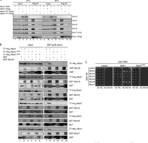
subunits of the MCM complex in a yeast two-hybrid assay and in anin vitropull-down assay (Fig. 2,AandB). Next, we tested whether the interaction between Mcb1 and the MCM complex is affected in themcb1L254Pandmcb1L363Pmutants.
Interest-ingly, neither the Mcb1L254P nor Mcb1L363P protein could
interact with any of the subunits of the MCM complex in the yeast two-hybrid assay and pull-down assay (Fig. 7,BandC). FIGURE 7.Analysis of the Mcb1䡠Mcm2–7 complex.A,mcm7-3HA,mcb1-5FLAG mcm7-3HA,mcb1L363P-5FLAG mcm7-3HA, andmcb1L254P-5FLAG mcm7-3HA
cells were grown in YES at 25 °C and then shifted up to 36 °C and incubated for a further 6 h. Soluble cell lysates were prepared using the glass bead method, immunoprecipitated with an anti-FLAG antibody, and immunoblotted for Mcm2 with an anti-Mcm2 antibody, for Mcm4 with an anti-Mcm4 antibody, for Mcm5 with an anti-Mcm5 antibody, for Mcm6 with an anti-Mcm6 antibody, for Mcm7-3HA with an anti-HA antibody, or for Mcb1-5FLAG with an anti-FLAG antibody.B, Mcb1 interacted with any of the individual MCM subunits, but Mcb1L254Pdid not. The interaction between bacterially expressed Mcb1tsmutant
proteins and Mcm2–7 is shown. T7-His6-tagged Mcb1 protein (T7-His6-Mcb1), T7-His6-tagged Mcb1
L254Pprotein (T7-His 6-Mcb1
L254P), T7-His 6-tagged
Mcb1L363Pprotein (T7-His 6-Mcb1
L363P), GST, and GST-tagged Mcm2–7 proteins were produced inE. coli. Purified GST or GST-tagged Mcm2–7 proteins, which
were specifically bound to glutathione-Sepharose, were mixed with soluble crude lysates ofE. coliexpressing T7-His6-Mcb1, T7-His6-Mcb1L254P, or T7-His6
-Mcb1L363Pprotein and subjected to pull-down by glutathione-Sepharose. GST pull-down samples were separated on 8% SDS-polyacrylamide gels and blotted
for T7-His6-Mcb1, T7-His6-Mcb1
L254P, or T7-His 6-Mcb1
L363Pwith an anti-His
6antibody and for GST fusions with an anti-GST antibody.C, two-hybrid interaction
between Mcb1 and Mcm2–7 or between Mcb1L254Pand Mcm2–7. Gal4-DBD-Mcb1 or Gal4-DBD-Mcb1L254Pin the AH109 strain was tested for interaction with
the Gal4-AD-Mcm2–7 plasmid in the two-hybrid assay. The interactions were monitored as described in the legend to Fig. 2A.
by guest on October 30, 2014
http://www.jbc.org/
These results suggest that the temperature-restricted pheno-types of themcb1tsmutants are not simply caused by the altered
interaction between the Mcb1 and MCM proteins.
Localization of the MCM Protein Is Compromised in mcb1ts Mutants—In fission yeast, Mcm2–7 proteins are constitutively localized in the nucleus throughout the cell cycle (5). Mutations that disrupt MCM complex formation, such asmcm4ts, cause
all of the MCM subunits to exit the nucleus (63). Therefore, the effect of themcb1⫹gene mutations on the nuclear localization of Mcm2–7 proteins was studied. The localization of all of the MCM complex subunits was observed in wild-type cells or mcb1tsmutants at both permissive and restrictive temperatures
using GFP or RFP (GFP for Mcm2, -3, -4, -6, and -7; RFP for Mcm5) (Fig. 8A). Mcm2-GFP showed clear nuclear localization at 25 °C in mcb1-5FLAG, mcb1L254P-5FLAG, and mcb1L363P
-5FLAGcells; however, at the restrictive temperature (34 °C), nuclear localization of Mcm2-GFP diminished, and cytoplas-mic staining increased in both mcb1L254P-5FLAG and
mcb1L363P-5FLAG cells (Fig. 8A). Similar observations were
made inmcb1L254P-5FLAGandmcb1L363P-5FLAGcells using
Mcm4-GFP, Mcm5-RFP, Mcm6-GFP, and Mcm7-GFP, whereas Mcm3-GFP showed no real change (Fig. 8A). The GFP or RFP signals observed inmcb1ts mutants at the restrictive
temperature were not completely excluded from the nucleus because there was residual signal in the nucleus. Afterward, we tested whether these phenotypes could be suppressed by a mul-ticopy plasmid containingmcm5⫹, which can partially sup-pressmcb1tsmutants as shown in Fig. 4C(Fig. 8B). Multicopy
plasmids containingmcm5⫹,mcb1⫹ as a positive control, or ded1⫹as a negative control were transformed intomcm2-GFP
mcb1L254P-5FLAGandmcm6-GFP mcb1L254P-5FLAGcells. As
expected, the mislocalization of Mcm2-GFP and Mcm6-GFP observed above was suppressed by the introduction of a multi-copy plasmid containingmcm5⫹. These results indicate that the mislocalization of MCM proteins is partially rescued by the overexpression ofmcm5⫹. Next, we examined whether these phenotypes were reflected in targeting to the nucleus, retention in the nucleus, or both.
MCM Proteins Are Actively Exported from the Nucleus in mcb1tsMutants—The loss of MCM proteins from the nucleus in mcb1ts mutants may reflect protein turnover and a failure to
import newly synthesized molecules, or it could indicate a defect in protein retention within the nucleus. Steady-state levels of MCM proteins were unchanged in cells arrested bymcb1tsmutations
(Fig. 8D). Because there was no evidence of any change in protein levels, the role of nuclear export in the redistribution of MCM proteins in mcb1ts mutants was examined. The crm1⫹ gene encodes a nuclear export receptor (64). To determine whether MCM relocalization requires active nuclear export, thecrm1and mcb1 genes were simultaneously inactivated. If MCM proteins requirecrm1-dependent active export, they should be trapped in the nucleus under these restrictive conditions.
Thecrm1mutant was used for this experiment. A tempera-ture-sensitivecrm1allele (crm1–11R) (65) was combined with
themcb1L254Pmutant. Interestingly, Mcm2-GFP and
Mcm6-GFP themselves remained in the nucleus at the restrictive tem-perature (Fig. 8C). These results suggest that an active nuclear export system is required for the relocalization of MCM
pro-teins when Mcb1 is inactivated. The localization of any one of the MCM proteins is influenced by the activity of all other members of the complex. When MCM function is abrogated, MCM subunits are removed from the nucleus via an active nuclear export system (63). Taking all this into consideration, these results suggest that MCM functions are abrogated in the
mcb1L254Pmutant.
DISCUSSION
The functions of MCM-BP during DNA replication have been extensively studied in several organisms; however, the mechanisms by which MCM-BP functions and associates with MCM complexes are not well understood, and there seems to be no unified view concerning the shared common function of MCM-BP across species. Therefore, the present study aimed to elucidate the molecular mechanisms underlying MCM-BP function in fission yeast through biochemical and genetic anal-yses. The results showed that the fission yeast MCM-BP, Mcb1, plays an important role as a regulator of MCM function in pre-RC formation during DNA replication.
Although Mcb1 is a novel binding partner of the MCM com-plex in fission yeast, the nature of the interaction between Mcb1 and MCM proteins is not yet clear. MCM proteins can be pres-ent in three forms: free monomers, subcomplexes, or a com-plete hexamer complex. In fission yeast (as in humans and Xenopus), Mcb1 interacts with all MCM subunits, except Mcm2 (29, 30). By contrast, ETG1 (AtMCM-BP) interacts with all MCM complex subunits inArabidopsis. Immunoprecipita-tion analysis showed that Mcb1 interacted with all MCM sub-units, including Mcm2, whereas the amount of Mcm2 co-pre-cipitated by Mcb1 was much lower than that of any other MCM. Two methods of preparing crude cell extracts for immu-noprecipitation were tested, the spheroplast method and the beads method; only a very small amount of Mcm2 was immu-noprecipitated by Mcb1-5FLAG using either method (Figs. 1B and 7A). Recently, Ding and Forsburg reported that an interac-tion between Mcm2 and Mcb1 can be observed when Mcb1 is overexpressed, suggesting that this interaction may be weak but not completely lacking (29). Our result is consistent with this observation. Furthermore, the results showed that the Mcb1 protein could bind to any individual subunit of the MCM com-plex and that the relative affinity of Mcb1 for Mcm2–7 in the immunoprecipitation analyses was variable. Therefore, the interaction profiles described above predict that Mcb1 might form complexes with the individual MCMs and/or subcom-plexes in addition to the complete MCM complex. Further studies usingin vitroreconstitution of the MCM complex will be required to clarify these points.
Two temperature-sensitivemcb1 gene mutants were used for genetic analysis of themcb1gene. Themcb1tscells showed a
cdcphenotype with an elongated cell shape and 2C DNA con-tent. Similarly, temperature-sensitive mutants of fission yeast mcmgenes arrested the cell cycle with acdcphenotype and showed the classic checkpoint-dependent arrest characteristic of other replication mutants. Thesemcb1tscells were sensitive
to MMS, HU, and CPT. Li et al. (30) recently reported the identification of additionalmcb1tsalleles that do not display
these sensitivities but share other phenotypes with the
by guest on October 30, 2014
http://www.jbc.org/
tions identified here. The difference in these two results may relate to the temperature utilized for the experiment rather than allele-specific differences in phenotype. There was a marked reduction in the interaction between Mcb1L254Pand
Mcm2–7in vivo, and the interaction between the Mcb1L254P
protein and individual MCM complex subunits was almost abolished both in the yeast two-hybrid assay and in thein vitro
pull-down assay. These reductions in thein vivo interaction between Mcb1 and Mcm2–7 were observed even at the permis-sive temperature, suggesting that the reduced level of the inter-action is still sufficient to support the viability ofmcb1L254Pcells at
the permissive temperature; however, the viability ofmcb1L254P
mutant cells was much lower than that of the wild-type cells, even at the permissive temperature, which is probably due to the
by guest on October 30, 2014
http://www.jbc.org/