NEW PYRAN OF AN ENDOPHYTIC FUNGUS
Fusarium
sp. ISOLATED
FROM THE LEAVES OF BROTOWALI (
Tinaspora crispa
)
Elfita
1,*, Munawar
2, Muharni
1, and Suprayetno
1 1Department of Chemistry, Faculty of Mathematics and Natural Sciences, Sriwijaya University, Jl. Raya Palembang Prabumulih Km 32, Indralaya, Ogan Ilir, South Sumatera 30662, Indonesia
2
Department of Biology, Faculty of Mathematics and Natural Sciences, Sriwijaya University, Jl. Raya Palembang Prabumulih Km 32, Indralaya, Ogan Ilir, South Sumatera 30662, Indonesia
Received May 21, 2013; Accepted October 20, 2013
ABSTRACT
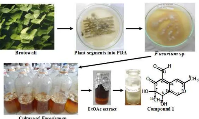
Endophytes are microorganisms that reside asymptomatically in the tissues of higher plants and are relatively unstudied and a promising source of novel organic natural metabolites exhibiting a variety of biological activities. As a part of our systematic search for new bioactive lead structures from endophytic, the endophytic fungus Fusarium sp. isolated from the leaves of Brotowali (Tinaspora crispa), was cultured for isolation of metabolite. The endophytic fungus was cultivated on 6 L of Potatos Dextose Broth (PDB) medium at room temperature (no shaking) for 8 weeks. The cultures were then extracted with ethyl acetate to afford 9.4 g of residue after removal of the solvent under reduced pressure. The extract was separated into the fractions by column chromatography (CC) on silica gel. The fractions were further separated by silica gel column chromatography to give one compound. The molecular structure was established on the basis of spectroscopic analysis including UV, IR, 1H-NMR, 13C-NMR, HMQC, HMBC, COSY, and MS. The compound was determined as a new pyran.
Keywords:new pyran; endophytic fungus; Fusarium sp.; Tinaspora crispa
ABSTRAK
Jamur endofitik merupakan mikroorganisme yang menempati jaringan tumbuhan tingkat tinggi dan masih terbatas kajiannya, serta merupakan sumber metabolit baru yang menunjukkan berbagai aktivitas biologis. Sebagai bagian dari penelitian sistimatis kami untuk menemukan senyawa bioaktif baru, maka telah diisolasi jamur endofitik Fusarium sp. dari daun Brotowali (Tinaspora crispa), dan telah dikultur untuk mengisolasi metabolit sekundernya. Jamur endofit dikultur dalam 6 L medium Potatos Dextose Broth (PDB) pada suhu kamar (keadaan statis) selama 8 minggu. Kultur kemudian diekstraksi dengan etil asetat untuk mendapatkan 9,4 g ekstrak pekat setelah penguapan pelarut pada tekanan rendah. Ekstrak difraksinasi dengan kromatografi kolom (CC) pada silika gel. Fraksi selanjutnya dikromatografi kolom silika gel hingga diperoleh satu senyawa. Struktur molekulnya ditentukan berdasarkan analisis spektroskopi UV, IR, 1H-NMR, 13C-NMR, HMQC, HMBC, COSY, dan MS. Senyawa hasil isolasi merupakan senyawa baru golongan piran.
Kata Kunci:piran baru; jamur endofitik; Fusarium sp.; Tinaspora crispa
INTRODUCTION
Endophytes are defined as fungi colonizing healthy tissue plant without overt symptoms or no apparent injury to the host. In the last decade, the exploration of secondary metabolites from endophytic fungi have proven that endophytes are a source of new compounds that have unique structures and a variety of biological activity such as anticancer, antimicrobial and other activities. The new compounds was potentially useful for human medicine, industrial, and agrochemical [1-2].
The selection of the host plant is important factor for the production of endophytic fungi and the isolation of
their bioactive secondary metabolites [2]. The strategi of plant selection to be a promising source of novel endophytes and their compound mainly on the basis of their unique environmental setting, ethnobotanical history (use by indigenous peoples), endemic, unusual longevity, and growing in areas of great biodiversity [3].
Indo. J. Chem., 2013, 13 (3), 209 - 215
210
practice of peoples living in Malaysia, Indonesia, and Thailand to treat ailments like fever, jaundice, hyperglycemia, wounds, intestinal worms, skin infections, tooth and stomach aches, coughs, asthma and pleurisy [7-8].
In our search for secondary metabolites of endophytic fungi, many bioactive and/or new compounds were isolated [4,9-11]. Elfita et al. [4] was reported the isolation of eight endophytic fungi from the stems and leaves of brotowali, but the fungusFusariumsp. was not found in the study. In this paper we report the isolation and structural identification a new pyran from Fusarium
sp. separated from the leaves of brotowali (T. crispa). Several Fusarium species isolated as endophytes have been reported to produce metabolites with antimicrobial, anticancer, and other activities, and also could be potential sources of novel natural products with industrial and agrochemical potential. There is speculation among the researchers about their nature of association in plants, as most Fusarium species are commonly encountered in soil as saprophytes, pathogens and are also associated with serious invasive mycoses in immunocompromised patients [2,12].
Bashyal et al. (2010) found tricinonoic acid and tricindiol, two irregular sesquiterpenes from an endophytic strain of Fusarium tricinctum, a fungus endophytic in the root tissue of the Sonoran desert plant,
Rumex hymenosepalus [13]. Wu et al. (2009) reported five compounds from endophytic fungus Fusarium sp. isolated from Trewia nudiflora. These compounds were including the three ent-trachylobane and two atisane-type diterpenoids [14].
EXPERIMENTAL SECTION
Materials
The leaves of brotowali were collected on March 2012 from the Indralaya, Ogan Ilir, South Sumatra. Material for isolation and cultivation of endophytic fungi using Oxoid: nutrient agar (NA), chloramphenicol, nutrient broth (NB), potato dextrose broth (PDB), and potato dextrose agar (PDA). Material for sterilization and separation by silica gel column chromatography using Merck: ethanol 70%, NaOCl, Si gel 60 (70-230 mesh.), thin layer chromatography (TLC), and silica gel 60 F254. Organic solvents (hexane, ethyl acetate, and methanol) were technical grade and distilled before use.
Instrumentation
The apparatus in the research were counter colony, autoclave, incubator, water bath, microscope, magnetic hotplate, UV lamp, column chromatography and generally apparatus in organic and microbiology
laboratory, Fisher Johns Melting Point, UV-Cary Varian 100 Conc I-UV Visible Spectrometer, FTIR-Perkin Elmer-Spectrum One, TOF MS ES+4.20e4, NMR spectra were recorded at 500 MHz (1H) and 125 MHz (13C) on JEOL JNM ECA-500 spectrometer, UV light at 254 nm and 365 nm.
Procedure
Isolation of endophytic fungi
The leaves sample was washed thoroughly in running tap water and air dried before it was processed. The materials were then surface sterilized by immersing them sequentially in 70% ethanol for 3 min and 0.5% NaOCl for 1 min and rinsed thoroughly with sterile distilled water. Then, with a sterile scalpel, outer tissues were removed and the inner tissues of 0.5 cm size were carefully dissected and placed on petri-plates containing potato dextrose agar medium (PDA) (200 g potato, 20 g dextrose, and 15 g agar in 1 L of H2O, supplemented with 100 mg/L of chloramphenicol to suppress bacterial growth). The plates were incubated at 25±2 °C until fungal growth appeared. The plant segments were observed once a day for the growth of endophytic fungi. Hyphal tips growing out the plated segments were immediately transferred into new PDA plates and then subcultured until pure cultures were obtained [15].
Identification of the endophyte
The endophytic fungal strain was identified by the morphological method. The morphological examination was performed by scrutinizing the fungal culture, the mechanism of spore production, and the characteristics of the spores. All experiments and observations were repeated at least twice [16].
Cultivation of pure fungal strain
The purified fungi (a small park) were transferred under sterile conditions to the PDB medium. For chemical investigations, the fungal strains were static cultivated into 15 flasks (1 L each) containing 400 mL of PDB medium for 8 weeks at room temperature [9-11].
Extraction, exploration, and structure elucidation
Fig 1.Isolation of the compound from ethyl acetate extract ofFusariumsp. from the leaves of brotowali
to give a pure compound.
The EtOAc fraction (9.4 g) was preabsorbed on silica gel and then chromatographed over a silica gel column using a gradient elution of hexane:EtOAc (10:0) to MeOH (100%). Based on detection by TLC using the eluent system, collected fractions was combined and rechromatographed over silica gel to give a pure compound. The molecular structure of compound was established on the basis of spectroscopic analysis including UV, IR, MS, 1H-NMR, 13C-NMR, HMQC, HMBC, and COSY.
RESULT AND DISCUSSION
The fungal strain was identified asFusarium sp by the Sekolah Ilmu dan Teknologi Hayati, Institute Teknologi Bandung, Indonesia. High colonization of the genus Fusarium were obtained from several plant species such as Mirabilis jalapa, Cephalotaxux mannii,
Sesbania grandiflora, familyAraceae, Leguminosaeand
Gramineae. This suggests allopatric existence of endophytic Fusarium based on different geographical regions and ecological niches and their ubiquity as endophyte among plant species [15, 17-20].
Then fungal Fusarium sp. was cultivated on 6 L of PDB medium for 8 weeks at room temperature. The culture was then extracted with ethyl acetate to afford 9.4 g of residue after removal of the solvent under reduced pressure. The extract (9.4 g) was separated over silica gel column (CC, 230–70 mesh, 30 × 4 cm) using a gradient of hexane:EtOAc (10:0) to MeOH (100%) to provide 6 fractions. Fraction 5 (3.7 g) was rechromatographed over silica gel (CC, 230–80 mesh, 30 × 3 cm) and using a gradient of EtOAc:MeOH (5:5) to MeOH (100%) to yield 3 fractions. Fraction 3 (2.9 g) was
rechromatographed again on silica gel (CC, 230–80 mesh, 30 × 2 cm) using the same gradient elution to yield compound1(2.3 g). The isolation of the compound from ethyl acetate extract of Fusarium sp. from the leaves of brotowali is described in Fig. 1.
(7R,8S)-7,8-dihydroxy-3,7-dimethyl-6-oxo-7,8-dihy dro-6H-isochromene-5-carbaldehyde (1), obtained as a
white crystal, mp. 230-231 °C; UV (MeOH) λmax nm:
255 and 381; UV (MeOH + NaOH) λmax nm: 255 and
381; IR (KBr) νmax cm-1: 3469.9 and 3375.4 (OH),
3099.6 (CH-vinilic), 2978.1 (CH-aliphatic), 1678.1 (C=O), 1608.3 and 1477.5 (C=C conjugation), 1043.5 (C-O ether); HRESI-MS ion peak at m/z 237.0762 [M + H]+; 1H-NMR (DMSO, 500 MHz) δH ppm: see Table 1 and Fig. 2; 13C-NMR (DMSO, 125 MHz)
δCppm: see Table 1.
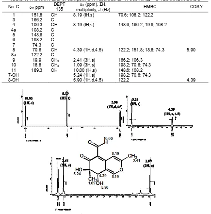
The HRESI-MS showed an [M+H]+ ion peak at m/z 237.0762 (calc. 237.0759), in accord with the molecular formula C12H12O5, implying seven degrees of unsaturation, and was supported by NMR 1D and 2D data (Table 1). The 1H-NMR spectrum, which was recorded in DMSO (Table 1) showed two hydroxy proton signal atH5.24 (1H,s) and 5.90 (1H,d,4.5 Hz), two methyl singlets (H 1.09 and 2.41), two vinilic proton signals which appeared at the same chemical shift at H8.19 (1H,s), and one aldehyde proton signal atH10.00 (1H,s).
Indo. J. Chem., 2013, 13 (3), 209 - 215
212
Table 1.The NMR data of compound1, recorded at1H-500 MHz;13C-125 MHz in DMSO
No. C Cppm DEPT 135
H (ppm), ΣH,
multiplicity, J (Hz) HMBC COSY
1 151.8 CH 8.19 (IH,s) 70.6; 108.2; 122.2
3 166.2 C
4 106.3 CH 8.19 (IH,s) 148.6; 166.2; 19.9; 108.2
4a 108.2 C
5 148.6 C
6 198.2 C
7 74.3 C
8 70.6 CH 4.39 (1H,d,4.5) 122.2; 151.8; 18.8; 74.3 5.90
8a 122.2 C
9 19.9 CH3 2.41 (3H,s) 166.2; 106.3 10 18.8 CH3 1.09 (3H,s) 198.2; 70.6; 74.3 11 189.3 CH 10.00 (IH,s) 148.6; 108.2
7-OH 5.24 (1H,s) 198.2; 70.6; 74.3
8-OH 5.90 (1H,d,4.5) 122.2 4.39
Fig 2.The1H-NMR spectrum of compound1
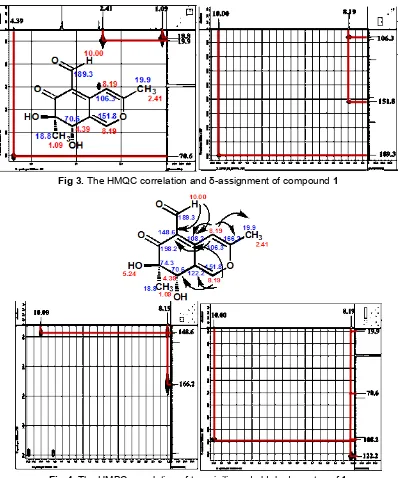
at C 151.8 (H 8.19,1H,s), 122.2, 166.2, 106.3 (H8.19,1H,s), 108.2, and 148.6, indicated the presence of three double bonds at 1,9, 3,4, and 5,10. The coupling constant between H-8 and 8-OH (J=4.5 Hz) indicated H-8, 8-OH configurations. The presence of cyclic ethers is shown by the presence of carbon and proton chemical shift at higher field atC151.8 (H8.19) and quarternary carbon at 166.2.
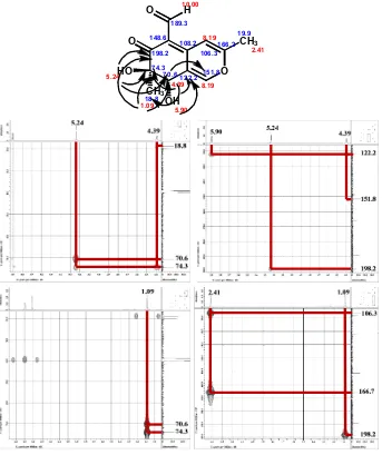
The HMBC correlations of isolated methyl protons H-10 with the oxygenated quarternary carbon C-7, the oxymethine carbon C-8, and the ketone carbon C-6 indicated that C-7 was connected to C-6, C-8, and C-10. Correlation from H-8 to C-8a, C-1, C-10, and C-7 led to the connection of C-8a to both C-1 and C-8, permitted
completion of the cyclohexenone subunit of 1. Further HMBC correlation from H-1 to C-3, C-8, C-4a, and C-8, and correlation from H-4 to C-5, C-1, C-9, and C-4a indicated that C-3 and C-4a was connected to C-1 and C-4 that have the same chemical shift. Correlation of the isolated methyl protons H-9 with the oxygenated quarternary carbon C-3 and the methine carbon C-4, thereby completing the pyran subunit of 1. Than the correlation of the aldehyde proton H-11 with C-5 established the connectivity between C-4 and C-5. The
HMBC correlation and δ-assignment of compound 1
Fig 3. The HMQC correlation and δ-assignment of compound 1
Fig 4.The HMBC correlation of two vinilic and aldehyde proton of1
Analysis of 1H–1H COSY spectrum (Fig. 6) led to the identification of two proton spin-system corresponding to the C-8–8-OH subunit of structure 1. All of the data showed that compound 1 has two rings that cyclohexenone and pyran has been called benzopyran.
Benzopyran is a very interesting class of heterocyclic compounds that results from the fusion of a benzene ring to a heterocyclic pyran ring. According to IUPAC nomenclature it is called chromene. Two classes of benzopyrans are 1-benzopyran and 2-benzopyran that vary by the orientation of the fusion of
the two rings compared to the oxygen. The 1-benzopyran has been called chromene and 2-benzopyran called as isochromene.
Base on Dictionary Natural Products data base (2010), compound 1 is new pyran. Previously, the similar pyran compounds have been found which has a ring of 7,8-dihydro-6H-isochromen-6-one that monapurfluor A and its isomers is monapurfluor B from red mold rice fermented by Monascus purpureus
Indo. J. Chem., 2013, 13 (3), 209 - 215
214
Fig 5.The HMBC correlation of hydroxyl, methyl, and methine proton of1
Fig 6.The COSY correlation of1
oxygenated at C-7. The difference is that compound1is substituted carbaldehyde group at C-5 and hydroxyl group at C-8, while others are not. The other three compounds cyclized at C-7 and C-8, respectively with a
furan ring to monapurfluor A and B, and the furanone ring for monasfluore B.
Exploration of the pyran has increased the types of secondary metabolites from endophytic fungi of brotowali. Exploration of secondary metabolites research needs to be done in order to get the profile of organic compounds produced by endophytic fungi of brotowali.
CONCLUSION
The endophytic fungi Fusarium sp. from the leaves of brotowali (T. crispa), has produced a new pyran. Based on spectroscopic analysis including UV, IR, MS,1H-NMR,13C-NMR, HMQC, HMBC and COSY, the compound is (7R,8S)-7,8-dihydroxy-3,7-dimethyl-6-oxo-7,8-dihydro-6H-isochromene-5-carbaldehyde.
O O
O H
CH3
CH3 OH HO
5 .2 4
5.90 1.09
4.39 10.00
2.41 8.19
8.19
106 .3
1 51.8 1 66.2
1 9.9
1 22.2 1 08.2 1 48.6
1 89.3
1 98.2
7 4.3 7 0.6
ACKNOWLEDGEMENT
The authors are grateful to the Directorate General of Higher Education for research funding through the Hibah Bersaing Grant in 2012.
REFERENCES
1. Li, H.J., Lin, Y.C., Vrijmoed, L.L.P., and Jones, E.B.G., 2004,Chin. Chem. Lett., 15, 4, 419–422. 2. de Souza, J.J., Vieira, I.J.C., Rodrigues-Filho, E.,
and Braz-Filho, R., 2011, Molecules, 16, 2, 10604–10618.
3. Strobel, G., Daisy, B., and Castillo, U., 2005, Plant Pathol. J., 4, 2, 161–176.
4. Elfita, Muharni, Munawar, Legasari, L., and Darwati, 2011,Indo. J. Chem., 11, 1, 53–58.
5. Kongkathip, N., Dhumma-upakorn, P., Kongkathip, B., Chawananoraset, K., Sangchomkaeo, P., and Hatthakitpanichakul, S., 2002, J. Ethnopharmacol., 83, 1-2, 95–99.
6. Haque, Md.A., Islam, S.M.A., and Mohammad, S., 2011,J. Pharm. Biomed. Sci., 13, 12, 1–4.
7. Najib, N.A.R.N., Furuta, T., Kojima, S., Takane, K., and Ali, M.M., 1999, J. Ethnopharmacol., 64, 3, 249–254.
8. Al-alusi, N.T., Kadir, F.A., Ismail, S., and Abdullah, M.A., 2010, Afr. J. Microbiol. Res., 4, 21, 2309–2312.
9. Elfita, Muharni, and Indah T., 2011,Makara Sci. Ser., 15, 2, 124–128.
10. Elfita, Muharni, Munawar, and Aryani, S., 2012,
Indo. J. Chem., 12, 2,195–200.
11. Elfita, Muharni, Munawar, and Rizki, 2012,Makara Sci. Ser., 16, 1, 46–50.
12. Tayung, K., Barik, B.P., Jagadev, P.N., and Mohapatra, U.B., 2011,Malays. J. Microbiol., 7, 1, 1–6.
13. Bashyal, B.P., and Gunatilaka, A.A.L., 2010, Nat. Prod. Res., 24, 4, 349–356.
14. Wu, X., Lu, C-H., and Shen, Y-M., 2009, Helv. Chim. Acta, 92, 12, 2783–2789.
15. Barik, B.P., Tayung, K., Jagadev, P.N., and Dutta, S.K., 2010,Eur. J. Biol. Sci., 2, 1, 8–16.
16. Guo, L., Wu, J-Z., Han, T., Cao, T., Rahman, K., and Qin, L-P., 2008,Molecules,13, 9, 2114–2125. 17. Powthong, P., Jantrapanukorn, B., Thongmee, A.,
and Suntornthiticharoen, P., 2012, Int. J. Pharm Biomed. Res., 3, 2, 132–136.
18. Saithong, P., Panthavee, W., Stonsaovapak, S., and Congfa, L., 2010,Maejo Int. J. Sci. Technol., 4, 3, 446–453.
19. Devaraju, R., and Satish, S., 2011, Asian J. Exp. Biol. Sci., 2, 1, 75–79.
20. Marciá-Vicente, J.G., Jansson, H.B., Abdullah, S.K., Descals, E., Salinas, J., and Lopez-Llorca, L.V., 2008,FEMS Microbiol. Ecol., 64, 1, 90–105. 21. Hsu, Y-W., Hsu, L-C., Chang, C-L., Liang, Y-H.,
Kuo, Y-H., and Pan, T-M., 2010,Molecules, 15, 11, 7815–7824.