Full Length Research Paper
The effect of storage and type of adhesive resin
on
microleakage of enamel margins in class V composite
restorations
Siavash Savadi Oskoee
1,2, Amir Ahmad Ajami
2, Soodabeh Kimyai
2*, Mahmood Bahari
2, Saeed
Rahimi
1,3, Parnian Alizadeh Oskoee
2, Elmira Jafari Navimipour
2and Shiva Solahaye
Kahnamouii
21
Dental and Periodontal Research Center, Tabriz University of Medical Sciences, Tabriz, Iran.
2
Department of Operative Dentistry, Faculty of Dentistry, Tabriz University of Medical Sciences, Tabriz, Iran.
3
Department of Endodontics, Faculty of Dentistry, Tabriz University of Medical Sciences, Tabriz, Iran.
Accepted 7 September, 2011
This study compared microleakage of enamel margins in class V cavities restored with two types of adhesives at three time intervals. A total of 120 bovine incisors were randomly divided into two groups (groups 1 and 2) according to the type of the adhesive used (dentin and enamel adhesives, respectively). Then, each group was divided into three subgroups (n = 20) (subgroups 1 to 3: evaluation of microleakage at 24 h, 6 months and 12 months intervals after restoration, respectively). Subsequent to restoration and immersion in fuschin, the teeth were sectioned and microleakage was evaluated. Kruskal-Wallis and Mann-Whitney U tests were used for comparison of microleakage of the three subgroups in each group and for two-by-two comparisons, respectively. Mann-Whitney U test was used for comparison of microleakage between enamel and dentin adhesives at each time interval. There were significant differences in the microleakage between the three time intervals in both adhesives (p < 0.001). The differences in microleakage between the two adhesives were significant at 12 month interval (P = 0.02), whereas there were no significant differences in the microleakage at other intervals between the two adhesives (p > 0.05). Dentin adhesive showed a better durability of the bond to enamel when compared to enamel adhesive subsequent to 12 months of storage in water.
Key words: Enamel adhesive resin, dentin adhesive resin, microleakage, water storage.
INTRODUCTION
In recent years, adhesive techniques which preserve tooth structures have gained popularity over techniques which provide mechanical retention for restorative materials. The principal purpose of bonding to tooth structures is to produce a durable and appropriate bond between the restorative material and tooth structures (Perdigao and Swift, 2006). Several adhesive resins have been introduced to improve the bond strength, facilitate and simplify bonding procedure steps (Van Meerbeek et al., 2006). Bond strength of adhesive restorations is of utmost importance for their clinical success (De Munck et
*Corresponding author. E-mail: [email protected]. Tel: 0098411- 3340310. Fax: 0098411-3346977.
al., 2005).
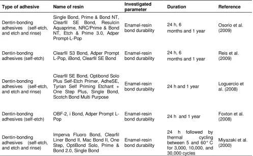
Table 1. Previous studies conducted on enamel-resin bond durability.
Type of adhesive Name of resin Investigated
parameter Duration Reference
Dentin-bonding adhesives (self-etch, and etch and rinse)
Single Bond, Prime & Bond NT, Clearfil SE Bond, Resulcin Aquaprime, NRC/Prime & Bond NT, Etch & Prime 3.0, Adper L-Pop, iBond, Clearfil SE Bond
Enamel-resin Plus Self-Etch Primer, AdheSE, Tyrian Self Priming Etchant +
Imperva Fluoro Bond, Clearfil Liner Bond II, Mac Bond II, One
microleakage result in tooth hypersensitivity, marginal discoloration, recurrent caries and pulp irritation (Perdigao and Swift, 2006; Van Meerbeek et al., 2006). At present, two kinds of adhesive resins are available for bonding to tooth structures. They include enamel-bonding and dentin-bonding adhesive systems (Perdigao and Swift, 2006; Van Meerbeek et al., 2006).
Enamel-bonding systems are hydrophobic and can only be applied to the enamel; however, dentin-bonding systems are hydrophilic and can be applied to both the enamel and dentin. As a result of the hydrophilic nature of dentin surfaces, only hydrophilic bonding systems are capable of producing an appropriate bond with dentin. Nevertheless, such systems are more susceptible to hydrolysis (Van Meerbeek et al., 2006; Powers and Sakaguchi, 2006). Previous studies conducted on enamel-resin bond durability using dentin-bonding adhe-sives are summarized in Table 1 (Osorio et al., 2009; Reis et al., 2009; Loguercio et al., 2008; Foxton et al., 2008; Miyazaki et al., 2000).
Since no studies have to date evaluated enamel margin microleakage of enamel-bonding adhesive systems in the long run, the aim of this in vitro study was to compare enamel margin microleakage in class V cavities of bovine teeth restored with two adhesive resins (enamel- and dentin-bonding adhesives) at 24 h, 6 months and 12 months intervals post-operatively.
MATERIALS AND METHODS
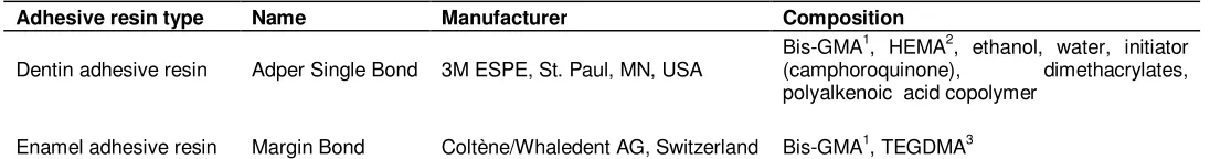
Table 2. Chemical composition of the adhesive resins used in the study.
Adhesive resin type Name Manufacturer Composition
Dentin adhesive resin Adper Single Bond 3M ESPE, St. Paul, MN, USA
Bis-GMA1, HEMA2, ethanol, water, initiator (camphoroquinone), dimethacrylates, polyalkenoic acid copolymer
Enamel adhesive resin Margin Bond Coltène/Whaledent AG, Switzerland Bis-GMA1, TEGDMA3
1
Bisphenol-glycidyl methacrylate; 2hydroxyethyl methacrylate; 3triethylene glycol dimethacrylate.
Table 3. Study groups and subgroups.
Group Subgroup
Group 1 (Dentin adhesive resin)
Subgroup A: evaluation of microleakage 24 h subsequent to restoration Subgroup B: evaluation of microleakage 6 months subsequent to restoration Subgroup C: evaluation of microleakage 12 months subsequent to restoration
Group 2 (Enamel adhesive resin)
Subgroup D: evaluation of microleakage 24 h subsequent to restoration Subgroup E: evaluation of microleakage 6 months subsequent to restoration Subgroup F: evaluation of microleakage 12 months subsequent to restoration
resin (3M ESPE, St. Paul, MN, USA) were used in the three subgroups of group 1 (A, B and C) and Margin Bond (Coltene Whaledent, Switzerland) adhesive resin was used in the three subgroups of group 2 (D, E and F); both resins were used according to the manufacturer’s instructions. Astralis 7 light-curing unit (Ivoclar Vivadent, FL-9494 Schaan, Liechtenstein) was used to light-cure the resins; the probe was 8 mm in diameter and the light intensity was 400 mW/cm2. The tip was placed perpendicular to the surface and curing continued for 20 s according to the manufacturer’s instructions. To restore cavities in all the groups, ValuxTM Plus (3M ESPE, St. Paul, MN, USA) composite was used with incremental technique and each layer was cured for 40 s using Astralis 7 light-curing unit at a light intensity of 400 mW/cm2.
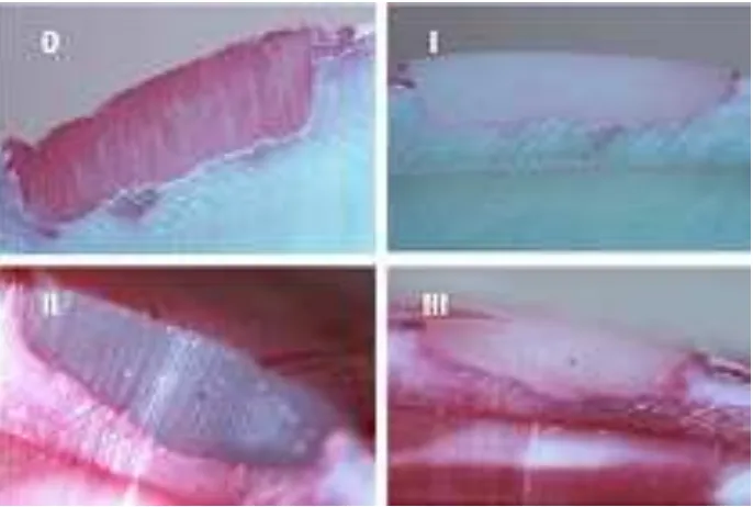
After restoration, the samples were polished with diamond polishing burs (Diamant Gmbh, D & Z, Goerzallee 307, 14167 Berlin, Germany) and polishing disks (Sof-LexTM, 3M ESPE, St. Paul, MN, USA). To simulate oral cavity conditions, a thermocycling procedure was undertaken, which consisted of 500 cycles at 5 ± 2°C / 55 ± 2°C with a dwell time of 30 s and transfer time of 10 s. Then the samples in subgroups A and D, B and E, and C and F were incubated for 24 h, 6 months and 12 months, respectively (Table 3), in distilled water at 37°C (Crim and Chapman, 1994; Okuda et al., 2002). After each group’s specific interval, the teeth were retrieved from distilled water in the incubators and dried. Then the teeth were covered with two layers of nail varnish up to 1 mm from the margins of restorations. The apex of each tooth was covered with sticky wax. The teeth were subsequently immersed in 2% basic fuschin solution (Hashimoto et al., 2000). The teeth were then divided into two halves in a bucco-lingual direction using a diamond disk (Diamant Gmbh, D & Z, Goerzallee 307, 14167 Berlin, Germany). The samples were evaluated under a stereomicroscope (Nikon, Japan) at a magnification of ×16 by two examiners so that dye penetration at gingival or occlusal margins could be classified as follows (Borges et al., 2007): 0: No dye penetration; I: Dye penetration along the gingival or occlusal wall without axial wall involvement; II: Dye penetration along the gingival or occlusal wall with axial wall involvement; III: Dye penetration beyond the gingival, occlusal or axial wall toward the pulp.
After evaluation of the depth of the dye’s penetration at the two margins, the score of the margin (gingival or occlusal) with the greatest dye penetration was recorded as the microleakage score of that specimen.
The non-parametric Kruskal-Wallis test was used to compare microleakage at the three intervals and between the two adhesive resins. Mann-Whitney U test was used for the two-by-two comparison of the groups. This latter test was also used to compare microleakage between the three time intervals and between the two adhesive resins. Statistical significance was defined at p < 0.05.
RESULTS
The frequency of microleakage scores in the subgroups in this study are demonstrated in Table 4. Kruskal-Wallis test demonstrated statistically significant differences in microleakage at the three time intervals between the two adhesive resins (p < 0.001). Two-by-two comparison of the groups demonstrated statistically significant diffe-rences in microleakage between 24 h and 6 months intervals, and between 24 h and 12 months intervals (p ≤
0.001); however, there were no significant differences in microleakage between the 6 months and 12 months intervals (p > 0.05).
Table 4. Microleakage scores in the subgroups.
Subgroup Microleakage score
0 I II III Total
A 6 9 5 0 20
B 0 6 8 6 20
C 1 3 14 2 20
D 6 6 7 1 20
E 0 4 7 9 20
F 0 0 14 6 20
Table 5. Kruskal-Wallis and Man-Whitney U test results for comparison of microleakage at the three intervals in each adhesive.
Type of adhesive Type of statistical test Comparison Test statistics
Dentin adhesive
Kruskal-Wallis Three time intervals X2 = 16.59, df = 2, p < 0.001 Mann-Whitney U 24 h and 6 months U = 77.00, p < 0.001 Mann-Whitney U 24 h and 12 months U = 80.50, p < 0.001 Mann-Whitney U 6 months and 12 month U = 183.00, P = 0.61
Enamel adhesive
Kruskal-Wallis Three time intervals X2 = 18.16, df = 2, p < 0.001 Mann-Whitney U 24 h and 6 months U = 80.00, P = 0.001 Mann-Whitney U 24 h and 12 months U = 66.50, p < 0.001 Mann-Whitney U 6 month and 12 months U = 198.00, P = 0.95
Table 6. Man-Whitney U test results for comparison of microleakage between two adhesives in each time interval.
Time interval Type of statistical test Comparison Test statistics
24 h Mann-Whitney U Dentin and enamel adhesives U = 176.50, P = 0.50 6 months Mann-Whitney U Dentin and enamel adhesives U = 165.00, P = 0.31 12 months Mann-Whitney U Dentin and enamel adhesives U = 132.00, P = 0.02
DISCUSSION
Longevity of the bond is an important factor in the clinical efficacy of adhesive restorations. In this study, the teeth restored with both the adhesive resins exhibited increased microleakage at enamel margins with time. In the same context, various in vitro studies have shown that long-term storage in water results in defects in bonded interfaces (Amaral et al., 2004; Koshiro et al., 2004; Malacarne et al., 2006; Armstrong et al., 2001; De Munck et al., 2003). In addition, it has been reported that sealing ability of total-etch and self-etch adhesive resins at enamel margins decreases with time (Malacarne et al., 2006; Osorio et al., 2009), which might be attributed to the absorption of water into the polymer networks with time and the subsequent hydrolytic degradation of adhesive resins.
The results of this study showed that the differences between the microleakage of Adper Single Bond and Margin Bond adhesive resins at 24 h and 6 months intervals were not statistically significant; however, at 12
Figure 1. Microleakage scores (0 to III) in the selected samples.
glycidyl methacrylate (Bis-GMA) monomers,hydroxyethyl methacrylate (HEMA) and dimethacrylates inside the polymer bed of the prefabricated polyalkenoic acid, leading to the formation of intertwined polymer networks called interpenetrated networks. These networks improve polymer strength (Nakabayashi, 2008), decrease water permeability (Abbasi et al., 2001) and increase longevity and resistance of the polymer to hydrolytic degradation (Kim et al., 2005).
On the other hand, Margin Bond contains Bis-GMA and triethylene glycol dimethacrylate (TEGDMA) monomers (Table 2). Both monomers are dimethacrylates and produce polymer networks with cross-linkings. They produce more pores and cavities for water penetration and are more susceptible to hydrolysis as compared to linear polymers (Malacarne et al., 2006; Ferracane, 2006; Rivera-Torres and Vera-Graziano, 2008). It seems that the presence of more pores and cavities in the polymer structure of Margin Bond as compared to Adper Single Bond had a role in its wear behavior in water. In addition, no intertwined polymer networks were produced in the polymer structure of Margin Bond; therefore, the presence of a protective effect against hydrolysis was not possible.
In this study, it was not possible to evaluate the solu-bility parameters of the adhesive resins used because manufacturers do not disclose the exact chemical composition and quantity of each component, which is necessary for such evaluations. Since the rate and polymer hydrolysis percentage are under the influence of pH and temperature, in addition to the influence of polymer nature and the percentage of the constituent polymers, it is suggested that future microleakage studies be carried out in vivo. It is also suggested that the
composite-enamel interface should be evaluated by electron microscopy.
According to the results of this study, Adper Single Bond adhesive resin demonstrated a better durability of bonding to enamel when compared to Margin Bond adhesive resin after a year of storage in water.
ACKNOWLEDGEMENT
The authors extend their sincere appreciation to the office of the Vice Chancellor for Research, Tabriz University of Medical Sciences, for the financial support of this research.
REFERENCES
Abbasi F, Mirzadeh H, Katbab AA (2001). Modification of the silicone rubber for biomedical application: a review. Polym. Int. 50(12): 1279-1287.
Amaral CM, Peris AR, Ambrosano GM, Pimenta LA (2004). Microleakage and gap formation of resin composite restorations polymerized with different techniques. Am. J. Dent. 17(3): 156-160. Armstrong SR, Keller JC, Boyer DB (2001). The influence of water
storage and C-factor on the dentin-resin composite microtensile bond strength and debond pathway utilizing a filled and unfilled adhesive resin. Dent. Mater. 17(3): 268-276.
Borges MA, Matos IC, Dias KR (2007). Influence of two self-etching primer systems on enamel adhesion. Braz. Dent. J. 18(2): 113-118. Crim GA, Chapman KW (1994). Reducing microleakage in Class II
restorations: an in vitro study. Quintessence. Int. 25(11): 781-785. De Munck J, Van Meerbeek B, Yoshida Y, Inoue S, Vargas M, Suzuki
K, Lambrechts P, Vanherle G (2003). Four-year water degradation of total-etch adhesives bonded to dentin. J. Dent. Res. 82(2): 136-140. De Munck J, Van Landuyt K, Peumans M, Poitevin A, Lambrechts P,
118-132.
Ferracane JL (2006). Hygroscopic and hydrolytic effects in dental polymer networks. Dent. Mater. 22(3): 211-222.
Finer Y, Santerre JP (2004). Salivary esterase activity and its association with the biodegradation of dental composites. J. Dent. Res. 83(1): 22-26.
Foxton RM, Melo L, Stone DG, Pilecki P, Sherriff M, Watson TF (2008). Long-term durability of one-step adhesive-composite systems to enamel and dentin. Oper. Dent. 33(6): 651-657.
Hashimoto M, Ohno H, Kaga M, Endo K, Sano H, Oguchi H (2000). In vivo degradation of resin-dentin bonds in humans over 1 to 3 years. J. Dent. Res. 79(6): 1385-1391.
Hashimoto M, Ohno H, Sano H, Tay FR, Kaga M, Kudou Y, Oguchi H, Araki Y, Kubota M (2002). Micromorphological changes in resin-dentin bonds after 1 year of water storage. J. Biomed. Mater. Res. 63(3): 306-311.
Hashimoto M, Ohno H, Sano H, Kaga M, Oguchi H (2003). In vitro degradation of resin-dentin bonds analyzed by microtensile bond test, scanning and transmission electron microscopy. Biomaterials, 24(21): 3795-3803.
Hashimoto M, Fujita S, Kaga M, Yawaka Y (2007). In vitro durability of one-bottle resin adhesives bonded to dentin. Dent. Mater. J. 26(5): 677-686.
Jaffer F, Finer Y, Santerre JP (2002). Interactions between resin monomers and commercial composite resins with human saliva derived esterases. Biomaterials, 23(7): 1707-1719.
Kim HW, Chung CW, Kim YB, Rhee YH (2005). Preparation and hydrolytic degradation of semi-interpenetrating networks of poly(3-hydroxyundecenoate) and poly(lactide-co-glycolide). Int. J. Biol. Macromol. 37(5): 221-226.
Koshiro K, Inoue S, Tanaka T, Koase K, Fujita M, Hashimoto M, Sano H (2004). In vivo degradation of resin-dentin bonds produced by a self-etch vs. a total-self-etch adhesive system. Eur. J. Oral. Sci. 112(4): 368-375.
Loguercio AD, Moura SK, Pellizzaro A, Dal-Bianco K, Patzlaff RT, Grande RH, Reis A (2008). Durability of enamel bonding using two-step self-etch systems on ground and unground enamel. Oper. Dent. 33(1): 79-88.
Malacarne J, Carvalho RM, de Goes MF, Svizero N, Pashley DH, Tay FR, Yiu CK, Carrilho MR (2006). Water sorption/solubility of dental adhesive resins. Dent. Mater. 22(10): 973-980.
Miyazaki M, Sato M, Onose H (2000). Durability of enamel bond strength of simplified bonding systems. Oper. Dent. 25(2): 75-80. Nakabayashi N (2008). Contribution of polymer chemistry to dentistry:
development of an impermeable interpenetrating polymer network to protect teeth from acid demineralization. Polym. Int. 57(2): 159-162. Nakamichi I, Iwaku M, Fusayama T (1983). Bovine teeth as possible
substitutes in the adhesion test. J. Dent. Res. 62(10): 1076-1081. Okuda M, Pereira PN, Nakajima M, Tagami J, Pashley DH (2002).
Long-term durability of resin dentin interface: nanoleakage vs. microtensile bond strength. Oper. Dent. 27(3): 289-296.
Osorio R, Monticelli F, Moreira MA, Osorio E, Toledano M (2009). Enamel-resin bond durability of self-etch and etch & rinse adhesives. Am. J. Dent. 22(6): 371-375.
Perdigao J, Swift Jr. EJ (2006). Fundamental concepts of enamel and dentin adhesion. In Roberson TM, Heymann HO, Swift EJ (eds) Sturdevant's art and science of operative dentistry, Mosby, USA, pp 245-271.
Powers JM, Sakaguchi RL (2006). Bonding to dental substrates. In Powers JM, Sakaguchi RL (eds) Craig's restorative dental materials, Mosby, USA, pp. 214-231.
Reis A, Moura K, Pellizzaro A, Dal-Bianco K, de Andrade AM, Loguercio AD (2009). Durability of enamel bonding using one-step self-etch systems on ground and unground enamel. Oper. Dent. 34(2): 181-191.
Rivera-Torres F, Vera-Graziano R (2008). Effects of water on the long-term properties of Bis-GMA and silylated-(Bis-GMA) polymers. J. Appl. Polym. Sci. 107(2): 1169-1178.
Santini A, Ivanovic V, Ibbetson R, Milia E (2004). Influence of marginal bevels on microleakage around Class V cavities bonded with seven self-etching agents. Am. J. Dent. 17(4): 257-261.