323
Suppression of Androgen Action and the Induction of Gross
Abnormalities of the Reproductive Tract in Male Rats
Treated Neonatally With Diethylstilbestrol
C. McKINNELL,* N. ATANASSOVA,† K. WILLIAMS,* J. S. FISHER,* M. WALKER,* K. J. TURNER,* P. T. K. SAUNDERS,* AND R. M. SHARPE*
From the*MRC Human Reproductive Sciences Unit, Centre for Reproductive Biology, Edinburgh, Scotland, United Kingdom; and the †Institute of Experimental Morphology and Anthropology, Bulgarian Academy of Science, Sofia, Bulgaria.
ABSTRACT: This study evaluated whether androgen action is
al-tered in rats treated neonatally with diethylstilbestrol (DES) at a dose that induced reproductive tract abnormalities. Rats were treated on alternate days 2–12 with 10g DES and studied on Day 18. DES-induced abnormalities included a 70% reduction in testis weight, dis-tension and overgrowth of the rete, disdis-tension and reduction in ep-ithelial height of the efferent ducts, underdevelopment of the epidid-ymal duct epithelium, reduction in epithelial height in the vas defer-ens, and convolution of the extra-epididymal vas. In DES-treated rats, androgen receptor (AR) immunoexpression was virtually absent from all affected tissues and the testis, whereas AR expression in controls was intense in epithelial and stromal cells. The DES-in-duced change in AR immunoexpression was confirmed by Western analysis for the testis. In rats treated neonatally with 1g DES, reproductive abnormalities were absent or minor, except for a 38% reduction in testis weight; loss of AR immunoexpression also did not occur in these rats. Treatment-induced changes in AR expression were paralleled by changes in Leydig cell volume per testis (91% reduction in the 10-g DES group; no change in the 1-g DES group). To test whether suppression of androgen production or ac-tion alone could induce comparable reproductive abnormalities to 10 g DES, rats were treated neonatally with either a potent gonado-tropin-releasing hormone antagonist (GnRHa) or with flutamide (50 mg/kg/day). These treatments reduced testis weight (68% for GnRHa, 40% for flutamide), and generally retarded development of the reproductive tract but failed to induce the abnormalities induced by 10g DES. GnRHa and flutamide caused no detectable change
in AR immunoexpression in target tissues, with the exception of mi-nor changes in the testes of flutamide-treated males. GnRHa treat-ment caused a reduction (83%) in Leydig cell volume comparable to that caused by 10g DES. Immunoexpression of estrogen re-ceptor alpha (ER␣) in the efferent ducts and of ERin all tissues studied were unaffected by any of the above treatments. Neonatal coadministration of testosterone esters (TE; 200g) with 10g DES prevented most of the morphological abnormalities induced by 10 g DES treatment alone, though testis weight was still subnormal (46% reduction in DES⫹TE vs 72% in DES alone and 49% with TE alone) and some lumenal distension was still evident in the ef-ferent ducts. Coadministration of TE with DES prevented DES-in-duced loss of AR immunoexpression (confirmed for testis by West-ern blot analysis). It is concluded that 1) reproductive tract abnor-malities induced in the neonatal male rat by a high (10g) dose of DES are associated with reduced AR expression and Leydig cell volume; 2) these changes are largely absent with a lower dose of DES (1g); 3) treatments that interfere with androgen production (GnRHa) or action (flutamide) alone failed to induce reproductive tract abnormalities or alter AR expression as did 10g DES; 4) a grossly altered androgen:estrogen balance (low androgen⫹ high estrogen) may underlie the reproductive tract abnormalities, other than reduced testis weight, induced by high doses of DES.
Key Words: Testis, rete testis, efferent ducts, epididymis, vas de-ferens, androgen receptor.
J Androl 2001;22:323–338
T
here is substantial evidence in the literature that ex-posure of male rodents to high levels of exogenous estrogens such as diethylstilbestrol (DES), either in utero or neonatally, results in major morphological and func-tional abnormalities of the testis and reproductive tractSupported in part by the European Centre for the Ecotoxicology of Chemicals (ECETOC) and by a Strategic Research Fund Award from AstraZeneca plc. N.A. received a Royal Society/NATO fellowship.
Correspondence to: Dr R.M. Sharpe, MRC Human Reproductive Sci-ences Unit, Centre for Reproductive Biology, 37 Chalmers Street, Edin-burgh EH3 9ET, Scotland (e-mail: [email protected]).
Received for publication August 21, 2000; accepted for publication September 28, 2000.
(Arai et al, 1983; Newbold and McLachlan, 1985; Fisher et al, 1998; Khan et al, 1998; Sharpe et al, 1998). Many of these effects can have life-long consequences (Khan et al, 1998; Atanassova et al, 1999, 2000). Furthermore, studies on the sons of women who were exposed to DES during pregnancy show that a similar range of abnormal-ities can also be induced in human males exposed to high levels of exogenous estrogens in utero (Stillman, 1982; Toppari et al, 1996).
→
Figure 1. Effect of neonatal treatment with vehicle (control), DES (10g), GnRHa, or flutamide on gross morphology of the testis and reproductive tract at Day 18. Note that only DES treatment results in overgrowth and distension of the rete (arrows; top row); distension (asterisks) and reduction in epithelial cell height of the efferent ducts (note loss of apical cytoplasm from epithelial cells; 2nd row); relative undergrowth of the epididymal duct with relative overgrowth of the stroma (note bulb shape of cauda; 3rd row); underdevelopment of the epithelium in the vas deferens (black lines show epithelial height; bottom row). Further examples of these abnormalities, at varying magnification, are shown in other figures. In contrast to the DES-treated group, note that both GnRHa and flutamide treatments caused retardation of development of the efferent ducts (smaller cross-sectional diameter than in controls) and epididymis (proportionately normal but smaller than in 18-day controls) without causing the changes observed in the DES-treated animals. Note that efferent ducts have been immunostained for ER␣to highlight the cell nuclei and to show that the levels of immunoexpression are unchanged. Scale bar
⫽500m (top and 3rd rows) or 100m (2nd and bottom rows). secretion by the pituitary gland during estrogen treatment (Brown-Grant et al, 1975; Bellido et al, 1990). However, this conclusion is difficult to reconcile with evidence from our own recent studies comparing the effects of neonatal DES treatment and those of gonadotropin suppression us-ing a potent gonadotropin-releasus-ing hormone (GnRH) an-tagonist (GnRHa; Sharpe et al, 1998; Atanassova et al, 1999). On the other hand, DES-induced effects such as cryptorchidism, hypospadias, reduced sperm production in adulthood, and abnormal development of the epididy-mis, bear striking similarities to those induced by in utero exposure to antiandrogens (Imperato-McGinley et al, 1992; van der Schoot, 1992; Silversides et al, 1995; Sharpe et al, 2000), or in which fetal Leydig cell function is compromised (Mylchreest et al, 1999, 2000).
The similarity between the phenotypes of males ex-posed developmentally to either estrogens or to antian-drogens suggested to us the possibility that both pheno-types might be the result, at least in part, of a similar underlying mechanism. This hypothesis is supported by evidence that in utero exposure of male rats to exogenous estrogens reduces expression of the messenger RNA for 17␣-hydroxylase/C17-20 lyase, a key enzyme in testos-terone production (Majdic et al, 1996), while neonatal administration of DES reduces both testicular and plasma androgen levels in 12-day-old rats (Keel and Abney, 1985). Furthermore, neonatal treatment with DES can cause life-long suppression of plasma testosterone levels (Atanassova et al, 1999), and postpubertal DES treatment clearly results in reduced androgen production (Cigorraga et al, 1980; Abney and Keel, 1986). At the very least, these various findings suggest an inter-relationship be-tween raised estrogen exposure and reduced androgen production. The primary purpose of the present study was therefore to assess whether the effects of neonatal admin-istration of DES to rats, at a dose that induces widespread reproductive tract abnormalities, was associated with al-tered androgen action. In planning and undertaking these studies, we also became aware that in our own and most (possibly all) published studies that have described DES-induced abnormalities in males during perinatal life, ex-tremely high doses have to be administered to induce such effects—generally equivalent to, or in excess of, 100g/ kg. In view of the extremely high affinity of estrogen
receptors (ERs) for potent estrogens such as DES (Kuiper et al, 1997), it seemed incongruous that such high doses were necessary if the induction of reproductive abnor-malities was mediated solely via an ER receptor-mediated mechanism. Therefore, another aspect of the present stud-ies was to evaluate whether only an extremely high dose of DES could induce reproductive tract abnormalities and, if so, whether this dose-dependence coincided with alter-ations in androgen action.
Materials and Methods
Animals, Treatments, Sample Collection, and Processing
Wistar rats bred in our own animal house were maintained under standard conditions and diet (rat and mouse breeding diet no. 3; SDS, Dundee, Scotland) that contains 15.5% soy-meal. All-male litters of 8–12 pups were generated by cross-fostering pups on day 1 (⫽day of birth). Rats were subjected to one of the fol-lowing treatments:
1) Subcutaneous injection of DES (Sigma Chemical Company, Poole, Dorset, United Kingdom) at a dose of 10g or 1g in 20L corn oil on days 2, 4, 6, 8, 10, and 12.
2) Subcutaneous injection of 10g DES plus 200g testoster-one esthers (TE; Sustanon; Organon Labs, Cambridge, United Kingdom) in 20L corn oil on days 2, 4, 6, 8, 10, and 12. The dose of TE administered was based on 3 considerations. First, our previous experience in restoring intratesticular and blood tes-tosterone levels in adult rats by injection of TE (Sharpe et al, 1990), scaled down appropriately. Second, the aim of restoring a balance between androgen and estrogen levels (ie, the dose of TE administered had to considerably exceed the dose of DES that was coadministered). Third, avoidance of doses of TE that have been shown to cause severe impairment when administered neonatally to male rats (see Kincl et al, 1965). The dose of TE was a compromise, taking account of these factors.
3) Injection as in (2) but with 200g TE alone.
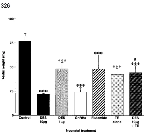
Figure 2. Effect of neonatal treatment with vehicle (control), DES (10 or 1g), GnRHa, flutamide, TE alone (200g), or 10g DES⫹200g TE on testis weight at day 18. Results are the mean⫾standard deviation for 8–23 rats per group. ***P⬍.001, in comparison with control group.
aP⬍.001, in comparison with DES-alone (10g) group.
→
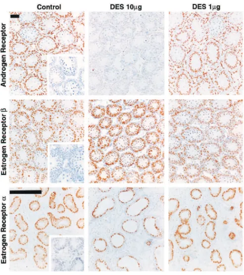
Figure 3. Effect of neonatal treatment with vehicle (control), DES (10g), GnRHa, or flutamide on immunoexpression of the AR in the testis (top row), efferent ducts (2nd row), caput epididymis (3rd row), and cauda epididymis/proximal vas deferens (bottom row) on day 18. Note the virtually complete loss of AR immunoexpression in the testis (particularly in Sertoli cells), and in both epithelial and stromal cells of the efferent ducts, caput epididymis, and cauda epididymis/proximal vas deferens of animals treated with DES. In contrast, note that treatment with GnRHa or flutamide causes little if any loss of immunoexpression of AR in these tissues, with the exception of the testis after flutamide treatment. Insets show negative controls in which the antiserum was substituted by (normal) blocking serum. Asterisks indicate lumenal distension of the efferent ducts. Scale bar⫽100m.
4, 6, 8, 10, and 12; this dose was shown by Imperato-McGinley et al (1992) to cause major reproductive tract abnormalities in male offspring when administered to pregnant rats.
6) Subcutaneous injection of 20L corn oil alone (control). Rats from the various treatment groups described above were sampled on Day 18, a time at which we have shown that major abnormalities of the excurrent ducts (Fisher et al, 1998, 1999) and reproductive tract (unpublished data) are evident in DES-treated rats. Animals were anesthetized with flurothane and the right testis was dissected out, weighed, and fixed for⬃5 hours in Bouins. The left testis was removed with the epididymis and proximal vas deferens still attached and similarly fixed. From some animals, the right testis was not fixed, but was used instead for protein extraction as described below.
After fixation, tissue was transferred into 70% ethanol before being processed for 17.5 hours in an automated Shandon pro-cessor and embedded in paraffin wax. Sections (5m thickness) were cut and floated onto slides coated with 2% 3-aminopro-pyltriethoxy-silane (Sigma) and dried at 50⬚C overnight before being used for immunohistochemistry as described below. All of the studies of the rete testis and reproductive tract described below utilized tissue sections of the left testis with the epidid-ymis attached, in order that minimal artefactual distortion was caused to the excurrent duct system.
For protein extraction, small pieces of unfixed testis tissue were snap-frozen in liquid nitrogen and stored at⫺70⬚C. The tissue was subsequently ground with a pestle in a mortar under
liquid nitrogen, and then resuspended in ice cold buffer consist-ing of 10 mM HEPES pH 7.8, 0.1 mM EDTA, 0.1 mM EGTA, 1 mM dithiothreitiol (all from Sigma) and a protease inhibitor cocktail (Complete; Roche, Lewes, United Kingdom). Protein concentration was measured by absorbance at 280 nm and the protein extracts were aliquoted and stored at⫺70⬚C. At least 2 separate experiments were performed for each of the treatments specified; comparable results were obtained in each experiment.
Antibodies used for Immunohistochemistry
Immunolocalization of AR was performed with the use of a rab-bit polyclonal antibody (Santa Cruz Biotechnology Inc, Santa Cruz, Calif) raised against an epitope at the N-terminus of human AR, and was used at a dilution of 1:200. ER␣was immunolo-calized using a mouse monoclonal antibody raised against a full-length human ER␣recombinant protein (Novocastra, Newcastle Upon Tyne, United Kingdom), and used at a dilution of 1:20. ERwas immunolocalized using an affinity-purified, polyclonal antipeptide immunoglobulin G (IgG) raised in sheep against a specific peptide in the hinge (D) domain of human ER, as pre-viously described in detail (Saunders et al, 2000); it was used at a dilution of 1:1000. The specificity of the ER antibodies has been detailed in our previous studies (Saunders et al, 2000; Wil-liams et al, 2000).
Immunohistochemistry
Unless otherwise stated, all incubations were performed at room temperature. Sections were deparaffinized in Histoclear (Nation-al Diagnostics, Hull, United Kingdom), rehydrated in graded eth-anols, and washed in water. At this stage, sections were subjected to a temperature-induced antigen retrieval step (Norton et al, 1994) in either 0.01 M citrate buffer pH 6.0 (for AR and ER␣), or 0.05 M glycine pH 3.5, and 0.01% EDTA (for ER). After pressure-cooking for 5 minutes at full pressure, sections were left to stand, undisturbed, for 20 minutes, then cooled under running tap water before being washed twice (5 minutes each) in Tris-buffered saline (TBS; 0.05 M Tris-HCl pH 7.4, 0.85% NaCl). Endogenous peroxidase activity was blocked by immers-ing all sections in 3% (v/v) H2O2in methanol (both from BDH
Figure 4. Comparative effect of neonatal treatment with vehicle (control) or with DES at a dose of 10 or 1g on immunoexpression of AR (top row) and ER
(middle row) in the testis, and of ER␣(bottom row) in the efferent ducts, on day 18. Note the lack of effect of treatment with the lower dose of DES on AR immunoexpression and the lack of effect of either DES dose on ERor ER␣immunoexpression. Insets show negative controls in which the antiserum was substituted by (normal) blocking serum (AR) or in which the antiserum was preabsorbed with recombinant peptide (ER␣and ER). Scale bars⫽100m.
two 5-minute washes in TBS, sections were incubated with a secondary antibody; namely, a 1:500 dilution in the appropriate blocking serum of biotinylated swine anti-rabbit IgG for AR (DAKO, High Wycombe, United Kingdom), biotinylated rabbit
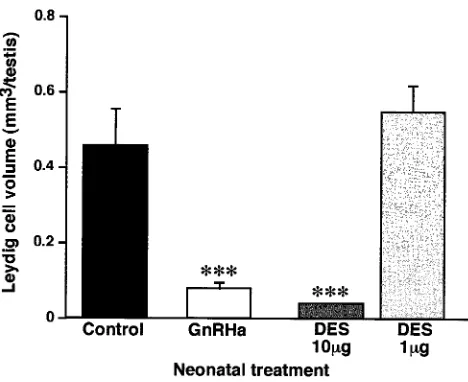
Figure 5. Effect of neonatal treatment with vehicle (control), GnRH antag-onist (GnRHa), 10g DES, or 1g DES on Leydig cell volume per testis at day 18. Values shown are the means⫹standard deviations for 6 rats per group. ***P⬍.001, in comparison with control group.
0.05 M Tris-HCl pH 7.4, according to the manufacturer’s instruc-tions. Sections were washed twice (5 minutes each) in TBS and immunostains were developed using 0.05% 3,3⬘ -diaminobenzi-dine (Sigma) in 0.05 M Tris-HCl pH 7.4, containing 0.01% (v/ v) H2O2, until staining in controls was optimal, when the reaction
was stopped by immersing all sections in distilled water. All sections were then lightly counterstained with hematoxylin, de-hydrated in graded ethanols, cleared in xylene, and coverslips placed on them using Pertex mounting medium (CellPath plc, Hemel Hempstead, United Kingdom).
To ensure the reproducibility of findings, tissue sections from a minimum of 3–6 animals in each treatment group were eval-uated, and this was performed for at least 2 separate experiments. Further confirmation was obtained by undertaking immunohis-tochemistry with tissue sections from control and treated animals on the same slide. For AR, changes in immunoexpression in treated animals compared with control animals were also con-firmed by Western blotting as described below. Specificity of immunostaining was checked for each antibody using previously established procedures. For AR, this involved substitution of the primary antibody by normal (blocking) serum, and for ER␣and ER, it involved preabsorption of the primary antibody with the respective recombinant protein (Saunders et al, 2000; Williams et al, 2000).
Immunostained sections were examined and photographed us-ing an Olympus Provis microscope (Olympus Optical, Honduras Street, London, United Kingdom) fitted with a Kodak DCS330 camera (Eastman Kodak, Rochester, NY). Captured images were stored on a G4 computer (Apple Macintosh) and compiled using Photoshop 5.0 software before being printed using an Epson Sty-lus 750 color printer (Seiko Epson Corp, Nagano, Japan).
Western Blotting
Testis protein extracts from control and treated animals were analyzed by sodium dodecyl sulfate-polyacrylamide gel electro-phoresis (SDS-PAGE) and Western blotting. Gels contained 6%
acrylamide and comprised 4 mL 30% acrylamide/bis (Anachem, Luton, United Kingdom), 5 mL 1.5 M Tris-HCl pH 8.8, 10.9 mL distilled water, and 0.1% SDS (Sigma). The gel mixture was degassed and polymerized by addition of 100 L 10% ammo-nium persulphate and 5 L TEMED (both from Sigma). Gels were loaded with 100g protein per lane, in denaturing/loading buffer comprising 50 mM Tris-HCl pH 6.8, 100 mM dithiothre-itol (Sigma), 2% SDS (Sigma), 10% glycerol (BDH, Poole, Unit-ed Kingdom), and 0.002% bromophenol blue (Bio-Rad Labo-ratories, Hemel Hempstead, United Kingdom). One lane was loaded with prestained molecular weight markers (Bio-Rad). The gels were run for approximately 1 hour at a constant current of 120 mA. After SDS-PAGE, gels were transferred to blotting buffer comprising 12 mM Tris base, 96 mM glycine, and 20% methanol (NuPAGE buffer; Novex, San Diego, Calif) and blot-ted onto a PVDF membrane (Immobilon-P; Millipore, Watford, United Kingdom) using the XCell-II system (Novex) run at 100 mA for 90 minutes. Membranes were blocked for 2–3 hours in NRS/TBS containing 0.05% (v/v) Tween-20 (Sigma), and 5% (w/v) skim milk powder (Marvel; Premier Brands Ltd, Moreton, United Kingdom). The AR antibody was diluted 1:200 in TBS and incubated with the membranes overnight at 4⬚C. To confirm the specificity of detection, AR antibody was preabsorbed over-night with a 10-fold excess of the AR N-20 peptide (Santa Cruz). The membranes were washed twice (15 minutes each) and then 4 times (5 minutes each) in TBS, and then incubated for 1 hour at room temperature with peroxidase-conjugated donkey anti-rabbit IgGs (Amersham Pharmacia Biotech, Little Chalfont, United Kingdom) diluted 1:4000 in TBS. The membranes were again washed twice (15 minutes each) then 4 times (5 minutes each) in TBS, and bound antibodies were detected using an ECL system (Amersham Pharmacia Biotech) according to the manu-facturer’s instructions.
Measurement of Leydig (3-Hydroxysteroid Dehydrogenase–Positive) Cell Volume per Testis
To evaluate whether neonatal treatment with DES or GnRHa altered Leydig cell volume in 18-day-old animals, sections from Bouins-fixed testes were immunostained for 3-hydroxysteroid dehydrogenase (3-HSD) as described previously (Majdic et al, 1996; Sharpe et al, 1998) and the volume of 3-HSD-positive cells per testis was determined using a point-counting method previously described in detail (Atanassova et al, 1999). One sec-tion from each of 3 blocks per animal was examined, and all points falling over 3-HSD-positive cytoplasm or over the nuclei of cells with 3-HSD-positive cytoplasm were scored and ex-pressed as percent volume per testis. These data were then con-verted to absolute volume per testis by multiplication by testis weight (⫽volume).
Statistics
←
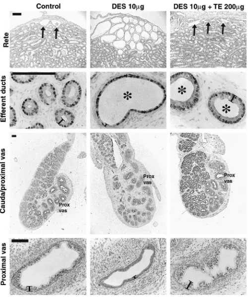
of DES⫹TE partially prevented lumenal distention of the efferent ducts (asterisks) but restored normal epithelial cell height (indicated by black lines in 2nd row) compared with DES treatment alone. Efferent ducts were immunostained for ER␣and the cauda epididymis/proximal vas for ERto highlight cell nuclei and illustrate the absence of any major change in immunoexpression of the two ERs in the 3 treatment groups. Scale bar⫽
200m (top and 3rd rows) or 100m (2nd and bottom rows).
Results
Induction of Gross Abnormalities of the Rete Testis and Reproductive Tract
Observations on DES-induced changes in gross mor-phology are summarized in the Table. The rete testis of all animals treated with 10g DES exhibited considerable overgrowth, with gross distention of the rete lumen and apparent invasion of the rete into the testicular parenchy-ma (Figure 1). The epithelium of the efferent ducts in all animals treated with 10g DES was reduced substantially in height compared with the control group, whereas the duct lumens were highly distended compared with con-trols (Figure 1); these findings confirm those we described previously (Fisher et al, 1999). Similar, though less pro-nounced, distension and reduction in epithelial height was also found in the caput epididymal duct of some animals (not shown). The cauda epididymis of rats treated neo-natally with 10 g DES exhibited pronounced underde-velopment of the epididymal duct (ie, fewer duct cross-sections), reduced height of the epithelium, and relative overgrowth of the periductal stroma (Figure 1). The latter changes also extended out into the vas deferens and, in addition, in⬃70% of animals the initial region of the vas outside of the epididymis was transformed from a straight to a convoluted duct structure more reminiscent of the epididymal duct and the region of proximal vas present within the caudal bulb (not shown, but see Table).
In contrast to the gross morphological changes induced by neonatal injection of 10 g DES, administration of a 10-fold lower dose of DES (1 g) caused only minor changes to the rete and efferent ducts and had no dis-cernible effect on the caput or cauda epididymis and vas deferens (not shown, but summarized in Table). Neonatal treatment with either GnRHa or flutamide did not cause any changes to the gross morphology of the rete, efferent ducts, caput or cauda epididymis, or vas that were com-parable to those induced by treatment with 10 g DES, though it was noticeable that the reproductive tract struc-tures in both of these groups were smaller when compared with controls, indicating that development was retarded (Figure 1, Table).
Testicular Morphology and Weight
Neonatal treatment with 10g DES resulted in an aver-age 72% reduction in testicular weight on day 18 (Figure 2, Table) and, at the microscopic level, there was clear retardation of seminiferous cord/tubule development, as
has been detailed elsewhere (Sharpe et al, 1998; Atan-assova et al, 2000). However, comparable retardation of development and reduction in testis weight was induced by neonatal treatment with GnRHa (Figure 2, Table), as reported previously (Sharpe et al, 1998). Treatment with the lower dose of DES (1 g) caused only minor retar-dation of testis development and a correspondingly small-er reduction (38%) in testis weight, whsmall-ereas neonatal treatment with flutamide caused a similar reduction (Fig-ure 2, Table).
Immunoexpression of AR and ERs in the Testis and Reproductive Tract
←
Figure 7. Effect of neonatal treatment with vehicle (control), 10g DES or cotreatment with 10g DES and testosterone (DES⫹TE) on immunoexpression of AR in the testis (top row), caput epididymis (middle row), and cauda epididymis and proximal vas deferens (bottom row) at 18 days of age. Note that coadministration of DES⫹TE was able to completely prevent loss of AR immunoexpression in all of the tissues when compared with animals treated with DES alone. Note also the cytoplasmic AR staining in epithelial cells of the cauda/proximal vas in the group treated with DES alone (bottom row, middle panel). Insets show negative controls in which the antiserum was substituted by (normal) blocking serum. Scale bar⫽100m.
testes of flutamide-treated animals was consistently weak-er than in controls (Figure 3). Treatment with the lowweak-er dose (1 g) of DES was without effect on AR immu-noexpression either in the testis (Figure 4) or any other tissue (results not shown, but summarized in Table).
To demonstrate the specificity of the observed changes in AR immunoexpression, the effect of neonatal DES treatment on immunoexpression of AR and ERs was com-pared. Neonatal treatment with either 10g or 1g DES had no detectable effect on immunoexpression of ERin the testis (Figure 4), and ER immunoexpression in all other tissues examined was also unaffected by DES or any other treatment (results not shown). Similarly, im-munoexpression of ER␣ in the efferent ducts was unaf-fected by DES (Figure 4) or by any other treatment (not shown).
Leydig (3-HSD-Positive) Cell Volume per Testis
The studies described above showed fundamental differ-ences in the effect of DES (10g) and GnRHa treatment on AR immunoexpression in reproductive tract tissues. To establish whether these differences could be explained by differential effects of the 2 treatments on Leydig cell de-velopment, the volume of Leydig (3-HSD-positive) cells per testis was measured. This showed that treatment with either 10 g DES or GnRHa resulted in substantial (⬎80%) and significant (P ⬍ .001) reduction in Leydig cell volume per testis, whereas treatment with 1g DES had no effect on this parameter (Figure 5). It is presumed that this reduction will have led to similar reductions in the blood levels of testosterone in rats treated neonatally with DES (10g) or with GnRHa, however, our attempts to measure plasma testosterone levels accurately in these groups was prevented by nonspecific interference in the assay (perhaps from plasma binding proteins).
Effect of Coadministration of DES and
Testosterone on Reproductive Tract Abnormalities, AR Immunoexpression, and Testis Weight
As induction of gross morphological abnormalities of the reproductive tract was only observed in animals in which loss of AR immunoexpression was also induced by the high dose of DES, it was reasoned that maintenance of AR expression by testosterone (ie, TE) administration (Bremner et al, 1994) might prevent the DES-induced ab-normalities. Animals were therefore treated simultaneous-ly with 10g DES⫹200g TE or with either treatment
alone. With one exception, all of the morphological changes resulting from DES (10g) treatment were com-pletely prevented by coadministration of DES⫹TE (Fig-ure 6), and this coincided with restoration of normal im-munoexpression of AR in all of the tissues examined (Figure 7). The one DES-induced abnormality that was not entirely prevented by coadministration of TE was lu-menal distention of the efferent ducts; this change was only partially prevented (Figure 6). In addition, testis weight in DES⫹TE-treated rats was not restored to con-trol levels, though weights were significantly higher than in the group treated with DES (10 g) alone and were comparable to animals treated with TE alone (Figure 2). The TE treatment alone induced no gross abnormalities of the reproductive tract, though it was noted that epidid-ymal and vas deferens development in this group were advanced compared with controls, having a morphologi-cal appearance similar to that of an animal aged 22–25 days (not shown). This advance was equally evident in the DES ⫹TE-treated group (Figures 6 and 7).
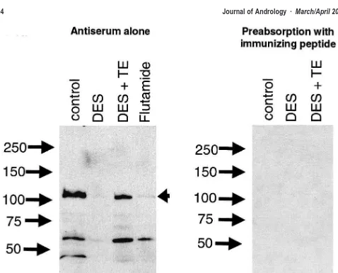
AR Protein Expression in the Testis—Western Analysis The changes in testicular AR immunoexpression in con-trol, DES-treated (10 g), DES ⫹TE–treated, and fluta-mide-treated animals were confirmed by Western blotting of testis protein extracts. The AR antibody recognized a protein band of ⬃113 kilodaltons (kd), corresponding to the molecular weight of the full-length AR protein, which was prominently expressed in extracts from control and DES ⫹ TE–treated animals (Figure 8). This band was virtually absent in extracts from DES-treated animals, and was only weakly expressed in extracts from flutamide-treated animals (Figure 8). Prominent bands at ⬃45 and
⬃60 kd, presumed to be AR breakdown products, were also absent in extracts from DES-treated animals but were present in the other treatment groups. Preabsorption of the antibody with the immunizing peptide resulted in the complete loss of detection of the 113-kd band, as well as the lower molecular weight bands (Figure 8).
Discussion
repro-Figure 8. Western analysis of AR expression in the testis of 18-day-old rats treated neonatally with vehicle (control), DES (10g), DES (10g)⫹TE (200g), or with flutamide. Numbers show the position of molecular weight markers in kd. The panel to the left shows a blot probed with the AR antiserum while the panel to the right shows a blot probed after preabsorption of the antiserum with the immunizing peptide. Arrowhead points to the expected position of the full length AR protein; lower molecular weight bands in the left-hand panel are presumed to be breakdown products of the full-length AR as they are reduced markedly in the DES-treated group and are absent after preabsorption with the immunizing peptide (right-hand panel).
ductive tract at 18 days of age. These changes were as-sociated with almost complete loss of AR immunoex-pression in the affected tissues as well as in the testis. All of the affected tissues also expressed ER (Saunders et al, 1997, 1998; Williams et al, 2000; unpublished data) and, in a few instances, ER␣(Fisher et al, 1997; Hess et al, 1997). However, no change was found in ER im-munoexpression in any of the tissues, nor of ER␣ im-munoexpression in the efferent ducts, from DES-treated animals. Together with the loss of AR immunoexpression there was major retardation of Leydig cell development in rats treated with the higher dose of DES, and it is reasonable to presume that this led to a drop in blood levels of testosterone. At face value, these findings are therefore consistent with the DES-induced reproductive tract abnormalities resulting from interference with an-drogen action. However, parallel studies in which Leydig cell development and androgen production (GnRHa) or
androgen action (flutamide) were inhibited, without ad-ministering exogenous estrogen, failed to reproduce the reproductive tract abnormalities or the widespread loss of AR expression seen in animals treated with the higher dose of DES. This suggests that although suppression of androgen action appears to be closely associated with the induction of DES-induced abnormalities of the reproduc-tive tract in males, the effects cannot be explained by
anti-androgenic activity alone.
Summary of the relationship between gross morphological changes to the reproductive tract induced by various neonatal treatments and the intensity of androgen receptor (AR) immunoexpression in the same tissue relative to control (not shown, but scored as⫹⫹⫹for all tissues examined)*
Retardation of development Major ⫺/⫹† Minor ⫹⫹⫹ Major ⫹⫹⫹ Minor ⫹⫹/⫹
Size reduction 72% 38% 68% 40%
Rete testis
Distension and overgrowth Major ⫺/⫹ Minor ⫹⫹⫹ Normal‡ ⫹⫹⫹ Normal‡ ⫹⫹
Efferent ducts
Distension/cell height reduction Major ⫺/⫹ Minor ⫹⫹⫹/⫹⫹ Normal‡ ⫹⫹⫹ Normal‡ ⫹⫹⫹
Epididymis/vas deferens
Relative stromal overgrowth⫹relative epithelial undergrowth
Major ⫺/⫹ Normal ⫹⫹⫹ Normal‡ ⫹⫹⫹ Normal‡ ⫹⫹⫹
Convoluted proximal vas Present§ Absent Absent Absent
* Summary data are based on a minimum of 6–12 animals per group from at least 2 separate experiments. DES indicates diethylstilbestrol. † Most animals were immunonegative for AR, but occasional animals showed sporadic positive immunostaining of some cells.
‡ Morphological development retarded so that appearance is comparable to that of a (normal) younger animal. § Present in most, but not all, animals.
flutamide merely retarded normal development of the re-productive tract without inducing major abnormalities, with the exception of cellular changes in the seminiferous tubules of the testis (see below); 2) simply raising estro-gen levels by injection of a lower (but still substantial) dose of DES (1 g) that did not suppress Leydig cell development was not sufficient to induce reproductive tract abnormalities and also had no effect on AR immu-noexpression; 3) major reproductive tract abnormalities were induced only after injection of a very high dose (10
g) of DES, which was also capable of suppressing Ley-dig cell development and AR immunoexpression; and 4) simultaneous treatment with high doses of both DES and TE prevented nearly all of the effects induced by DES alone, and also restored normal AR immunoexpression. This interpretation also fits with other pieces of published data that demonstrate that disturbance of the androgen: estrogen balance in males results in various abnormalities, such as gynecomastia in adult men (reviewed in Sharpe, 1998).
The male reproductive tract abnormalities induced by neonatal DES treatment in the present studies are consis-tent with earlier studies in which DES or other poconsis-tent estrogens have been administered neonatally (Arai et al, 1983; Bellido et al, 1990; Fisher et al, 1998; Khan et al, 1998; Sharpe et al, 1998) or in utero (McLachlan et al, 1975; Thomson et al, 1981; Newbold et al, 1985) to mice, rats, hamsters, or monkeys. The underdevelopment of ep-ithelial cells/tissue observed in the efferent ducts, caput and cauda epididymis, and vas deferens of DES-treated (10 g) rats in the present studies is also matched by
similar changes reported in the prostate (Rajfer and Cof-fey, 1979; Chang et al, 1999) and seminal vesicles (New-bold and McLachlan, 1985; Williams et al, 2000) of neo-natally estrogen-treated rats. Furthermore, our finding that DES-induced abnormalities are associated with the sup-pression of AR immunoexsup-pression in the affected tissues is consistent with studies showing a similar association between morphological effects and AR immunoexpres-sion in the prostate of rats treated neonatally with estro-gens (Prins, 1992; Prins and Birch, 1995). Examination of the doses administered in these various experiments indicates that induction of major reproductive tract ab-normalities in the male requires administration of ex-tremely high doses (⬎100 g/kg) of potent estrogens, such as DES or ethinyl estradiol. Data from human males exposed in utero to DES, in whom similar reproductive tract abnormalities have been reported as in rodents, also involved administration of similarly high doses (5–150 mg/day or ⬃100–3000g/kg/day) to the pregnant moth-ers (Stillman, 1982; Toppari et al, 1996). Our own find-ings in male rats exposed neonatally to DES show that only these very high doses are able to induce major re-productive tract abnormalities (see also Sharpe et al, 1998; Atanassova et al, 1999; Fisher et al, 1999). Such evidence seems at odds with the high affinity of both ER␣
estrogen action that results in the reproductive tract ab-normalities.
It is unclear from our study why only a very high dose of DES was able to suppress AR expression in repro-ductive tract tissues; the mechanism of this induction is also unclear. The most obvious mechanism is that the suppression of Leydig cell development induced by 10
g DES led to reduced testosterone levels, which in turn led to a reduction in AR expression, based on findings in adult males in which testosterone levels are experi-mentally lowered (Rajfer and Coffey, 1979; Blok et al, 1992; Bremner et al, 1994). Although we were unable to measure plasma testosterone in the present study, it is well established that exposure of males to estrogens, including to DES, can suppress both Leydig cell devel-opment and steroidogenesis (Abney, 1999). However, this explanation would fail to explain why AR immu-noexpression was unaffected in males treated neonatally with GnRHa in which we found a reduction in Leydig cell development of similar magnitude to that induced by DES (10g). We also have no ready explanation as to why 10g, but not 1g, DES can affect Leydig cell development and AR immunoexpression elsewhere, when either of these doses will elevate endogenous es-trogen levels to supranormal levels.
A notable exception to the above discussion concern-ing DES-induced reproductive abnormalities are the changes induced in testis weight. Neonatal treatment with GnRHa was able to induce a similar reduction in testis weight to that induced by the high dose (10 g) of DES, and coadministration of DES ⫹ TE only par-tially ameliorated the DES-induced decrease. Indeed, ad-ministration of TE alone also significantly reduced testis weight to the same extent as did DES ⫹ TE, and this reduction was similar in magnitude to that induced by flutamide or by the lower dose (1g) of DES. Detailed comparison of testis cell composition and numbers has not yet been undertaken for all of these treatment groups, though we have previously established that numbers of Sertoli cells, germ cells, and apoptotic germ cells are very similar at day 18 in rats treated with DES (10g) or with GnRHa (Sharpe et al, 1998). In the latter 2 groups there is comparable retardation of pubertal de-velopment that may be secondary to lowered FSH as well as, or instead of, lowered testosterone levels (Sharpe et al, 1998). It is therefore reasoned that some of the effects of DES (10g) treatment on the testis are not attributable to suppression of testosterone action and that, correspondingly, they are not preventable by co-administration of DES⫹TE.
There are alternative interpretations of the present findings. For example, although our results point to in-volvement of suppressed androgen action in the adverse reproductive effects induced by DES in males, this does
not exclude the possibility of direct effects of DES on the target tissues, all of which express ERs (Hess et al, 1997; Saunders et al, 1997, 1998; Williams et al, 2000). In addition, it is probable that the TE treatment elevated endogenous testosterone levels above normal, at least in some of the reproductive tract tissues. Evidence for this included advancement of epididymal development and increase in size of the seminal vesicles (unpublished) in TE-treated and DES ⫹ TE–treated animals. This ‘‘ac-celerated’’ development, although not pronounced, might somehow protect against the effects of DES treat-ment. We are unable to discount this possibility. Based on the doses of TE and DES that were coadministered we restored an androgen:estrogen ratio of 20:1, though factors such as different half-lives and so on mean that this is very much an approximation. Nevertheless, this value is markedly lower than the ratio of 1000:1 or greater reported in the literature for adult rats (there are no data for neonates; de Jong et al, 1973, 1974). It there-fore seems highly unlikely that we have grossly elevated the androgen:estrogen ratio compared with normal. Fu-ture studies in which different doses of TE are coadmin-istered with DES or in which TE administration is de-layed with respect to DES treatment may provide further insight on these issues. Other options are to combine treatment with an antiandrogen with a dose of DES that is ineffective on its own.
possess both estrogenic and anti-androgenic activity in the same molecule.
Acknowledgments
We are grateful to Professor Ian Mason (Edinburgh) for antiserum to 3 -HSD and to Dr R Deghenghi and Europeptides for their generous gift of GnRH antagonist. We are indebted to Jim MacDonald for expert assis-tance.
References
Abney TO. The potential roles of estrogens in regulating Leydig cell development and function: a review. Steroids. 1999;64:610–617. Abney TO, Keel BA. Temporal effects of diethylstilbestrol administration
in vivo on testosterone production in Leydig cells. Arch Androl. 1986; 17:79–86.
Arai Y, Mori T, Suzuki Y, Bern HA. Long-term effects of perinatal ex-posure to sex steroids and diethylstilbestrol on the reproductive sys-tem of male mammals. Int Rev Cytol. 1983;84:235–268.
Atanassova N, McKinnell C, Turner KJ, et al. Comparative effects of neonatal exposure of male rats to potent and weak (environmental) estrogens on spermatogenesis at puberty and the relationship to adult testis size and fertility; evidence for stimulatory effects of low estro-gen levels. Endocrinology. 2000;141:3898–3907.
Atanassova N, McKinnell C, Walker M, et al. Permanent effects of neo-natal estrogen exposure in rats on reproductive hormone levels, Sertoli cell number, and the efficiency of spermatogenesis in adulthood.
En-docrinology. 1999;140:5364–5373.
Bellido C, Pinilla L, Aguilar R, Gaytan F, Aguilar E. Possible role of changes in postnatal gonadotrophin concentrations in permanent im-pairment of the reproductive system in neonatally oestrogenized male rats. J Reprod Fertil. 1990;90:369–374.
Blok LJ, Bartlett, JMS, Bolt-de Vries J, Themmen APN, Brinkmann AO, Weinbaur GF, Nieschlag E, Grootegoed JA. Effect of testosterone dep-rivation on expression of the androgen receptor in rat prostate, epi-didymis and testis. Int J Androl. 1992;15:182–198.
Bremner WJ, Millar MR, Sharpe RM, Saunders PT. Immunohistochem-ical localisation of androgen receptors in the rat testis: evidence for stage-dependent expression and regulation by androgens.
Endocri-nology. 1994;135:1227–1234.
Brown-Grant K, Fink G, Greig F, Murray MA. Altered sexual develop-ment in male rats after estrogen administration during the neonatal period. J Reprod Fertil. 1975;44:25–42.
Chang WY, Wilson MJ, Birch L, Prins GS. Neonatal estrogen stimulates proliferation of periductal fibroblasts and alters the extracellular ma-trix composition in the rat prostate. Endocrinology. 1999;140:405– 415.
Cigorraga SB, Sorrel S, Bator J, Catt JK, Dufau ML. Estrogen depen-dence of a gonadotropin-induced steroidogenic lesion in rat testicular Leydig cells. J Clin Invest. 1980;65:699–705.
Fisher JS, Millar MR, Majdic G, Saunders PTK, Fraser HM, Sharpe RM. Immunolocalisation of oestrogen receptor-␣(ER␣) within the testis and excurrent ducts of the rat and marmoset monkey from perinatal life to adulthood. J Endocrinol. 1997;153:485–495;
Fisher JS, Turner KJ, Brown D, Sharpe RM. Effect of neonatal exposure to estrogenic compounds on development of the excurrent ducts of the rat testis through puberty to adulthood. Environ Health Perspect. 1999;107:397–405.
Fisher JS, Turner KJ, Fraser HM, Saunders PTK, Brown D, Sharpe RM. Immunoexpression of aquaporin-1 in the efferent ducts of the rat and marmoset monkey during development, its modulation by estrogens,
and its possible role in fluid resorption. Endocrinology. 1998;139: 3935–3945.
Hess RA, Gist DH, Bunick D, Lubahn DB, Farrell A, Bahr J, Cooke PS, Greene GL. Estrogen receptor (␣and) expression in the excurrent ducts of the adult male rat reproductive system. J Androl. 1997;18: 602–611.
Imperato-McGinley J, Sanchez RS, Spencer JR, Yee B, Vaughan ED. Comparison of the effects of the 5␣-reductase inhibitor finasteride and the antiandrogen flutamide on prostate and genital differentiation: dose-response studies. Endocrinology. 1992;131:1149–1156. de Jong FH, Hey AH, van der Molen HJ. Effect of gonadotrophins on
the secretion of oestradiol-17and testosterone by the rat testis. J
Endocrinol. 1973;57:277–284.
de Jong FH, Hey AH, van der Molen HJ. Oestradiol-17and testosterone in rat testis tissue: effect of gonadotrophins, localization and produc-tion in vitro. J Endocrinol. 1974;60:409–419.
Keel BA, Abney TO. Estrogen regulation of testicular androgen produc-tion during development in the rat. J Endocrinol. 1985;105:211–218. Khan SA, Ball RB, Hendry WY III. Effects of neonatal administration of diethylstilbestrol in the male hamster: disruption of reproductive function in adults after apparently normal pubertal developmental.
Biol Reprod. 1998;58:137–142.
Kincl FA, Pi AF, Macquero M, Lasso LH, Oriol A, Dorfman RI. Inhi-bition of sexual development in male and female rats treated with various steroids at the age of five days. Acta Endocrinol. 1965;49: 193–206.
Kuiper GG, Carlsson B, Grandien K, Enmark E, Haggblad J, Nilsson S, Gustafsson JA. Comparison of the ligand binding specificity and tran-script tissue distribution of estrogen receptors␣and. Endocrinology. 1997;138:863–870.
Majdic G, Sharpe RM, O’Shaughnessy PJ, Saunders PT. Expression of cytochrome P450 17␣-hydroxylase/C17-20 lyase in the fetal rat testis is reduced by maternal exposure to exogenous estrogens.
Endocri-nology. 1996;137:1063–1070.
McLachlan JA, Newbold RR, Bullock B. Reproductive tract lesions in male mice prenatally exposed to diethylstilbestrol. Science. 1975;190: 991–992.
Mylchreest E, Sar M, Cattley RC, Foster PMD. Disruption of androgen-regulated male reproductive development by di (n-butyl) phthalate during late gestation in rats is different from flutamide. Toxicol Appl
Pharmacol. 1999;156:81–95.
Mylchreest E, Wallace DG, Cattlet RC, Foster PMD. Dose-dependent alterations in androgen-regulated male reproductive development in rats exposed to di (n-butyl)-phthalate during late gestation. Toxicol
Sci. 2000;55:143–151.
Newbold RR, Bullock B, McLachlan JA. Lesions of the rete testis in mice exposed prenatally to diethylstilbestrol. Cancer Res. 1985;45: 5145–5150.
Newbold RR, McLachlan JA. Diethylstilbestrol associated defects in mu-rine genital tract development. In: McLachlan JA, ed. Estrogens in
the Environment II. New York: Elsevier; 1985:288–318.
Norton AJ, Jordan S, Yeomans P. Brief, high-temperature heat denatur-ation (pressure cooking): a simple and effective method of antigen retrieval for routinely processed tissues. J Pathol. 1994;173:371–379. Prins GS. Neonatal estrogen exposure induces lobe-specific alterations in adult rat prostate androgen receptor expression. Endocrinology. 1992; 130:3703–3714.
Prins GS, Birch L. The developmental pattern of androgen receptor ex-pression in rat prostate lobes is altered after neonatal exposure to estrogen. Endocrinology. 1995;136:1303–1314.
Rajfer J, Coffey DS. Effects of neonatal steroids on male sex tissues.
Invest Urol. 1979;17:3–8.
oestro-gen receptor beta (ER) occurs in multiple cell types, including some germ cells, in the rat testis. J Endocrinol. 1998;156:R13–R17. Saunders PTK, Maguire SM, Gaughan J, Millar MR. Expression of
es-trogen receptor beta (ER beta) in multiple rat tissues. J Endocrinol. 1997;154:R13–R16.
Saunders PTK, Millar MR, Williams K, et al. Differential expression of estrogen receptor-alpha and -beta and androgen receptor in the ovaries of marmoset and human. Biol Reprod. 2000;63:1098–1105. Sharpe RM. The roles of estrogen in the male. Trends Endocrinol Metab.
1998;9:371–377.
Sharpe RM, Atanassova N, McKinnell C, et al. Abnormalities in func-tional development of the Sertoli cells in rats treated neonatally with diethylstilbestrol: a possible role for estrogens in Sertoli cell devel-opment. Biol Reprod. 1998;59:1084–1094.
Sharpe RM, Maddocks S, Kerr JB. Cell-cell interactions in the control of spermatogenesis as studied using Leydig cell destruction and tes-tosterone replacement. Am J Anat. 1990;188:3–20.
Sharpe RM, McKinnell C, Atanassova N, et al. Interaction of androgens and estrogens in development of the testis and male reproductive tract. In: Jegou B, Pineau C, Saez J, eds. Testis, Epididymis and
Technol-ogies in the Year 2000. Berlin: Springer; 2000:173–195.
Silversides DW, Price CA, Cooke GM. Effects of short-term exposure to hydroxyflutamide in utero on the development of the reproductive tract in male mice. Can J Physiol Pharmacol. 1995;73:1582–1588. Stillman RJ. In utero exposure to diethylstilbestrol: adverse effects on the
reproductive tract and reproductive performance in male and female offspring. Am J Obstet Gynecol. 1982;142:905–921.
Thompson RS, Hess DL, Binkerd PE, Hendrickx AG. The effects of prenatal diethylstilbestrol exposure on the genitalia of pubertal
Ma-caca mulatta. II. Male offspring. J Reprod Med. 1981;26:309–316.
Toppari J, Larsen JC, Christiansen P, et al. Male reproductive health and environmental xenoestrogens. Environ Health Perspect. 1996; 104(Suppl 4):741–803.
van der Schoot P. Disturbed testicular descent in the rat after prenatal exposure to the anti-androgen flutamide. J Reprod Fertil. 1992;96: 483–496.