CLINICAL RESEARCH
Clinical Trials
Percutaneous Left Atrial Appendage
Transcatheter Occlusion (PLAATO
System) to Prevent Stroke in High-Risk
Patients With Non-Rheumatic Atrial Fibrillation
Results From the International Multi-Center Feasibility Trials
Stefan H. Ostermayer, MD,* Mark Reisman, MD, FACC,† Paul H. Kramer, MD, FACC,‡ Ray V. Matthews, MD, FACC,§ William A. Gray, MD, FACC,† Peter C. Block, MD, FACC,储
Heyder Omran, MD,¶ Antonio L. Bartorelli, MD, FACC,# Paolo Della Bella, MD,# Carlo Di Mario, MD, FACC,** Carlo Pappone, MD,†† Paul N. Casale, MD, FACC,‡‡
Jeffrey W. Moses, MD, FACC,§§ Athena Poppas, MD, FACC,储 储 David O. Williams, MD, FACC,储 储
Bernhard Meier, MD, FACC,¶¶ Allan Skanes, MD,## Paul S. Teirstein, MD, FACC,*** Michael D. Lesh, MD,††† Toshiko Nakai, MD,††† Yves Bayard,* Kai Billinger, MD,* Thomas Trepels, MD,* Ulrike Krumsdorf, MD,* Horst Sievert, MD, FACC*
Frankfurt and Bonn, Germany; Seattle, Washington; Shawnee Mission, Kansas; Los Angeles, La Jolla, and San Francisco, California; Atlanta, Georgia; Milan, Italy; London, United Kingdom; Lancaster, Pennsylvania; New York, New York; Providence, Rhode Island; Bern, Switzerland; and London, Ontario, Canada
OBJECTIVES These studies were conducted to evaluate the feasibility of percutaneous left atrial appendage (LAA) occlusion using the PLAATO system (ev3 Inc., Plymouth, Minnesota).
BACKGROUND Patients with atrial fibrillation (AF) have a five-fold increased risk for stroke. Other studies have shown that more than 90% of atrial thrombi in patients with non-rheumatic AF originate in the LAA. Transvenous closure of the LAA is a new approach in preventing embolism in these patients.
METHODS Within two prospective, multi-center trials, LAA occlusion was attempted in 111 patients (age 71 ⫾ 9 years). All patients had a contraindication for anticoagulation therapy and at least one additional risk factor for stroke. The primary end point was incidence of major adverse events (MAEs), a composite of stroke, cardiac or neurological death, myocardial infarction, and requirement for procedure-related cardiovascular surgery within the first month.
RESULTS Implantation was successful in 108 of 111 patients (97.3%, 95% confidence interval [CI] 92.3% to 99.4%) who underwent 113 procedures. One patient (0.9%, 95% CI 0.02% to 4.9%) experienced two MAEs within the first 30 days: need for cardiovascular surgery and in-hospital neurological death. Three other patients underwent in-hospital pericardiocentesis due to a hemopericardium. Average follow-up was 9.8 months. Two patients experienced stroke. No migration or mobile thrombus was noted on transesophageal echocardiogram at one and six months after device implantation.
CONCLUSIONS Closing the LAA using the PLAATO system is feasible and can be performed at acceptable risk. It may become an alternative in patients with AF and a contraindication for lifelong anticoagulation treatment. (J Am Coll Cardiol 2005;46:9 –14) © 2005 by the American College of Cardiology Foundation
Atrial fibrillation (AF) is responsible for more than 15% of all strokes (1–3). Besides irregular heart rate and possible lowered endurance, AF leads to insufficient contraction of the left atrium. Stagnation of blood flow within the left atrium leads to
hypercoagulability and thus to an increased risk for thrombus formation (4). Several surgical, echocardiographic, and autopsy studies have shown that more than 90% of all thrombi in patients with non-rheumatic AF forming in the left atrium
*From the CardioVascular Center Frankfurt, Sankt Katharinen, Frankfurt, Ger-many; †Swedish Cardiovascular Research Institute, Seattle, Washington; ‡Shawnee Mission Medical Center, Shawnee Mission, Kansas; §Good Samaritan Hospital, Los Angeles, California;储Emory University Hospital, Atlanta, Georgia; ¶Sankt Marien Hospital, Academic Center of the University of Bonn, Bonn, Germany; #Centro Cardiologico Monzino Istituto di Ricerca e Cura a Carattere Scientifico, Milan, Italy; **Royal Brompton Hospital and Imperial College, London, United Kingdom; ††Electrophysiology and Arrhythmology Unit, San Raffaele Hospital, Milan, Italy; ‡‡Lancaster General Hospital, Lancaster, Pennsylvania; §§Columbia University,
New York, New York;储 储Rhode Island Hospital, Providence, Rhode Island; ¶¶Swiss Cardiovascular Center Bern, University Hospital, Bern, Switzerland; ##London Health Sciences Center, London, Ontario, Canada; ***Scripps Clinic Medical Group, La Jolla, California; and the †††University of California, San Francisco, California. This research was supported by ev3 Inc., Plymouth, Minnesota. Dr Lesh is the inventor of the PLAATO device; he was founder, CEO, and a shareholder of APPRIVA Medical, the company that was taken over by ev3.
originate in the left atrial appendage (LAA) (5–7). Therefore, occluding the LAA would seem to be a logical approach to preventing thrombus formation and subsequent cardioembolic events in these patients.
A new device has been designed and developed that al-lows percutaneous LAA transcatheter occlusion (PLAATO System, ev3 Inc., Plymouth, Minnesota) via a trans-septal approach. It has been shown to be safe and effective in occluding the LAA in animal experiments (8).
Two concurrent feasibility studies were conducted, one in Europe and the other in North America. The purpose of these studies was to evaluate the feasibility, safety, and performance of the PLAATO system for LAA occlusion in patients with non-rheumatic AF who were at high risk for ischemic stroke and not candidates for long-term anticoag-ulation with warfarin.
A small subset of this patient population has been pre-viously reported (9,10).
METHODS
Study design. These were prospective, non-randomized, multi-center studies. Enrollment started in August 2001 and finished in November 2003.
The primary study end point was the occurrence of major adverse events (MAEs) within one month of the index procedure; MAEs were defined as new major or minor stroke, cardiac or neurological death, myocardial infarction, or requirement for cardiovascular surgery related to the PLAATO procedure.
The secondary end points were to assess the ability of the PLAATO system to occlude the LAA and avoid MAEs during the initial hospitalization as well as MAEs and formation of mobile left atrial thrombus within six months of the procedure.
There also were two device performance end points, including: 1) implantation success, defined as successful delivery and deployment of the PLAATO implant into the LAA and the absence of MAEs within one month of the index procedure; and 2) treatment success, defined as successful delivery and deployment of the PLAATO im-plant into the LAA and LAA occlusion, as visualized by the investigator with angiography, immediately after placement of the implant.
Serious adverse events (SAEs) were defined as any events that were fatal or life threatening, resulted in persistent or
significant disability, required surgical intervention or inten-sive care unit treatment to prevent permanent impairment or damage, or resulted in re-admission or prolongation of hospitalization.
Patients. The LAA occlusion was attempted in 111 pa-tients with non-rheumatic AF of at least three months’ duration.
None of them were candidates for anticoagulation ther-apy, defined as having a contraindication to warfarin on the basis of the warfarin product label.
To be eligible, the patients were required to have a history of transient ischemic attack (TIA) or stroke or at least one (in Europe) or two (in North America) of the following risk factors that put them at high risk for thromboembolic events: presence of congestive heart failure or a left ventric-ular ejection fraction (LVEF) ⬍40%; a history of systolic hypertension ⬎160 mm Hg; diabetes; age ⱖ65 years; history of myocardial infarction or known coronary stenosis
ⱖ50%; and moderate or dense spontaneous echocardio-graphic contrast or blood flow velocityⱕ20 cm/s within the LAA.
Key criteria for exclusion were thrombus formation in the left atrium or LAA as well as complex aortic plaque, mitral or aortic stenosis or regurgitation, left atrial diameter⬎6.5 cm, acute myocardial infarction or unstable angina, and recent stroke (⬍2 months) as well as symptomatic carotid disease. Patient characteristics at baseline are summarized in
Tables 1and2.
Device and implantation technique. The PLAATO de-vice consists of a self-expanding nitinol cage (range of diameter 15 to 32 mm) covered with expanded polytetra-fluoroethylene in order to close off blood flow into the remaining part of the LAA. Three rows of anchors along the struts help stabilize the occluder in the appendage.
All patients received aspirin (300 to 325 mg) twice a day and clopidogrel (75 mg) twice a day 48 h before the procedure as well as antibiotic therapy 1 h before the intervention. After venous and trans-septal puncture, the device was delivered under transesophageal echocardiogram (TEE) and fluoroscopic guidance through a specially de-signed 12-F trans-septal sheath into the LAA. Heparin was
Table 1. Patient Characteristics at Baseline
Patients (nⴝ111)
Mean age (yrs) 71
Standard deviation 9
Minimum 42
Maximum 90
Male 66 (59%)
Duration of atrial fibrillation
⬍1 yr 21 (19%)
1–3 yrs 34 (31%)
⬎3 yrs 56 (50%)
Mean left atrial appendage orifice diameter (mm) 22
Standard deviation 4
Minimum 11
Maximum 30
Abbreviations and Acronyms
AF ⫽atrial fibrillation CI ⫽confidence interval LAA ⫽left atrial appendage
LVEF⫽left ventricular ejection fraction MAE ⫽major adverse event
SAE ⫽serious adverse event
administered in order to keep the activated clotting time above 250 s. The initial device chosen was 20% to 50% larger than the ostium of the LAA measured by angiogra-phy and TEE. Both distal (through a special lumen in the device) and proximal (through the distal sheath in the left atrium) dye injections as well as TEE imaging assessed the adequacy of occlusion of the LAA. If an inadequate seal or sub-optimal position of the device was identified, the device was collapsed and re-positioned or exchanged for a device of different size while trans-septal access was maintained.
The degree of sealing after device release was assessed by proximal dye flow and subdivided into four categories: well-defined flow of dye completely filling the appendage (grade 1, “severe leak”); filling two-thirds (grade 2, “mod-erate leak”); filling one-third (grade 3, “mild leak”); and barely detectable or no detectable blush of dye flowing into the appendage (grade 4, “trace leak” to “absent leak”). The LAA occlusion was defined as a leak of grade 3 or 4. The present study was the first to use the aforementioned angiographic grading to evaluate LAA occlusion. Addition-ally, transesophageal Doppler color flow was assessed and graded on a five-point scale: multiple jets of free flow (grade 1, “severe leak”); ⬎3 mm diameter jet (grade 2, “moderate leak”); 1 to 3 mm diameter jet (grade 3, “mild leak”); ⬍1 mm diameter jet (grade 4, “trace leak”); no jet (grade 5, “absent leak”). Successful LAA occlusion was defined as a grade of 3 or higher.
Follow-up. After LAA closure, patients were placed on aspirin (300 to 325 mg/day) indefinitely. In North America,
clopidogrel (75 mg/day) was prescribed for four to six weeks as well as sub-acute endocarditis prophylaxis for the follow-ing six months because of a possible increased risk for infective endocarditis (11). In the European protocol, clo-pidogrel and prophylactic antibiotic therapy were left to the investigators’ discretion. Patients were followed with chest X-ray, transthoracic echocardiography, and clinical exami-nations after procedure, at 1 week (in Europe), 1 month, 3 months (in North America), 6 months, and 12 months.
In Europe, a TEE was obtained in all patients at one month. The first 20 patients enrolled in North America received a TEE after one month and after six months as well. Transesophageal echocardiogram also was performed at other times at the discretion of the investigator, if clinically indicated. All echocardiograms were evaluated by a core-laboratory.
Statistical analysis. Analyses were conducted on an intent-to-treat basis. Continuous variables are summarized by mean, standard deviation, and minimum and maximum values. Estimates for frequency of occurrence of events are expressed as percentages or rates with 95% confidence intervals (CIs).
Ethics. Subjects’ written informed consent was obtained, and the procedures were performed in accordance with the ethical standards of the responsible committee and with the Helsinki Declaration of 1975, revised in 1983.
RESULTS
Percutaneous LAA occlusion was successful in 108 of 111 patients (97.3%, 95% CI 92.3% to 99.4%). Three patients did not receive a PLAATO device because of the presence of a left atrial thrombus at the time of procedure (n ⫽1), because of vessel perforation of the right femoral artery during the attempt to access the right femoral vein (n⫽1), and because of a cardiac tamponade after trans-septal puncture (n ⫽ 1). The last-mentioned patient underwent pericardiocentesis, median sternotomy, and ligation of the LAA 4 h after the index procedure. During hospitalization the patient developed right leg deep vein thrombosis and died at 27 days after the procedure owing to probable cerebral hemorrhage after anticoagulation had been instituted. The average LAA orifice diameter was 22⫾4 mm, and the median implant size was 29 mm. Two patients received an 18-mm device; 1 received a 20-mm; 7 received a 23-mm; 15 received a 26-mm; 33 received a 29-mm; and 50 received a 32-mm occluder.
Fifty-five procedures (48.7%) were performed with local anesthesia, and 58 (51.3%) were performed under general anesthesia. The average procedure time was 68 ⫾ 28 min. The average fluoroscopy time was 18⫾ 9 min.
Of the 108 patients in which device implantation was successful, 100 (92.6%) received aspirin and 82 (75.9%) received clopidogrel after the procedure.
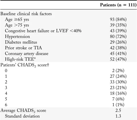
Duration of follow-up. All of the 108 patients completed their one-month follow-up; 97 (89.8%), their six-month Table 2. Pre-Procedural Stroke Risk Factors
Patients (nⴝ111)
Baseline clinical risk factors
Ageⱖ65 yrs 93 (84%)
Age⬎75 yrs 39 (35%)
Congestive heart failure or LVEF⬍40% 43 (39%)
Hypertension 80 (72%)
Diabetes mellitus 29 (26%)
Prior stroke or TIA 42 (38%)
Coronary artery disease 45 (41%)
High-risk TEE* 52 (47%)
Average CHADS2score 2.5
Standard deviation 1.3
*Defined as moderate or dense spontaneous echo contrast or blood flow velocityⱕ20 cm/s within the left atrial appendage. †The CHADS2index calculates the individual stroke risk by assigning one point each for the presence of congestive heart failure, hypertension, age⬎75 years, and diabetes, and two points for history of stroke or transient ischemic attack. Thus, CHADS2is an acronym for risk factors and their scoring. The stroke rate per 100 patient-years under aspirin therapy rises with increasing score: 0.8 with 0 points, 2.2 with 1 point, 4.5 with 2 points, 8.6 with 3 points, 10.9 with 4 points, 12.3 with 5 points, and 13.7 with 6 points (23).
follow-up; and 74 (68.5%), their one-year visit. One patient was lost to follow-up. The average follow-up was 9.8 months, a total of 90.7 documented implant years.
Primary study end points. Of the 111 enrolled patients, 1 patient (0.9%, 95% CI 0.02% to 4.9%) experienced two MAEs within the first 30 days after procedure: need for emergent cardiovascular surgery owing to cardiac tampon-ade, and neurological death in-hospital as previously described.
Secondary study end points. By six months after the procedure, the rate of patients that experienced no MAEs and/or mobile left atrial thrombus was 97.9% (95% CI 92.2% to 99.8%) on the basis of 95 of 97 patients with six-month follow-up. Two patients did not meet this end point: again, the aforementioned patient with two MAEs during the index hospitalization, and one patient who experienced an ischemic stroke 173 days after procedure.
Device performance end points. Implantation and treat-ment success were achieved in 108 of 113 procedures (95.6%, 95% CI 90.0% to 98.6%) performed in 111 patients (97.3%, 95% CI 92.3% to 99.4%). Complete recapture and retrieval was successful whenever necessary. Of the 108 implanted devices, 94 (87.0%) showed “trace leak” to “absent leak” after LAA occlusion; 14 patients (13.0%) showed “mild leak.”
Echocardiographic results. Immediately after the proce-dure, 86 of 88 patients (97.7%, 95% CI 92.0% to 99.7%) with assessable TEE showed successful occlusion of their appendage. At one month, 60 of 60 patients (100%) met this criterion (grade 3: 8 patients [13.3%]; grade 4: 31 patients [51.7%]; grade 5: 21 patients [35.0%]). At six months, 49 of 50 patients (98.0%, 95% CI 89.4% to 100%) showed successful LAA occlusion (grade 2: 1 patient [2.0%]; grade 3: 5 patients [10.0%]; grade 4: 27 patients [54.0%]; grade 5: 17 patients [34.0%]). In one patient, TEE revealed a laminar layer along the superior edge of the occluder at the six-month follow-up. With continuing administration of aspirin and clopidogrel this laminar growth resolved over the following six months, showing no remaining material in the 12-month TEE. Echocardio-graphic examinations showed no mobile thrombus and no disruption of mitral valve function or pulmonary vein inflow in any patient. The echocardiographic results matched the
outcome of chest X-ray in excluding device migration or dislodgement from the LAA. The concurrence of trans-esophageal Doppler color flow and angiography regarding evaluation of LAA occlusion is presented inTable 3.
Adverse events. There were seven MAEs in five patients (Table 4). A total of nine procedure-related SAEs occurred in seven patients. None of the SAEs were adjudicated to be device-related. Procedure-related SAEs included pleural effusion (n⫽1) and dyspnea requiring reintubation (n⫽1) in one patient, pericardial effusion in two patients (n⫽2), and cardiac tamponade in two patients (n⫽2). In three of them, pericardiocentesis was performed. One patient also developed a left-sided hemothorax (n ⫽1) that was noted on the first day after the procedure and was treated by thoracocentesis. One patient experienced right leg deep vein thrombosis (n⫽1), and another patient developed brachial plexus palsy (n ⫽1).
Incidence of stroke and TIA. Of the 111 enrolled pa-tients, 2 (1.8%, 95% CI 0.2% to 6.4%) experienced a stroke 173 and 215 days after the implant procedure. In both patients, TEE performed at the one- and six-month follow-up showed the device in stable position with no thrombogenic layer on the device surface. Doppler color flow at six months showed “trace leak” and “absent leak” in these two patients. Three TIAs occurred in two patients. The observed annual stroke rate was 2.2% (two events during 90.7 documented implant years).
Deaths. There were six deaths in 111 patients (5.4%). None were adjudicated as device- or procedure-related. The causes of death included cardiac or neurological death (n⫽
4), secondary complications after gastrointestinal bleeding (n⫽ 1), and an incarcerated hernia (n⫽ 1).
DISCUSSION
Atrial fibrillation, whether intermittent or sustained, in-creases the risk for cardioembolic events leading to an overall annual stroke rate of 4.5%/year (12–14). Anticoag-ulation is highly effective in preventing embolic events in these patients. With a risk reduction of almost 70%, it is superior to aspirin and aspirin plus low-intensity, fixed-dose warfarin treatment (13,15,16). Moreover, the use of warfa-rin has a narrow therapeutic range. Restrictions in everyday life, pharmacological interactions, and the potential risk of major hemorrhage limit this method of treatment (15,17). These concerns lead to an extensive anticoagulation under-Table 3. Left Atrial Appendage Occlusion Verified by
Transesophageal Doppler Color Flow and Angiography
Transesophageal
Transesophageal echocardiographic images were available from 88 patients after procedure. These two methods showed concurrence in 63 of 88 patients (71.6%) (5 patients ⫹ 58 patients). Two patients had a “Moderate Leak” (grade 2) in transesophageal Doppler color flow, whereas the angiography showed “Trace Leak” to “Absent Leak” (grade 4).
pts⫽patients; other abbreviations as inTable 2.
Table 4. Occurrence of MAEs During 90.7 Documented Implant Years
MAE Patients (nⴝ111)
Major or minor stroke 2 Myocardial infarction 0 Requirement for cardiovascular surgery related to
PLAATO procedure
1
Cardiac or neurological death 4 Any MAE 7 (in 5 patients)
use in general clinical practice (18,19). Furthermore, the benefit of anticoagulation as compared with aspirin treat-ment is more substantial in patients who are at higher risk for stroke (15,16). Yet these patients are more likely to have relative or absolute contraindications to warfarin treatment. Percutaneous LAA occlusion is an improved method of a known approach (7,20,21), excluding the LAA from blood flow and thus preventing thrombus formation and subse-quent thromboembolic complications. The advantages of this technique include a less invasive procedure, a faster recovery as compared with surgical ligation, and the reduced risk of potential bleeding owing to the absence of long-term anticoagulation therapy.
In our cohort of 111 patients, device implantation was successful in all but 3 patients. Five patients (4.5%) experi-enced pericardial effusions or hemopericardia that were without sequelae in four patients. In two of the four patients that required pericardiocentesis, however, this incidence occurred due to catheter manipulation during the attempt to access the LAA after trans-septal puncture. They were the first treated patients at a new site. It can be assumed that this number will decrease with increased experience with this new technique.
Five patients experienced MAEs during follow-up (5.5%/ year). In the Second Copenhagen Atrial Fibrillation, Aspi-rin and Anticoagulation Study (22), the risk of stroke, myocardial infarction, and vascular death in patients with AF receiving only aspirin therapy was 4.7%/year (intention-to-treat analysis).
Although the intention of closing the LAA is stroke prevention, the primary aim of this study was to show that LAA occlusion using the new PLAATO device is feasible and can be performed at acceptable risk. In order to make a more definitive statement on reduction of stroke, longer follow-up is needed. As of December 2003, the annual stroke rate of the 108 patients that underwent successful occlusion of their LAA was 2.2%. The cause of stroke in these patients remained uncertain. Transesophageal echo-cardiogram performed shortly before and after these events showed sufficient occlusion of the LAA with no thrombus formation on the device surface. This would seem to suggest a non-cardioembolic origin and, more importantly, shows that LAA occlusion cannot protect from all ischemic events.
On the basis of the patients’ CHADS2score (an acronym
for congestive heart failure, hypertension, age ⬎75 years,
diabetes mellitus, and stroke or transient ischemic attack) (2.5 points) (23), the estimated annual stroke rate for this patient population was 6.3%, assuming they were taking aspirin; this means the relative stroke reduction was 65%. The expected stroke rate may be even higher, since the
CHADS2 index does not take echocardiographic results
such as dense spontaneous echo contrast, a powerful stroke predictor (24), into its calculation.
Study limitations. This was a non-randomized study with a relatively selected patient population. Calculating their
stroke risk by using the CHADS2score may only give an
idea of what the expected risk for stroke in these patients may be. Furthermore, the follow-up period of the present study was limited to a maximum of 17 months after device implantation. Further studies are necessary to evaluate the performance of the PLAATO device regarding stroke prevention and the long-term tolerance of the device. Further limitations include the limited numbers of assess-able TEEs during follow-up.
Conclusions. The results of the present study show that percutaneous LAA occlusion using the new PLAATO system can be performed at acceptable risk. Because it is less invasive, this technique is more practical for a large number of patients compared with surgical closure. Longer-term studies are needed to confirm long-term safety of the device and a reduction in thromboembolic events. This technique may become an alternative therapeutic strategy for patients with AF who are at increased risk for ischemic stroke and, therefore, should receive anticoagulation therapy, but have a contraindication for long-term warfarin treatment.
Acknowledgments
The authors greatly appreciate the assistance of Franziska Buescheck and Madlen Reschke of the CardioVascular Center Frankfurt, as well as Becky Rorke, Kristine Zurowski, MA, and Linda Johnson, PhD, of ev3.
Reprint requests and correspondence: Dr. Horst Sievert, Car-dioVascular Center Frankfurt, Seckbacher Landstrasse 65, 60389 Frankfurt, Germany. E-mail: [email protected].
REFERENCES
1. Sandercock P, Bamford J, Dennis M, et al. Atrial fibrillation and stroke: prevalence in different types of stroke and influence on early and long term prognosis (Oxfordshire Community Stroke Project). BMJ 1992;305:1460 –5.
2. Wolf PA, Benjamin EJ, Belanger AJ, Kannel WB, Levy D, D’Agostino RB. Secular trends in the prevalence of atrial fibrillation: the Framingham Study. Am Heart J 1996;131:790 –5.
3. Kannel WB, Wolf PA, Benjamin EJ, Levy D. Prevalence, incidence, prognosis, and predisposing conditions for atrial fibrillation: population-based estimates. Am J Cardiol 1998;82:2N–9N. 4. Li-Saw-Hee FL, Blann AD, Lip GY. Effects of fixed low-dose
warfarin, aspirin-warfarin combination therapy, and dose-adjusted warfarin on thrombogenesis in chronic atrial fibrillation. Stroke 2000; 31:828 –33.
5. Aberg H. Atrial fibrillation. I. A study of atrial thrombosis and systemic embolism in a necropsy material. Acta Med Scand 1969;185: 373–9.
6. Stoddard MF, Dawkins PR, Prince CR, Ammash NM. Left atrial appendage thrombus is not uncommon in patients with acute atrial fibrillation and a recent embolic event: a transesophageal echocardio-graphic study. J Am Coll Cardiol 1995;25:452–9.
7. Blackshear JL, Odell JA. Appendage obliteration to reduce stroke in cardiac surgical patients with atrial fibrillation. Ann Thorac Surg 1996;61:755–9.
8. Nakai T, Lesh MD, Gerstenfeld EP, Virmani R, Jones R, Lee RJ. Percutaneous left atrial appendage occlusion (PLAATO) for prevent-ing cardioembolism—first experience in canine model. Circulation 2002;105:2217–22.
10. Hanna IR, Kolm P, Martin R, Reisman M, Gray W, Block PC. Left atrial structure and function after percutaneous left atrial appendage transcatheter occlusion (PLAATO): six-month echocardiographic follow-up. J Am Coll Cardiol 2004;43:1868 –72.
11. Dajani AS, Taubert KA, Wilson W, et al. Prevention of bacterial endocarditis: recommendations by the American Heart Association. Circulation 1997;96:358 – 66.
12. Wolf PA, Abbott RD, Kannel WB. Atrial fibrillation as an indepen-dent risk factor for stroke: the Framingham Study. Stroke 1991;22: 983– 8.
13. Atrial Fibrillation Investigators. Risk factors for stroke and efficacy of antithrombotic therapy in atrial fibrillation. Analysis of pooled data from five randomized controlled trials. Arch Intern Med 1994;154: 1449 –57.
14. Hart RG, Pearce LA, Rothbart RM, McAnulty JH, Asinger RW, Halperin JL. Stroke with intermittent atrial fibrillation: incidence and predictors during aspirin therapy. Stroke Prevention in Atrial Fibril-lation Investigators. J Am Coll Cardiol 2000;35:183–7.
15. Van Walraven C, Hart RG, Singer DE, et al. Oral anticoagulants vs aspirin in nonvalvular atrial fibrillation—an individual patient meta-analysis. JAMA 2002;288:2441– 8.
16. Hart RG, Halperin JL, Pearce LA, et al. Lessons from the stroke prevention in atrial fibrillation trials. Ann Intern Med 2003;138: 831– 8.
17. Levine MN, Raskob G, Landefeld S, Kearon C. Hemorrhagic complications of anticoagulant treatment. Chest 2001;119:108S–21S. 18. Brass LM, Krumholz HM, Scinto JM, Radford M. Warfarin use
among patients with atrial fibrillation. Stroke 1997;28:2382–9. 19. Bungard TJ, Ghali WA, Teo KK, McAlister FA, Tsuyuki RT. Why
do patients with atrial fibrillation not receive warfarin? Arch Intern Med 2000;160:41– 6.
20. Madden J. Resection of the left auricular appendix. JAMA 1948;140: 769 –72.
21. Johnson WD, Ganjoo AK, Stone CD, Srivyas RC, Howard M. The left atrial appendage: our most lethal human attachment! Surgical implications. Eur J Cardiothorac Surg 2000;17:718 –22.
22. Gullov AL, Koefoed BG, Petersen P, et al. Fixed minidose warfarin and aspirin alone and in combination vs adjusted-dose warfarin for stroke prevention in atrial fibrillation: Second Copenhagen Atrial Fibrillation, Aspirin, and Anticoagulation Study. Arch Intern Med 1998;158:1513–21.
23. Gage BF, van Walraven C, Pearce L, et al. Selecting patients with atrial fibrillation for anticoagulation—stroke risk stratification in pa-tients taking aspirin. Circulation 2004;110:2287–92.
24. The Stroke Prevention in Atrial Fibrillation Investigators Committee on Echocardiography. Transesophageal echocardiographic correlates of thromboembolism in high-risk patients with nonvalvular atrial fibrillation. Ann Intern Med 1998;128:639 – 47.
APPENDIX