Research Report
Dental Journal
(Majalah Kedokteran Gigi)2015 December; 48(4): 183–187
Levels of crystalline silica dust in dental laboratorium of Dental
Health Technology Study Program of Vocational Faculty,
Universitas Airlangga
Eny Inayati,1 Sherman Salim,2 Sonya Harwasih,1and Sri Redjeki Indiani1
1Dental Health Technology Diploma Study Program, Faculty of Vocational, Universitas Airlangga 2Department of Prosthodontics, Faculty of Dental Medicine, Universitas Airlangga
Surabaya-Indonesia
abstract
Background: Silicosis is an occupational lung disease caused by inhaling particles of crystalline silica in a long time. The disease then results in inflammation and defects in lung tissue. Prosthesis construction is usually conducted in dental laboratory using a lot of materials containing crystalline silica, such as gypsum, ceramics, planting material, sandblast and others. Purpose: This research aims to determine levels of crystalline silica dust in the dental laboratory of Dental Health Technology Diploma Study Program, Vocational Faculty, Universitas Airlangga. Method: Three measurement points was determined, namely point 1, point 2 and point 3 in each dental laboratory space (I and II). Suctioning dust was performed at those points using Low Volume Dust Sampler (LVDS). Samples taken were divided into two groups, namely X and Y. Taking dust samples were carried out for 30 minutes. Elements of crystalline silica contained in the dust were quantitatively measured using XR Defractometry tool, while size and morphology of silica were measured using SEM EDX tool. Data obtained were statistically analyzed by paired t test. Result: The results showed significant differences in the levels of the total dust measured and crystalline silica in the form of quartz and cristobalite among those two dental laboratory spaces. Conclusion: It can be concluded that the levels of the total dust and silica quartz dust in the dental laboratory spaces I and II were greater than the Threshold Limit Value (TLV) determined.
Keywords: dust; silica; crystalline; dental laboratory
Correspondence: eny Inayati, c/o: Program Studi D3 Teknik Kesehatan Gigi, Fakultas Vokasi Universitas Airlangga. Jl. Srikana 65 Surabaya 60286, Indonesia. e-mail: [email protected]
introduction
Workers in dental laboratory, often known as dental technicians, during their work are always exposed to a variety of factors that can affect their health condition, such as physical exposure (noise, vibration, illumination, electrics), chemical exposure (gas, dust), radiation exposure (microwave, infrared, ultraviolet) and biological exposure (bacteria, fungi, viruses).
However, pathogenic effects still depend on the concentration of toxins in the air and the duration of the exposure. There are three ways how toxic agents penetrate into the organism, namely inhalation (gas, vapor, dust), direct contact with the skin or mucous membranes and
gastrointestinal process (when the norms of hygiene and labor protection are ignored).1
Silicosis is an occupational lung disease caused by inhaling crystalline silica mostly common used in denture manufacture. Silicosis is a respiratory illness that causes inflammation and defects in lung tissue. Crystalline silica dust inhaled will be stored in the respiratory system. Deposition point, however, depends on particle size. The largest size of the particle will be saved in the nasopharynx (the upper respiratory tract, nose and throat) and eliminated by the organisms. Meanwhile, the smallest particles (respirable) will be penetrated into the trachea, bronchi and alveolar duct (throat, lung area above and below). This condition is considered as the early development of silicosis.3 During 1994-2000, the Center for Disease
Control and Prevention (CDC) in Atlanta had conducted a research reporting nine cases of silicosis suffered by dental technicians.4 The CDC claims that dental technicians have
a high risk for exposure to silicosis dust through air.5
Dental Health Technology Diploma Study Program of Vocational Faculty, Universitas Airlangga is an educational institution that graduates dental technicians. Students take this study program for 3 years or more. During their study, students learn how to make prosthesis of teeth in the dental laboratory using materials containing crystalline silica, such as gypsum, ceramics, planting material, sandblast and others. As a result, those students are exposed to crystalline silica through breathing during denture making process causing dust in the air, such as stirring the powder, removing the results of mold casting, cutting, polishing casting and ceramics, as well as using sandblaster. Therefore, students, dental technicians and teaching staffs who daily work in the lab will have a high risk for exposure to crystalline silica material. Nevertheless, levels of total dust and silica in the laboratory of Dental Health Technology Diploma Study Program of Vocational Faculty, Universitas Airlangga had never been measured.
Therefore, this research aims to determine the levels of crystalline silica in the dust in the dental laboratory of Dental Health Technology Diploma Study Program, Vocational Faculty, Universitas Airlangga. Finally, the significance of this research is to control and prevent air pollution optimally against harmful silica material impact on those students, dental technicians and teaching staffs working in there.
materialsandmethod
Samples used were dust aspirated or collected from the dental laboratory spaces I and II using a LVDS (Low Volume Dust Sampler, Hitachi type 35 RC-20SC5). Filtration technique was used in this research to collect dust particles. Before collecting the dust, measurement points were determined. There were three measurement points, namely point 1, point 2 and point 3 in the dental laboratory spaces I and II. Suctioning dust was conducted on such points, each of which was measured 2 times, and the pump
speed used was 10-30 lpm.6 This technique used a circular
filter (round) with a porosity of 0.3-0.45 µm.
The samples were divided into two groups, namely Group X and Group Y. Filters for both X and Y were previously weighed before stored in petri discs, and then put in a desiccator for 24 hours. There are two filters used in Group X, namely X1 set on holder before practicum and X2 set on holder during practicum. Meanwhile, in Group Y as a control group, there were also two filters used, namely Y1 put in petri disc and Y2 placed in the open space in the dental laboratory.
Afterwards, the pump was turned on, and the speed of air flow was checked again. Collecting dust was performed for 30 minutes. The indoor air temperature and pressure then were recorded when dust sampler was turned on. The indoor air temperature and air pressure were also measured when the pump was turned off. The filters were removed from the holders by using tweezers, put in petri disc, and put back into the desiccator for 24 hours. Level of total dust collected then was weighed using an analytical balance (with a minimum sensitivity of 0.01mg). Data obtained were put into the following formula:7
C = (X2-X1) – (Y2-Y1) × 1000
F x T
Note:
C = total dust concentration (mg/m3) x1 = the weight of the filter prior to exposure x2 = the weight of the filter after exposure
y1 = the weight of filter without treatment before exposure y2 = the weight of filter without treatment after exposure T = time (minutes)
F = flowrate (liters per minute).
elements of crystalline silica in dust were measured quantitatively using X-Ray Defractometry tool (xPertPro PANalytical), while the size and morphology of silica were measured using a scanning electron microscope, energy Dispersive X-ray (eDX SeM, Inspect S50). The result data obtained in the scale ratio were tabulated and statistically analyzed by t test.
results
The results of the statistical test using paired T-test, moreover, showed that there were significant differences in the total dust found at those three points in the dental laboratory space I between before and during practicum with p: 0.003<α 0:05. Similarly, there were significant differences in the total dust found at those three points in the dental laboratory space II between before and during practicum with p: 0.000 <α 0:05.
The results of the statistical test using Independent T-test, furthermore, showed that there were significant differences in the total dust between the dental laboratory
Dental Journal (Majalah Kedokteran Gigi) p-ISSN: 1978-3728; e-ISSN: 2442-9740. Accredited No. 56/DIKTI/Kep./2012. Open access under CC-BY-SA license. Available at http://e-journal.unair.ac.id/index.php/MKG
space I and the dental laboratory space II during the practicum with p: 0.015<α 0:05. The mean results indicate that the total dust in the dental laboratory space I during practicum was currently greater than in the dental laboratory space II. The focus of this research was crystalline silica dust in the form of quartz and cristobalite.
5
Figure 1. The distribution of Si (purple color) in the total dust found in the dental laboratory space II at point 2 (with 40.000x magnification).
5
Figure 2. Si with various morphology and size found in the dental laboratory space II at point 2 (with 40.000x magnification).
discussion
The results showed that the total dust in both dental laboratory spaces was higher than the Threshold Limit Value (TLV) about 10 mg/m3.8 As a result, it indicates that
the condition of those two dental laboratory spaces was not good and healthy for both dental technicians and students working there since the total dust can make them discomfort and trigger several potential diseases. For instance, it can reduce vision, cause unpleasant sediment on eyes, nose and ear as well as lead to skin damage.
eDX of SeM examination results obtained on the dust, moreover, indicated that the level of silica (Si) contained was quite high (Figure 1). This was due to the use of lab materials in the form of planting materials, gypsum, as well as ceramic materials for the manufacture of dental crowns, permanent dentures and removable dentures. Materials contained in dental ceramics are kaolin and feldspar Table 1. The mean and standard deviations of the total dust
measured before and during the practicum in the dental laboratory spaces I and II in Dental Health Technology Diploma Study Program, Vocational Faculty, Universitas Airlangga
Activities Mean
Standard Deviations (mg/m3)
p
The dental laboratory space I
Before the practicum 0.90 0.70
During the practicum 22.02 2.53 0.003 The dental laboratory space II
Before the practicum 0.58 0.02
During the practicum 10.42 0.28 0.000 p = probability
Table 2. The mean and standard deviations of the total dust measured during the practicum in the dental laboratory spaces I and II in Dental Health Technology Diploma Study Program, Vocational Faculty, Universitas Airlangga
Activities Mean
(mg/m3) DeviationsStandard p The dental laboratory
space I
22.02 2.53 0.015
The dental laboratory space II
10.42 0.28
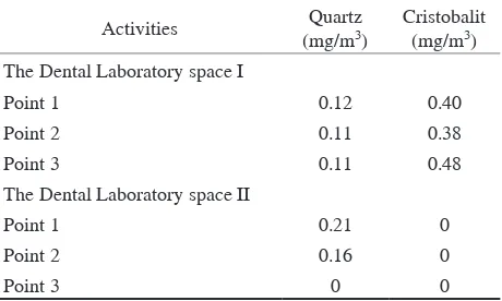
Table 3. Levels of crystalline silica dust (quartz and cristobalite) in the dental laboratory spaces I and II in Dental Health Technology Diploma Study Program, Vocational Faculty, Universitas Airlangga
Activities Quartz
(mg/m3) Cristobalit (mg/m3) The Dental Laboratory space I
Point 1 0.12 0.40
Point 2 0.11 0.38
Point 3 0.11 0.48
The Dental Laboratory space II
Point 1 0.21 0
Point 2 0.16 0
(K2O-Al2O3.SiO2) on build-up, polishing and finishing processes, which can be a source of spread of silica dust in room.9 Similarly, Figure 1 depicts how silica spread at all
points 1, 2 and 3 in the dental laboratory spaces I and II. The results of Xray Defractometry examination, furthermore, showed that there was quartz dust found at all sampling points in the dental laboratory space I. Meanwhile, in the dental laboratory space II, quartz dust was only found at point I and II. On the other hand, cristobalite dust was found at all sampling points in the dental laboratory space I. Nevertheless, there was no cristobalite dust found at all sampling points in the dental laboratory space II. These results indicate that both the dental laboratory spaces I and II had been exposed to silica dust, either quartz or cristobalite although quartz dust was only found at point I and II in the dental laboratory space II, while both quartz and cristobalite were not found at point 3 in the dental laboratory space II.
In addition, levels of silica found in this research were greater than the specified threshold limit value determined, about 0.1 mg/m3 for quartz and 0.05 mg/m3 for
cristobalit.10,11 This situation was triggered by a condition
that during practicum there were no specific rooms divided based on kinds of laboratorial work. All of the rooms are interconnected and not separated by a partition. The rooms are also cramped and lack of exhaust systems and ventilation (especially dental laboratory I). As a result, students get a high risk of being exposed or contaminated with air containing hazardous materials, such as metal, resin or silica causing pneumoconiosis.
Some cases of pneumoconiosis due to silica actually have been found.12 Silica dust can cause silicosis. Silica
contained in dental ceramics can be spread when stirring the ceramic powder or during grinding or polishing. Consequently, the surrounding environment can be exposed to it. exposure can also occur when stirring planting material and divesting (demolishing casting results). Planting materials often contain cristobalite, silica crystalline material that is the most toxic. During 1994-2000, occupational disease surveillance program in five countries had identified nine cases of silicosis in people working in dental laboratories.4 Silicosis arising from
inhalation of fine dust containing crystalline silica through the respiratory tract. Silica particles larger than 0.6 µ will be retained in the upper respiratory tract, while silica particles between 0.3 µ s/d 0.6 µ will arrive at the alveoli. Silica particles below 0.3 µ will follow brown movement, which is that dust particles can be inhaled and exhaled again.13
Size of silica dust particle found in this research was in a variety of sizes ranging from 0.5 s/ d 7.3 µ (Figure 2). The size and morphology of silica particles found were various due to grinding, divesting, polishing, sandblasting processes during practicum. As a result, dust can enter through the upper respiratory tract to pulmonary alveoli parts, and this can cause silicosis.
Silica dust found in the dental laboratory spaces, however, was not primarily lead to health problems of the students, dental technicians and teaching staff since there are many factors influencing health problems caused by Silica dust exposure. The health problems may arise when there is an interaction of several factors, such as high level of silica dust, frequency of exposure time, condition and endurance of those students, dental technicians and teaching staffs. A health problem caused by silica dust is known as silicosis disease. Symptoms of silicosis can be started by short breath, mid cough and chest tightness. Silicosis can get worse even though the cause has been terminated since silicosis cannot be cured, but the severity can only be prevented by avoiding silica dust exposure. Silicosis disease is often associated with other diseases, such as tuberculosis, kidney disease, lung cancer, fever and weight loss, even leading to death. If exposed to organisms that cause tuberculosis, mycobacterium tuberculosis, silicosis patients will have a risk three times more likely to suffer tuberculosis.14
The level of silica in the dental laboratory spaces I and II was quite high since materials used in the laboratory contained a lot of silica. Besides that, a range of laboratory processes, such as investing, sandblasting, grinding, polishing and others, can cause silica dust flying in the laboratory. Laboratory conditions that are not too wide with the big number of students and dental technicians and inadequate ventilation have significantly made silica dust exposure high enough. Fortunately, students and dental technicians work in the laboratory less than 8 hours and not every day. This is in accordance with Regulation of Minister of Labor and Transmigration No. 1311 stating that based on threshold limit values (TLV) as the standards of working environmental factors recommended in workplace, workers can still tolerate bad condition of the workplace causing disease or illness for not more of 8 hours a day or 40 hours a week. Nevertheless, those students, dental technicians and teaching staffs working in the dental laboratory still need to be protected form silicosis disease.
Prevention of silicosis disease in the dental laboratory spaces I actually can be conducted by organizing the spaces of the laboratory to prevent dust produced from flying anywhere. For example, adequate ventilation needs to be set by installing exhaust fan or vacuum cleaner connected to a pipe, which ends can get into the water reservoir so that dust does not fly, but directly go into the water. Besides, students, dental technicians and teaching staffs must wear personal protective equipment (PPe), such as gloves, goggles, gowns and masks during in the laboratory in order to maintain health. Personal Protective equipment used should meet the standards of OSHA, namely: can protect against hazards, can be worn comfortably, cannot restrict movement, as well must be durable and can easily be cleaned.15 Finally, in conclusion the levels of total dust
and silica quartz dust in both the dental laboratory spaces I and II were greater than TLV determined.
Dental Journal (Majalah Kedokteran Gigi) p-ISSN: 1978-3728; e-ISSN: 2442-9740. Accredited No. 56/DIKTI/Kep./2012. Open access under CC-BY-SA license. Available at http://e-journal.unair.ac.id/index.php/MKG
references
1. Kartaloglu Z, Ilvan A, Aydilek R. Dental technician’s pneumoconiosis: mineralogical analysis of two cases. Yonsei Med J 2003; 44: 169-73.
2. Hu SW, Lin YY, Wu TC, Hong CC, Lung SC. Workplace air quality and lung function among dental laboratory technician. Am J Ind Med 2006; 49(2): 85-92.
3. Maynard AD, KuempeleD. Airbone nanostructured particle and occupational health. J of Nanoparticle Researach 2005; 7: 587-614.
4. Rosenman KD, Petchere, Schill DP, Valiante DJ, BresnitzeA, Cummings KR, Sociee, Filios MS. Silicosis in dental laboratory technicians – Five States, 1994-2000. Mortality & Morbidity Weekly Report 2004; 53(9): 195-7.
5. Alavi A, Shakiba M, Nejad AT, Massahnia S, Shiari A. Resporatory fendings in dental laboratory technicians in rasht (North of Iran). Tannafos 2011; 10(2): 44-9.
6. Badan Standarisasi Nasional. Pengukuran kadaar debu total di udara tempat kerja. SNI 16-7058-2004: 3.
7. Aditya SA, Denny A. Identifikasi kadar debu di lingkungan kerja dan keluhan subyektif pernafasan tenaga kerja bagian finish mill. Jurnal Kesehatan Lingkungan 2007; 3(2): 161-72.
8. Menteri Kesehatan RI. Persyaratan kesehatan lingkungan kerja perkantoran dan industri. Kemenkes No. 1405/MeN/XI/2002. 9. Kim TS, Kim HA, Heo Y, Park Y, Park CY, Roh YM. Level of
silica in the respirable dust inhaled by dental technicians with demonstration of respirable symptoms. Ind Health 2002; 40(3): 260-5.
10. Badan Standarisasi Nasional. Nilai Ambang Batas (NAB) zat kimia di udara tempat kerja. SNI19-0232-2005: 18.
11. Permernakertrans RI No 13.2011. Nilai ambang Batas Bahan Fisika dan Kimia di tempat kerja.
12. Morgenroth K, Kronenberger H, Michalke G, Scknabel R. Morphology and pathogenesis of pneumoconiosis in dental technicians. Pathol Res Pract 1985; 179(4-5): 528-36.
13. Teguh P, Susanto JP. Kualitas debu dalam udara sebagai dampak industri pengecoran logam ceper. Jurnal Teknologi Lingkungan 2001; 2(2): 168-74.
14. Farazi A, Jabbariasi M. Silico tuberculosis and associated risk factors in central province of Iran. Pan Afr Med J 2015; 20: 333.
15. Occuptional Safety and Health Administration (OSHA). Personal protective equipment. Available from: www.osha.gov. 2003. Accesed June 11, 2015.