N
itrogen is often the major limiting nutrient for plant growth. Most plants obtain their nitrogen from soil nitrate, which is largely derived from either fertilizers or the mineralization of indigenous organic matter. The nitrate is converted to NH4+
by the sequential reduc-tive action of the plant enzymes nitrate reductase and nitrite reductase. Some plants, most notably many legumes, can also obtain their nitrogen from atmospheric N2. These legumes form a symbiotic
association with rhizobia, which are able to reduce N2to NH4
+
by the action of nitrogen-ase (Fig. 1). Ammonia is then transferred from the microbe to the plant. Plants also produce significant amounts of NH4
+
from photorespiration, phenylpropanoid biosyn-thesis and amino acid catabolism. Conse-quently, the reduced form of nitrogen ultimately available to higher plants for assimilation is NH4
+
, irrespective of the pri-mary nitrogen source1
. However, because NH4
+
is toxic to plant cells, because of its ability to uncouple respiration at low con-centrations, it must be rapidly assimilated into non-toxic organic compounds. This necessitates strict regulatory control of the nitrogen-assimilation pathway.
Prior to 1970 it was generally assumed that ammonia was assimilated by the direct amination of a-ketoglutarate to produce glutamate in a single reaction catalyzed by glutamate dehydrogenase. However, the plant enzymeÕs high Kmfor NH4
+
(>1 mM)
suggested that glutamate dehydrogenase did not have a major role in nitrogen assimilation2
. Subsequent studies using radiolabeled (13
N) and stable (15
N) nitrogen isotopes, enzyme inhibitors and mutants of plant nitrogen metabolism indicated that the primary assimilation of NH4
+
into amino acids occurs via the joint action of glutamine synthetase and glutamate synthase [also termed glu-tamate 2-oxoglutarate aminotransferase (GOGAT)] (Fig. 1)3
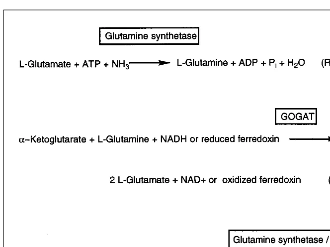
. The reaction catalyzed by glutamine synthetase involves the ATP-dependent amination of glutamate to yield glutamine. GOGAT then catalyzes the transfer of the amide group from glutamine to a-ketoglutarate to yield two molecules of glutamate. These two reactions, collectively referred to as the glutamine syn-thetase/GOGAT cycle, are now generally agreed to be the primary
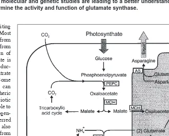
route of nitrogen assimilation in plants (Fig. 2). The synthesized glutamate can be used either to replenish the pool of glutamate for subsequent glutamine synthetase catalysis or to donate its amino group to form other nitrogen-containing compounds. One impor-tant fate of glutamate and glutamine is the synthesis of aspartate and asparagine, produced in reactions catalyzed by aspartate aminotransferase and asparagine synthetase (Fig. 1). These amino acids are important nitrogen-transport compounds in many plants. Carbon skeletons required for these initial NH4
+
-assimilatory reactions are provided by a-ketoglutarate and oxaloacetate1. The requirement for and metabolism of these tricarboxylic acid cycle intermediates tightly link nitrogen assimilation and carbon metabolism.
Glutamate synthase and nitrogen
assimilation
Stephen J. Temple, Carroll P. Vance and J. Stephen Gantt
The assimilation of ammonia by a wide variety of organisms is the primary route for the intro-duction of nitrogen into the biosphere. The assimilatory enzymes glutamine synthetase and
glutamate synthase catalyze reactions that convert a-ketoglutarate and ammonia to
glu-tamate, which is then used in a wide variety of biosynthetic reactions. These enzymes also play a major role in the reassimilation of ammonia derived from photorespiration in C3plants. Recent biochemical, molecular and genetic studies are leading to a better understanding of the factors that determine the activity and function of glutamate synthase.
Fig. 1.Diagram showing nitrogen assimilation in legume root nodules and the cellular lo-cation of the enzymes involved. In the diagram, glutamine, asparagine and ureides derived from purines are the primary nitrogenous compounds exported to other cells and provided for transport throughout the plant. Photosynthate provides the carbon skeletons for amino acid biosynthesis via glycolysis and the tricarboxylic acid cycle. Substantial additional car-bon can also be derived from non-photosynthetic CO2fixation via phosphoenolpyruvate.
Biochemistry and enzymology of glutamate synthase
In higher plants, GOGAT occurs as two distinct isoforms, NADH-GOGAT (EC 1.4.1.14) and ferredoxin-dependent NADH-GOGAT (Fd-GOGAT) (EC 1.2.7.1); these differ in molecular mass, subunit composition, enzyme kinetics, antigenic and reductant specificity, and metabolic function4,5
. Fd-GOGAT is an ironÐsulphur flavo-protein with a subunit molecular mass of 130Ð180 kDa that is gen-erally considered to function as a monomer. The enzyme has a pH optimum of 6.9Ð7.5 and apparent Kmvalues for ferredoxin,
glut-amine and a-ketoglutarate of 2Ð6, 100Ð1000 and 7Ð70 mM, respectively6
. In combination with the plastid-localized isoform of glutamine synthetase, Fd-GOGAT catalyzes the assimilation of NH4
+
derived from both the light-dependent reduction of NO3
2
and the NH4 +
generated during photorespiration5
. Maize roots contain an Fd-GOGAT isoform that is immunologically distinct from the enzyme found in leaves, suggesting that the two forms are encoded by distinct genes. The root isoform has been impli-cated in the assimilation of NH4
+
derived from soil NO3
2
(Ref. 7). Recent studies indicate that Arabidopsisalso contains two distinct and apparently functional Fd-GOGAT genes (GLU1and GLU2). The GLU1gene is expressed predominantly in leaves, and GLU2
expression is more abundant in roots8,9
.
Although NADH-GOGAT, like Fd-GOGAT, is also an ironÐsulphur flavoprotein, this enzyme is found primarily in non-green tissues. NADH-GOGAT has been purified and character-ized from legume root nodules and rice cell cultures4,10,11
. In nitrogen-fixing legume nodules, NADH-GOGAT activity has been found to increase markedly during nodule development, and this activity is associated with a single form of the enzyme4
. In higher plants, NADH-GOGAT exists as monomers with a native subunit mass of approximately 225Ð230 kDa; has a pH-optimum
range from 7.5 to 8.5; and apparent Km
values for NADH, glutamine and a
-ketoglutarate of 4Ð13, 400Ð1000 and 39Ð 960 mM, respectively6. In root nodules, NH4+
is exported from the nitrogen-fixing bac-teroids to the host-plant cytoplasm, where it is rapidly assimilated via NADH-GOGAT and cytosolic glutamine syn-thetase into amino acids (Fig. 1). There is negligible NADH-GOGAT mRNA, enzyme activity or immunoreactive protein in alfalfa leaves and roots1
. In bean nod-ules, NADH-GOGAT appears to occur as two isoforms (I and II), with the observed increase in GOGAT activity during nodule development resulting primarily from an increase in activity of isozyme II (Ref. 12). The expression patterns of the genes encoding cytosolic glutamine synthetase and NADH-GOGAT appear to be coordi-nated in non-legumes, where the proteins function together in processes such as the primary assimilation of NH4
+
derived from soil NO3
2, the reassimilation of NH
4 +
released during amino acid catabolism, and/or the reassimilation of NH4
+
released during seed germination3,8
. In a study with rice, NADH-GOGAT protein and activity increased four- and sixfold, respectively, in the apical spikelets during the first 15 d after flowering; the levels reached a maxi-mum when seed storage protein accumu-lation was initiated13
. The majority of the NADH-GOGAT protein was found to be associated with the young grain tissue. Although changes in Fd-GOGAT activity par-alleled the changes in NADH-GOGAT activity, the relative abun-dance of NADH-GOGAT was about threefold higher than that of Fd-GOGAT (Ref. 13). These results suggest that in rice NADH-GOGAT is responsible for the synthesis of glutamate from the glutamine that is transported from senescing tissues to the spikelets.
Evolutionary and structural relationships among diverse glutamate synthase proteins
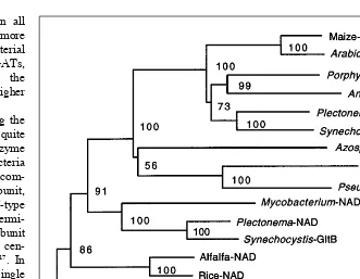
GOGAT is found in all types of organisms, and its amino acid sequence is remarkably well conserved. To examine the evolu-tionary relationships among the eubacterial and eukaryotic GOGAT proteins, a phylogenetic tree can be constructed based on the amino acid sequences of regions common to all eubacterial and eukaryotic GOGAT proteins (Fig. 3). With the exception of the Synechocystis sp. gltBgene product, all of the Fd-GOGAT proteins cluster together. This group contains Fd-GOGATs from higher plants (maize, rice and Arabidopsis), two red alga species (Porphyra purpureaand Antithamnionsp.) and two cyanobacteria (Synechocystis sp. and Plectonema boryanum). This analysis shows that eukaryotic Fd-GOGAT is closely related to bacterial Fd-GOGATs, and suggests that the genes encoding these enzymes are derived from the eubacterial precursors of chloroplasts. These data are consistent with an endosymbiotic origin of plastids. Addi-tional support for this conclusion comes from the finding that the Fd-GOGAT gene is found in the plastid genomes of red algae14
. Higher plant GOGAT genes are all located in the nuclear genome, into which they were presumably transferred from the endosym-biont genome15
. The phylogenetic analysis also suggests that the Fig. 2.The glutamate synthase cycle. The first reaction is catalyzed by glutamine synthetase
L-glutamate: ammonia ligase (ADP-forming) (EC 6.3.1.2). The second reaction is catalyzed by glutamate synthase (GOGAT), which can exist in two forms: L-glutamate: NAD+
gltB gene, which has been found in all cyanobacteria examined, is generally more similar to eukaryotic and eubacterial NAD(P)H-GOGATs than to Fd-GOGATs, and did not functionally replace the NADH-GOGAT genes found in higher plants.
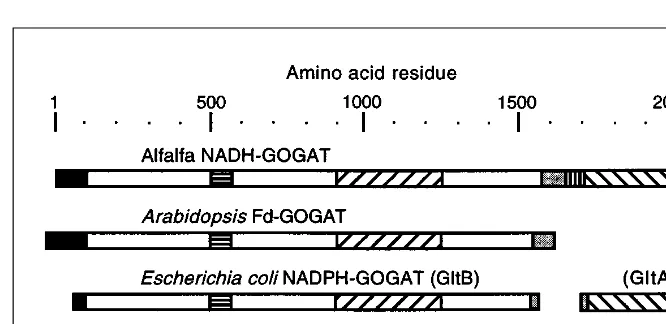
The structural relationships among the various GOGAT proteins are quite diverse16
(Fig. 4). In general, the enzyme found in non-photosynthetic eubacteria uses NADPH as a cofactor and is com-posed of two subunits. The large subunit, encoded by gltB, contains a PurF-type amidotransferase domain near its N-termi-nus and a 3Fe/4S center. The small subunit may contain additional ironÐsulphur cen-ters and an NADPH-binding region17
. In eukaryotes, which contain a single ironÐsulphur center18
, sequences similar to both of these subunits, separated by a short hydrophilic region, are found in the single NADH-GOGAT polypeptide. Fd-GOGAT is structurally similar only to the large sub-unit of the eubacterial NADPH-GOGAT protein. These data suggest that the NADPH-GOGAT small subunit serves pri-marily to couple the oxidation of NADPH to the reductive transfer of the amido group of glutamine to the a-keto position of
2-oxoglutarate.
The consensus view of GOGAT struc-ture and function has recently been chal-lenged. Two GOGAT genes, gltBand gltS, have been isolated and characterized from a Synechocystissp.19
. Both of these genes are similar to the large subunit gene of non-photosynthetic eubacteria, but one is much more similar to plant NADH-GOGAT than it is to any other Fd-GOGAT protein. This
indicates that cyanobacteria might, like plants, have both Fd- and
NAD(P)H-GOGAT enzymes. However, significant
Synecho-cystisFd-GOGAT activity was retained in mutants that contained insertionally mutagenized gltB and gltS genes. Additionally, Navarro et al.19
and other researchers have been unable to detect any NAD(P)H-GOGAT activity in this organism. Thus, based on both biochemical and mutational analyses, it appears that the two
Synechocystisgenes encode Fd-GOGAT. However, recent analy-sis of the complete Synechocystisgenomic sequence suggests that this organism contains a gene similar to gltA, which encodes the small subunit of all eubacterial NAD(P)H-GOGAT proteins.
Another test of the consensus view of GOGAT structure and function comes from comparisons of eubacterial and eukaryotic GOGAT sequences with those of archaebacteria. All examined eubacteria and eukaryotes contain genes encoding NAD(P)H-GOGAT proteins similar to E. coli gltAand gltBgene products. A recent report20
describes the isolation and characterization of a GOGAT gene from the archaebacterium Pyrococcussp. KOD1. This gene encodes a protein that is very similar to the eubacterial
gltA-encoded GOGAT small subunits. However, unlike the gltA
gene in eubacteria, it is not linked to the gltBgene. Furthermore, when the Pyrococcus gltAgene was expressed in E. coli, the puri-fied product was capable of catalyzing both glutamine- and ammonia-dependent, NADPH-coupled glutamate synthesis. This
result is reminiscent of evidence obtained with E. coli NADPH-GOGAT showing that the gltA-encoded subunit alone can cata-lyze the ammonia-dependent synthesis of glutamate21
. It is not known whether Pyrococcuscontains a gltB-like gene.
In contrast to the findings of Jongsareejit et al.20
, examination of the recently sequenced Methanococcus jannaschii genome revealed that this archaebacterium may not contain a gene encod-ing a gltAsubunit. Although several genes are similar to gltA, all have been assigned other functions, and none is nearly as similar to E. coli gltAas E. coli gltAis to Pyrococcus gltA. Furthermore, the structure of the protein encoded by the M. jannaschii gltB-like gene is unusual. Comparisons of eubacterial and eukaryotic GOGAT large subunits show that their deduced amino acid sequences are similar throughout most of the protein, with gener-ally more than 40% amino acid identity. This relationship does not hold true when the product of the M. jannaschii gltB-like gene is examined. Compared with the corresponding eubacterial and eukaryotic proteins, this protein is very small. Additionally, com-parison of this 510 amino acid (55 kDa) protein with other GOGAT proteins shows that it is composed of two distinct regions that are similar to the eubacterial GltB subunit (Fig. 4). The region from approximately amino acids 90 to 160 of the archaebacterium GltB-like protein is similar to a region from about 500 to 580 of the eukaryotic GOGATs, and contains no known functional properties. Fig. 3.An unrooted phylogenetic tree of NAD(P)H- and Fd-glutamate synthase (GOGAT) proteins. The neighbor-joining method32found in the CLUSTAL W suite of programs33was
used to calculate the tree. The numbers correspond to ÔbootstrapÕ percentages. The length of the bar represents 0.1 substitutions per site. GOGAT protein sequence information was obtained either from the GenBank protein database or from conceptual translations of GOGAT-encoding genes found in the GenBank nucleotide database. The accession num-bers used were: Maize-Fd, M59190; Arabidopsis-Fd, Y09667; Porphyra-Fd (P. purpurea), U38804; Antithamnion-Fd, Z21705; Plectonema-Fd (P. boryanum), 085735;
Synechocystis-GltS, X92480; Azospirillum-NADP (A. brasilense), L04300; Escherichia -NADP (E. coli), AE000400; Pseudomonas-NADP (P. aeruginosae), U81261;
Mycobacterium-NAD (M. tuberculosis), Z83864; Plectonema-NAD (P. boryanum), D85230; Synechocystis-GltB, X80485; Alfalfa-NAD, L01660; Rice-NAD, AB0011916;
The C-terminal 350 amino acids of the archaebacterium GOGAT are similar to amino acids 880 to 1240 of eukaryotic GOGATs. This region binds flavin mononucleotide and contains the 3Fe/4S center. Further research is needed to elucidate the structure and function of GOGAT in these ÔprimitiveÕ organisms.
Post-translational processing and intracellular localization of glutamate synthase
Fd-GOGAT proteins from Arabidopsisand maize contain a pre-sequence with many of the characteristics of plastid transit pep-tides, including a net positive charge and high proportion of threonine and/or serine residues. Consistent with the projected plastid localization of the plant enzyme is the plastid localization of its cofactor, ferredoxin, and the plastid location of the Fd-GOGAT genes in red algae. Similarly, both rice and alfalfa4
NADH-GOGATs contain presequences that are thought to be involved in plastid targeting. Interestingly, presequences are found in all characterized eukaryotic GOGAT proteins, including yeasts22
and nematodes, and all eubacterial GltB and cyanobac-terial Fd-GOGAT proteins. The yeast and nematode presequences do not have the appearance of mitochondrial transit peptides22
, and thus their function is unknown. The N-terminus of all charac-terized GltB-like proteins is a cysteine, which is thought to be a critical component of the glutamine amidotransferase activity of the enzyme16
. It is possible that one of the functions of the pre-sequence is to ensure the proper proteolytic processing needed to generate this N-terminal cysteine and activate the enzyme.
Mutants of glutamate synthase
Understanding of plant nitrogen metabolism has been greatly aided by the isolation and study of mutants defective in their ability to catalyze defined biochemical reactions. Arabidopsis
mutants that contained only 1Ð2% of wild-type Fd-GOGAT activ-ity in leaves were isolated in 198023
. These mutants exhibited severe stress symptoms in normal air, but grew normally when placed in an atmosphere that minimized photorespiration [0.7% (v/v) CO2]. The Fd-GOGAT mutants were part of a wide range of
mutant lines that were deficient in key enzymes of the
photorespiratory carbon and nitrogen cycles. This pioneering work confirmed the existence of the photorespiratory cycle; established the importance of Fd-GOGAT in photorespiratory NH4
+
reassimilation; and was instrumental in establishing
Arabidopsisas a model plant system. Sub-sequent analysis of one of these Fd-GOGAT mutants indicated that it contains undetectable Fd-GOGAT protein in either leaf or root tissue, although it does retain a low level of activity and transcript24
. This apparent conflict between genetic and mol-ecular data is at least partly resolved by the recent finding that Arabidopsishas a sec-ond Fd-GOGAT gene8
. The GLU2gene9
is expressed predominantly in roots, and has very low levels of expression in leaves;
GLU1 is expressed predominantly in
leaves. Presumably, GLU1expression, but not GLU2expression, was affected in the photorespiratory mutants.
Using a similar screening strategy, Fd-GOGAT mutants have been isolated from barley and pea25
. Fd-GOGAT activity is virtually undetectable in the leaves of the barley mutants. However, levels of NADH-GOGAT are present at wild-type levels, indicating that no compensatory mechanisms exist and confirming the distinct roles proposed for the two GOGAT isoforms. Immunoblot analysis of four barley Fd-GOGAT mutant lines indicated that three had no detectable cross-reacting Fd-GOGAT protein and that the fourth had significantly reduced levels of this protein26
. Photosynthetic CO2assimilation
in the barley Fd-GOGAT mutants is reduced on transfer from elevated CO2to air. This effect on photosynthesis is unlikely to be
caused solely by the accumulation of NH4 +
and may result from the unusual and complex interaction of carbon and nitrogen metab-olism that occurs in these mutants as a result of photorespiration27
. Genetic engineering techniques have recently been used to gen-erate transgenic tobacco plants that express an Fd-GOGAT cDNA fragment in the antisense orientation under the control of the CaMV-35S promoter. The five transgenic tobacco lines recovered were found to contain between 40 and 100% of wild-type levels of Fd-GOGAT activity and protein. Under ambient growth conditions, severe symptoms of ammonia toxicity were seen in the two lines that contain less than 60% of wild-type Fd-GOGAT activity28
. In contrast to the multiplicity of mutants for Fd-GOGAT, there are no plant mutants for NADH-GOGAT activity. The phenotype of such mutants is difficult to predict, but one might expect a sub-stantial reduction in growth and nitrogen content, particularly in nodulated legumes and in those plant species that reduce NO3
2 in their roots.
Regulation of GOGAT in the plant
Light and a variety of metabolites have been shown to exert major regulatory controls over many metabolic pathways. It is well established that light, mediated via phytochrome, has a positive effect on the expression of the chloroplast-localized isoform of glutamine synthetase29
. Recent evidence indicates that light also exerts a positive regulatory effect on the expression of Fd-GOGAT (GLU1) (Ref. 24). GLU2expression is also induced by light, although the induction of this gene by sucrose in the dark indicates that the light-induced expression may in part be caused by an increase in the concentration of carbon metabolites9
. A Fig. 4.Regions of sequence similarity among eukaryotic, eubacterial and archaebacterial
recently proposed model suggests that the regulation of nitrogen-assimilatory enzymes in Arabidopsisis related to light- and dark-mediated stimuli interconnected to levels of organic carbon and nitrogen8,9
. In another recent study, tobacco transformants with very low nitrate reductase activity were used to investigate poss-ible coordinated regulatory signals that influence carbon and nitrogen metabolism30
. Nitrate was shown to act as a signal, resulting in widespread changes in the expression of key genes in the pathways of nitrogen and carbon metabolism, including Fd-GOGAT. The induction of organic acid metabolism and repres-sion in starch metabolism by nitrate could increase the availability of carbon skeletons for nitrogen assimilation30
.
Despite the results from many biochemical studies, and the availability of cDNA clones and antibodies to all of the enzymes involved in plant nitrogen assimilation, the question as to which enzymes are limiting and thus restricting the effectiveness of efforts to enhance nitrogen-use efficiency remains unresolved. However, studies of plant gene expression in developing alfalfa nodules suggest that NADH-GOGAT is uniquely regulated as compared to the other genes involved in nitrogen and carbon metabolism4,11
. Although cytosolic glutamine synthetase, aspar-tate aminotransferase, phosphoenolpyruvate carboxylase and malate dehydrogenase transcripts readily accumulate in ineffec-tive (non-N2-fixing) nodules, NADH-GOGAT transcript accumu-lation remains low at root background levels. It is clear that NADH-GOGAT expression is strikingly different to that of either cytosolic glutamine synthetase or aspartate aminotransferase-1 (Refs 4 and 11) (Fig. 5). Although maximum expression of NADH-GOGAT occurred in effective nodules, that in ineffective nodules and roots was only 12Ð20% of the maximum. By
comparison, cytosolic glutamine synthetase transcript abundance in ineffective nodules is only slightly reduced (15%) as compared to effective nodules, and aspartate aminotransferase-1 is ex-pressed at high levels in roots and both effective and ineffective nodules. Thus, unlike other key plant genes encoding proteins involved in nitrogen assimilation, increased levels of NADH-GOGAT expression were associated only with effective nod-ules31
. These experiments show that active nitrogen fixation, and perhaps NH4
+
itself or a downstream product of its metabolism, is required for maximum NADH-GOGAT gene expression.
Although substantial progress has been made with the molecu-lar genetics of primary acquisition and assimilation of nitrogen, elucidating which genes are most crucial to enhancing nitrogen-use efficiency remains an important goal. A previously poorly studied area that may assist with this effort is the signal-trans-duction pathway involved in the activation of the genes involved in primary nitrogen assimilation, and the associated generation of carbon skeletons.
Acknowledgements
We wish to thank many colleagues for providing reprints and communicating recent results. The work was supported by National Science Foundation Grant No. IBN-9206890. This paper is a joint contribution from the Plant Sciences Research Unit, United States Dept of Agriculture, Agricultural Research Service, and the Minnesota Agriculture Experiment Station (Paper No. 971130039, Scientific Journal Series).
References
01Vance, C.P. (1997) The molecular biology of N metabolism, in Plant Metabolism(Dennis, D.T. et al., eds), pp. 449Ð477, Longman
02Stewart, G.R., Mann, A.F. and Fentem, P.A. (1980) Enzymes of glutamate formation: glutamate dehydrogenase, glutamine synthetase, glutamate synthase, in The Biochemistry of Plants(Vol. 5) (Miflin, B.J., ed.), pp. 271Ð327, Academic Press
03Lea, P.J., Robinson, S.A. and Stewart, G.R. (1990) The enzymology and metabolism of glutamine, glutamate and asparagine, inThe Biochemistry of Plants(Vol. 16) (Miflin, B.J. and Lea, P.J. eds), pp. 121Ð159, Academic Press
04Gregerson, R.G. et al. (1993) Molecular characterization of NADH-dependent glutamate synthase from alfalfa nodules, Plant Cell 5, 215Ð226
05Sakakibara, H. et al. (1991) Molecular cloning and characterization of complementary DNA encoding ferredoxin dependent glutamate synthase in maize leaf, J. Biol. Chem.266, 2028Ð2035
06Lea, P.J. et al. (1990) Enzymes of ammonia assimilation, in Methods in Plant Biochemistry(Lea, P.J., ed.), pp. 257Ð276, Academic Press
07Redinbaugh, M.G. and Campbell, W.H. (1993) Glutamine synthetase and ferredoxin-dependent glutamate synthase expression in the maize (Zea mays) root primary response to nitrate,Plant Physiol. 101, 1249Ð1255
08Lam, H-M. et al. (1996) The molecular-genetics of nitrogen assimilation into amino acids in higher plants, Annu. Rev. Plant Physiol. Plant Mol. Biol.47, 569Ð593
09Oliveira, I.C. et al. (1997) Molecular-genetic dissection of ammonium assimilation in Arabidopsis thaliana, Plant Physiol. Biochem.35, 185Ð198
10 Yamaya, T. et al. (1992) Tissue distribution of glutamate synthase and glutamine synthase in rice leaves, Plant Physiol. 100, 427Ð432
11 Vance, C.P. et al. (1995) Alfalfa NADH-dependent glutamate synthase: structure of gene and importance in symbiotic N2fixation, Plant J.8, 345Ð358
12 Chen, F-L. and Cullimore, J.V. (1988) Two isozymes of NADH-dependent glutamate synthase in root nodules of Phaseolus vulgarisL., Plant Physiol. 88, 1411Ð1417
13 Hayakawa, T. et al. (1993) Changes in the content of two glutamate synthase proteins in spikelets of rice (Oryza sativa) plants during ripening, Plant Physiol.101, 1257Ð1262
c vFig. 5.An example of the analysis of glutamate synthetase (GOGAT), glutamine synthetase (GS) and aspartate aminotrans-ferase transcripts (AAT-1) in alfalfa roots and root nodules induced by effective and ineffective Rhizobium melilotistrains. RNA was isolated from the roots and root nodules collected 12 d after the plants were inoculated with either effective wild-type
R. melilotistrain 102F51 or with ineffective strain 7154 (Nif2
14 Valentin, K., Kostrzewa, M. and Zetsche, K. (1993) Glutamate synthase is plastid-encoded in a red alga: implications for the evolution of glutamate synthase, Plant Mol. Biol.23, 77Ð85
15 Gantt, J.S. et al. (1991) Transfer of rpl22 to the nucleus greatly preceded its loss from the chloroplast and involved the gain of an intron, EMBO J.10, 3073Ð3078
16 Curti, B. et al. (1996) Glutamate synthase: a complex ironÐsulphur flavoprotein, Biochem. Soc. Trans.24, 95Ð99
17 Vanoni, M.A. et al. (1992) Characterization of the flavins and the ironÐsulfur centers of glutamate synthase from Azospirillum brasilenseby adsorption, circular dichroism, and electron paramagnetic resonance spectroscopies, Biochemistry31, 4613Ð4623
18 Knaff, D.B. et al. (1991) Spectroscopic evidence for a [3FeÐ4S] cluster in spinach glutamate synthase, J. Biol. Chem.266, 15080Ð15084
19 Navarro, F. et al. (1995) Existence of two ferredoxin-glutamate synthases in the cyanobacterium Synechocystissp. PCC 6803. Isolation and insertional inactivation of gltBand gltS genes, Plant Mol. Biol.27, 753Ð767
20 Jongsareetjit, B. et al. (1997) Gene cloning, sequence and enzymatic properties of glutamate synthase from the hyperthermophilic arcgaeon Pyrococcussp. KOD1,Mol. Gen. Genet. 254, 635Ð642
21 Maentsaelae, P. and Zalkin, H. (1976) Active subunits of Escherichia coli glutamate synthase, J. Bacteriol.126, 539Ð541
22 Filetici, P. (1996) Sequence of the GLT1gene from Saccharomyces cervisiae reveals the domain structure of yeast glutamate synthase, Yeast12, 1359Ð1366
23 Somerville, C.R. and Ogren, W.L. (1980) Inhibition of photosynthesis in Arabidopsismutants lacking leaf glutamate synthase activity, Nature286, 257Ð259
24 Suzuki, A. and Rothstein, S. (1997) Structure and regulation of ferredoxin-dependent glutamate synthase from Arabidopsis thaliana, Eur. J. Biochem. 243, 708Ð718
25 Lea, P.J. and Forde, B.G. (1994) The use of mutants and transgenic plants to study amino acid metabolism, Plant Cell Environ.17, 541Ð556
26Avila, C. et al. (1993) Cloning and sequence analysis of a cDNA for barley ferredoxin-dependent glutamate synthase and molecular analysis of photorespiratory mutants deficient in the enzyme, Planta189, 475Ð483
27Hausler, R.E. et al. (1994) Control of photosynthesis in barley leaves with reduced activities of glutamine synthetase or glutamate synthase, Planta194, 406Ð417
28Hirel, B. et al. (1997) Manipulating the pathway of ammonia assimilation in transgenic non-legumes and legumes,J. Plant Nutr. Soil Sci.160, 283Ð290
29Peterman, T.K. and Goodman, H.M. (1991) The glutamine synthetase gene family of Arabidopsis thaliana: light-regulation and differential expression in leaves, roots and seeds, Mol. Gen. Genet.230, 145Ð154
30Scheible, W-R. et al.(1997) Nitrate acts as a signal to induce organic acid metabolism and repress starch metabolism in tobacco, Plant Cell9, 783Ð798
31Vance, C.P. et al. (1994) Primary assimilation of nitrogen in alfalfa nodules: molecular features of the enzymes involved, Plant Sci.101, 51Ð64
32Saitou, N. and Nei, M. (1987) The neighbor-joining method: a new method for reconstructing phylogenetic trees, Mol. Biol. Evol. 4, 406Ð425
33Higgins, D.G. and Sharp, P.M. (1988) CLUSTAL: a package for performing multiple sequence alignments on a microcomputer, Gene 73, 237Ð244
Stephen J. Temple is at the Dept of Agronomy and Plant Genetics, University of Minnesota, St Paul, MN 55108, USA; Carroll P. Vance is at the US Dept of Agriculture, Agricultural Research Service, Plant Science Research Unit and the Dept of Agronomy and Plant Genetics, University of Minnesota, St Paul, MN 55108, USA; and J. Stephen Gantt*is at the Dept of Plant Biology, University of Minnesota, St. Paul, MN 55108, USA.
*Author for correspondence (tel +1 612 625 4763; fax +1 612 625 1738; e-mail [email protected]).
I
ndependent endosymbiotic uptake of ancient prokaryotes into an early eukaryotic host is thought to have produced two types of organelles, chloroplasts and mitochondria, that are each sur-rounded by a double-membrane system. Fundamental differences in structure and function reflect the different origins of the two organelles. A distinguishing feature of chloroplasts is their many subcompartments, which include the outer- and inner-envelopemembranes, the thylakoid-membrane network, the interenvelope space, the stroma and the thylakoid lumen. Newly evolved pro-teins, employed in refining and/or regulating chloroplast function, are dependent on a specific import mechanism Ð as are those pro-teins encoded by genes that have become displaced, during evolu-tion, to the nucleus. The major components that catalyse this protein translocation pathway Ð which starts in the cytosol, and
The protein translocation apparatus
of chloroplast envelopes
The evolution of the chloroplast from a photosynthetic prokaryote has resulted in the dis-placement of most of the prokaryote genes to the nucleus of the host eukaryote. Accordingly, the new organism has evolved targeting and translocation mechanisms on the organellar membranes for nuclear-encoded proteins. In plastids, the protein-import machinery is dis-tinct from that of other organelles, in both composition and mechanism. Recently, proteins homologous to several subunits of the chloroplast import machinery were identified in the
cyanobacterium Synechocystis PCC6803. It appears that parts of the protein-import
machin-ery of chloroplasts are derived from ancient transport systems in cyanobacteria. These observations open up new avenues for elucidating the origin of the chloroplast membranes and functional properties of the protein-import machinery.