www.elsevier.com/locate/jinsphys
Calcium transport by isolated anterior and posterior Malpighian
tubules of Drosophila melanogaster: roles of sequestration and
secretion
K. Dube
1, D.G. McDonald, M.J. O’Donnell
*Department of Biology, McMaster University, 1280 Main Street West, Hamilton, Ontario, Canada, L8S 4K1
Received 3 February 2000; accepted 6 April 2000
Abstract
Ca2+ transport was examined in isolated Malpighian tubules (MTs) of adult Drosophila melanogaster. All segments of both anterior and posterior MTs have substantial capacity to transport Ca2+and to play a role, therefore, in calcium homeostasis and
elimination of excess dietary Ca2+. Approximately 85% of Ca2+ which enters the tubule is sequestered, and
|15% is secreted in
soluble form into the tubule lumen. Tubules secreting fluid at maximal rates can remove an amount of Ca2+ equal to the whole
animal calcium content in |9 h. Distal segments of the pair of anterior MTs can sequester the same amount of Ca2+ in ,2 h.
Functional advantages of high Ca2+ turnover rates are discussed. Transepithelial Ca2+ secretion is increased by treatments which depolarize the transepithelial potential (thapsigargin, high K+), or acidify the secreted fluids (bicarbonate-free salines). The effects of pharmacological reagents and variations in bathing saline ionic composition indicate that the processes of secretion and seques-tration are controlled independently, and that diltiazem-sensitive Ca2+ channels are an important component of sequestration. The
contribution of some form of apical Ca2+ pump is evaluated. 2000 Elsevier Science Ltd. All rights reserved.
Keywords: Malpighian tubule; Ion transport; Drosophila; Ca2+secretion; Ca2+sequestration
1. Introduction
The Malpighian tubules (MTs) and hindgut together form the primary system for osmoregulation, ionoregul-ation and excretion in insects. A large number of studies (reviewed by Beyenbach, 1995; Dow et al., 1998; O’Donnell and Spring, 2000) have examined mech-anisms and control of transport of K+, Na+and Cl2by
insect MTs. However, there have been fewer studies of MT calcium transport and the contributions of MTs to whole animal and haemolymph calcium regulation.
Calcium excretion in insects involves both internal sequestration and elimination of calcium in soluble form (Taylor, 1986). In some species, only the former process is found. In Rhodnius, for example, there are high rates of exchange of calcium across the basolateral membrane
* Corresponding author. Tel.:+1-905-525-9140 ext. 23103; fax: +1-905-522-6066.
E-mail address: [email protected] (M.J. O’Donnell).
1 Present address: Nicholas Beaver Laboratory, 361 Southgate
Drive, Guelph, Ontario, N1G 3M5.
0022-1910/00/$ - see front matter2000 Elsevier Science Ltd. All rights reserved. PII: S 0 0 2 2 - 1 9 1 0 ( 0 0 ) 0 0 0 6 9 - X
of the tubules, but no calcium is detectable in the secreted fluid (Maddrell et al., 1991). In this species, then, all calcium appears to be sequestered in the tubules, and none is released in the urine. High calcium concen-trations in the fluid secreted by the MTs might impair the function of the rectal epithelium, and this might limit the amount of calcium which can be excreted in soluble form (Maddrell et al., 1991).
In other species, such as Drosophila, MTs both sequester calcium as insoluble concretions (Wessing and Zierold, 1992) and eliminate calcium in solution in the secreted fluid (O’Donnell and Maddrell, 1995). In adult tubules of Drosophila melanogaster, calcium is secreted by both the main and lower segments of the MTs; the latter transports calcium at higher rates per unit length (O’Donnell and Maddrell, 1995). Calcium- and phos-phate-rich concretions are found in the distal segment of the anterior tubules of the larvae of Drosophila hydei (Wessing and Zierold, 1992). MTs contain up to 92% of whole animal calcium in the latter species, and MT calcium content increases with that of the diet.
This study of Drosophila MTs addresses two ques-tions concerning Ca2+
1. What are the relative contributions of sequestration
of calcium within the tubule, and secretion of calcium in soluble form into the tubule lumen? Importantly, we
have examined both processes in tubules of a single species and the same stage of development, whereas pre-vious studies have examined either Ca2+ secretion by tubules of adult D. melanogaster or calcium seques-tration by larval tubules of D. hydei. Ca2+
transport has been measured across all segments of both anterior and posterior MTs of adult D. melanogaster. Both the flux of calcium into the tubule cells across the basolateral membrane (basolateral Ca2+flux) and the flux across the epithelium and into the lumen (transepithelial Ca2+flux) have been determined. A reduction in the value of trans-epithelial relative to basolateral Ca2+flux indicates that Ca2+ is retained or sequestered in some way within the tubule cells.
2. What are the mechanisms of MT Ca2+ transport?
Characterization of Ca2+transport involved measurement of basolateral and transepithelial Ca2+fluxes in response to changes in bathing saline ionic composition or the addition of pharmacological reagents such as putative inhibitors of calcium-transporting ATPases and cal-cium channels.
2. Methods and materials
2.1. Insects and fluid secretion assays
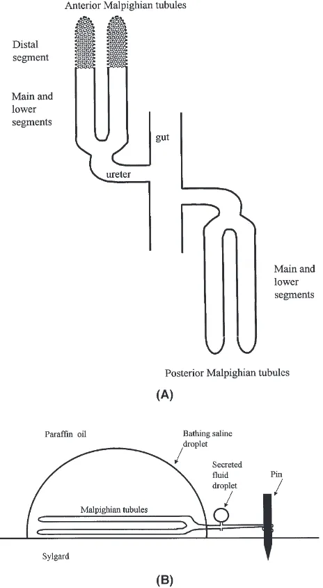
Adult female flies were selected from a laboratory col-ony maintained on standard fly medium containing inac-tivated yeast, sucrose and agar supplemented with fresh active yeast (Busto et al., 1999). Tegosept in ethanol and propionic acid were used to prevent mould growth. MTs were isolated and secreted fluid was analysed as described previously (O’Donnell and Maddrell, 1995). The four MTs consist of an anterior and posterior pair, and each pair is connected to the hindgut through a short ureter (Fig. 1A). For experiments examining transport across the whole MT, a preparation described in O’Don-nell and Maddrell (1995) was used (Fig. 1B). All four tubules, still connected to a very short length of the gut, were dissected and one pair of tubules was placed in a droplet of bathing saline under paraffin oil. One tubule of the other pair was cut away and discarded, and the remaining tubule was pulled out into the paraffin oil and wrapped around a short length (1–2 mm) of a fine steel minute pin (0.15 mm o.d.) stuck into the Sylgard bottom of the dish. Fluid was thus collected after it had passed through the entire length (i.e. all segments) of the two tubules upstream of their common ureter.
Control saline consisted of (in mmol l21
): 135 NaCl, 20 KCl, 2 CaCl2, 8.5 MgCl2, 10.2 NaHCO3, 4.3
Fig. 1. (A) Schematic diagram showing the segments of anterior and posterior Malpighian tubules. Stippling of the distal segment indicates the presence of lumenal Ca2+-rich concretions. (B) Schematic diagram
indicating arrangements for collection of fluid secreted by all segments of a pair of Malpighian tubules.
(concentrations in mmol l21) to control saline: 1.7 gly-cine, 7l-proline, 6.15l-glutamine, 0.95l-histidine, 0.55
l-leucine, 4.5 l-lysine, and 1.3 l-valine. The pH of
AARS was adjusted to 7.0 and calcium concentrations were varied by substitution with NaCl to maintain con-stant osmolality. Measurements with Ca2+-selective microelectrodes indicated that nominally Ca2+-free AARS contained 0.02 mmol l21 Ca2+.
Secreted fluid pH and Ca2+concentration were meas-ured with H+-selective and Ca2+-selective microelec-trodes based on the ionophores tridodecylamine and ETH 1001 (Fluka Chemical Corp, Ronkonkoma, NY), respectively. Procedures for electrode fabrication have been described previously (Maddrell et al., 1993). Briefly, borosilicate glass pipettes were acid-washed, dried on a hot plate and silanized by exposure to vapours of dimethyldichlorosilane. Silanization renders the glass surface hydrophobic and facilitates filling with, and retention of, the hydrophobic ionophore cocktail.
The Ca2+ concentration or the pH of secreted fluid droplets was measured under paraffin oil by positioning the ion-selective and reference microelectrodes in the drop and measuring the potential relative to that in drops of calibration solutions. A trial-and-error approach was used to determine the optimal extent of silanization. Insufficient silanization resulted in displacement of the cocktail by aqueous solutions, whereas excess silaniz-ation resulted in displacement of the cocktail by paraffin oil. Electrodes were calibrated in droplets of calibration solutions under paraffin oil before and after measure-ment of 3–6 secreted fluid droplets. Calibration solutions for pH microelectrodes were made from control saline adjusted to differ by 1 pH unit and bracketing the range of interest. Slopes of pH microelectrodes were 56–59 mV per pH unit. Selectivity of these electrodes for H+ relative to Na+, K+ and Ca2+
were 1010.4 , 109.8
and .1011.1, respectively (Ammann, 1986). Preliminary measurements indicated that pH of secreted fluid slowly (.1 h) became alkaline, presumably due to loss of CO2 into the paraffin oil. The pH of secreted droplets from isolated MTs was therefore measured as soon as possible (,30 min) after collection. Also, pH was measured alter-nately between sample groups so that there was no dif-ference in the average time between collection and analysis of samples from control and experimental groups.
Calibration solutions for Ca2+ microelectrodes were made from control saline containing 0.2 or 2 mmol l21 Ca2+
. Slopes of Ca2+
microelectrodes were 26–28 mV/10-fold change in calcium activity. Selectivity of these electrodes for Ca2+ relative to Na+, K+ and Mg2+ was 105.5, 105.4 and
.104.9, respectively (Ammann, 1986).
2.2. Drug preparation
Drugs were obtained from Sigma and were added directly to the medium bathing isolated MTs at times indicated for each set of experiments. Diltiazem, verapa-mil and cAMP were dissolved directly in the bathing medium. Stock solutions of ruthenium red were prepared in 0.01 mol l21 NaOH. Stock solutions of thapsigargin, A23187 and nifedipine were prepared in ethanol so that the final concentration of ethanol in saline bathing the tubules was ,1%. Previous studies have shown that tubules are unaffected by 1% ethanol (O’Donnell et al., 1996). Salines for control and experimental tubules con-tained the same concentration of ethanol.
2.3. Basolateral Ca2+ flux measurements
Pairs of MTs were dissected under saline and placed under liquid paraffin oil into 10 µl droplets of bathing medium containing 45Ca2+. Preliminary measurements showed that isolated MTs accumulated 45Ca2+ linearly over periods of 10–240 min, so exposure times$40 min were used in all subsequent experiments. MT pairs were removed and washed in “cold” Ca-free saline containing 2 mmol l21
EGTA. The binding affinity of EGTA is much higher than that of biological ligands, thus ensur-ing removal of 45Ca2+ bound non-specifically to the tubule surfaces. Analysis of 45Ca2+ levels in the wash droplets indicated that three washes of 5 s each were sufficient to remove .92% of surface-bound calcium. MTs were then lysed osmotically by placement in 10µl of deionized H20 under paraffin oil. Both the water and the MTs were then transferred by pipette to 4 ml of Beckman Ready Safe Liquid Scintillation Cocktail. 45Ca2+ content was determined by counting for 10 min
in a LKB Wallac 1217 liquid scintillation counter. Initial and final counts of the bathing medium were not signifi-cantly (.5%) different, and the initial value was used to determine the specific activity of the bathing saline. After subtraction of background counts, calcium flux (pmol min21
tubule21
) was calculated from the meas-ured cpm, using the specific activity (cpm mol21
) of the saline containing 45Ca2+ and the duration of exposure. Background values were determined by measuring blank vials of 4 ml liquid scintillation cocktail and 1 µl of “cold” saline.
2.4. Transepithelial Ca2+flux measurements
Methods for fly dissection and collection of fluid secreted by MTs isolated under paraffin oil are described in Dow et al. (1994). Droplets of secreted fluid were collected and their volumes were calculated as (p/6)d3, where d is the diameter of the spherical droplet measured by an ocular micrometer. Secretion rates (nl min21
by the interval (min) over which the droplet formed. Cal-cium concentration of secreted fluid droplets was meas-ured by a Ca2+-selective microelectrode, as described previously (O’Donnell and Maddrell, 1995). Transepi-thelial Ca2+ flux (pmol min21 tubule21) was calculated as the product of fluid secretion rate (nl min21tubule21) and secreted fluid Ca2+concentration (mmol l21)
2.5. Statistics
Where appropriate, data are presented as means±SEM. Statistical significance of differences between means was determined using Student’s t-test (two-tailed), ANOVA or Tukey Test, taking p,0.05 as the critical level.
3. Results
3.1. Effects of changes in bathing saline Ca2+
concentration and cyclic AMP on transepithelial Ca2+ flux
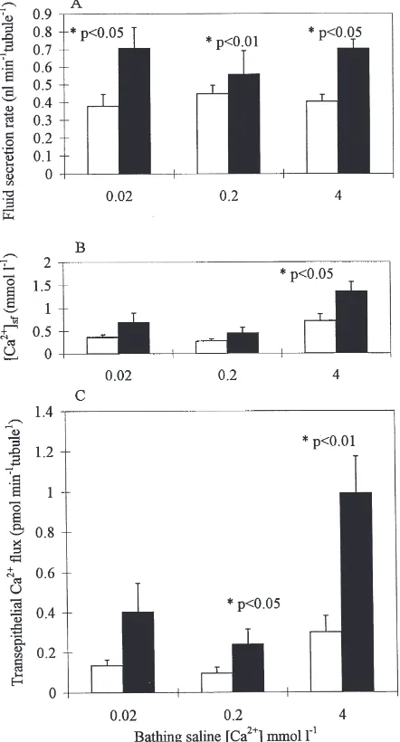
Posterior MTs bathed in saline containing 0.02, 0.2 or 4 mmol l21
Ca2+
secreted fluid at comparable rates (Fig. 2). Secretion rates were stable for.120 min. Reduction in bathing saline Ca2+ to
,0.02 mmol l21 by addition of 2 mmol l21 EGTA impaired cell viability; tubules secreted at low rates (,0.3 nl min tubule21) for a few minutes, then stopped secreting.
Fluid secretion rates increased significantly in response to cAMP in AARS containing 0.02, 0.2 or 4 mmol l21 Ca2+ (Fig. 2A). The concentration of Ca2+in the secreted fluid also increased in response to cAMP for tubules bathed in saline containing 4 mmol l21 Ca2+ (Fig. 2B).
Similar changes in secretion rates and secreted fluid calcium concentration in response to cAMP in 0.2 and 4 mmol l21 Ca2+ AARS were found in earlier studies in which tubules were bathed in SBM (O’Donnell and Maddrell, 1995). However, an unexpected result of the present study was that transepithelial Ca2+
flux across both stimulated and unstimulated tubules bathed in nom-inally Ca2+-free AARS containing 0.02 mmol l21 Ca2+ was similar to that of tubules bathed in AARS containing 0.2 mmol l21 Ca2+ (Fig. 2C). The Ca2+ concentration in fluid secreted by cAMP-stimulated tubules bathed in AARS containing 0.02 mmol l21 Ca2+ was nearly 35 times higher than that in the medium (Fig. 2B).
Basolateral45Ca2+flux across posterior MTs exceeded transepithelial Ca2+flux by 7-fold or more (Fig. 3 versus Fig. 2C), indicating that |85% of the Ca2+which enters the tubule cell is sequestered and |15% is transferred into the tubule lumen. The former figure includes the small quantity of 45
Ca2+
in the tubule lumen. However, calculations based on lumen dimensions (Dow et al.,
Fig. 2. Effects of changes in bathing saline calcium concentrations on transepithelial calcium transport. Fluid secretion rate (A), secreted fluid calcium concentration (B), and transepithelial calcium flux (C) of isolated pairs of whole posterior MTs bathed in nominally calcium-free (0.02 mmol l21Ca2+), 0.2 mmol l21 Ca2+and 4 mmol l21 Ca2+
AARS. Control values (open bars) determined after 40 min of secretion. Cyclic AMP (1 mmol l21) was then added and measurements
were repeated after a further 40 min (closed bars). Secretion rate, secreted fluid calcium concentration and transepithelial calcium flux all increased in response to cAMP for MTs isolated in 4 mmol l21
Ca2+AARS. Values are expressed as mean±SEM. n=4–8 MT pairs.
1994) indicate that the 45Ca2+content of the fluid in the tubule lumen was ,0.2% of the total45Ca2+ content.
Basolateral calcium flux across whole anterior MTs bathed in 4 mmol l21AARS was four times greater than that of whole posterior MTs (Fig. 3). Stimulation with 1 mmol l21cAMP doubled Ca2+flux across the basolateral membrane of posterior MTs, which lack a distal segment (Fig. 3). Stimulation with 1 mmol l21 cAMP did not increase Ca2+
Fig. 3. Basolateral calcium flux of isolated anterior and posterior MTs without (open bars) and with (closed bars) the addition of 1 mmol l21cAMP. Calcium flux of whole posterior MTs increased in response
to 1 mmol l21cAMP (p,0.01). Note that calcium flux by the whole
anterior MTs did not increase with stimulation of cAMP (1 mmol l21);
however, calcium flux across the distal segment decreased and that across the anterior main and lower segments increased. Pairs of whole MTs were placed in 4 mmol l21Ca2+SBM labelled with45Ca2+for
40 min. Values are expressed as mean±SEM. n=10 MT pairs.
due to the opposing effects of cAMP on Ca2+ transport by main and lower versus distal segments of the anterior MTs. When the distal segment was removed from the anterior MTs, Ca2+ flux increased 1.5-fold for the remaining main and lower segments after the addition of 1 mmol l21 cAMP (Fig. 3). In contrast, calcium flux across the distal segment of anterior MTs decreased 35% after stimulation with 1 mmol l21 cAMP (Fig. 3).
3.2. Effects of thapsigargin and A23187
Intracellular Ca2+ levels can be elevated by exposure of tubules to either A23187, a Ca2+ionophore, or thapsi-gargin, which blocks the Ca2+
-ATPase responsible for accumulation of Ca2+
within the endoplasmic reticulum at concentrations of 0.1–1 µmol l21 (Thastrup et al., 1990). Typical doses of A23187 used in studies of epi-thelia range from 0.4µmol l21(Clark et al., 1998) to 1– 10µmol l21(Peterson and Gruenhaupt, 1990). Transepi-thelial calcium flux of isolated whole posterior MTs in SBM increased 15-fold within 40 min of addition of 1
µmol l21 A23187, from 0.07±0.02 to 1.04±0.21 pmol min21tubule21(p
,0.01; n=5). Similarly, transepithelial Ca2+flux increased 10-fold within 40 min of addition of 0.2µmol l21 thapsigargin (from 0.08±0.02 to 0.85±0.25 pmol min21tubule21; p
,0.05; n=5). The increase in flux was associated with increases in both secretion rate (3.3-fold and 3.1-fold for A23187 and thapsigargin, respectively) and secreted fluid calcium concentration
(3.7-fold and 3.5-fold for A23187 and thapsigargin, respectively).
In contrast, calcium flux across the basolateral mem-brane of whole posterior MTs was not significantly changed within 40 min of addition of either 1µmol l21 A23187 or 0.2µmol l21thapsigargin, even though it was much larger than transepithelial calcium flux (.2.0 pmol min21 tubule21; n=12 control and 12 experimental tubules for each drug).
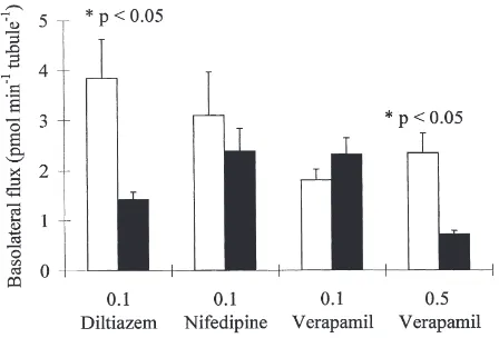
3.3. Effects of Ca2+ channel blockers
Verapamil, diltiazem and nifedipine are well-known blockers of Ca2+channels in neuronal and epithelial cells (Hille, 1992). Verapamil (0.5 mmol l21) and diltiazem (0.1 mmol l21) decreased calcium flux 3.3-fold and 2.7-fold, respectively, across the basolateral membrane of isolated MTs (Fig. 4). These concentrations are compa-rable to those used in studies of Ca2+channels in other epithelia (e.g. 0.1 mmol l21 verapamil, Zhuang and Ahearn, 1996; 0.1–1.0 mmol l21verapamil, Saunders et al., 1990; 0.05 mmol l21 diltiazem, Hanai et al., 1991). Nifedipine (0.1 mmol l21) or lower concentrations of verapamil (0.1 mmol l21
) did not inhibit calcium flux across the basolateral membrane (Fig. 4). None of the drugs at the concentrations indicated in Fig. 4 signifi-cantly inhibited either fluid secretion or transepithelial calcium transport as determined by analysis of secreted fluid droplets with calcium-selective microelectrodes (N=12 tubules for each drug).
3.4. Effects of ruthenium red
Ruthenium red at 0.1 mmol l21 is a putative inhibitor of Ca2+-ATPases in mosquito larvae (Barkai and Willi-ams, 1983) and also binds to voltage-dependent Ca2+
Fig. 4. Basolateral calcium flux in the absence (open bars) and pres-ence (solid bars) of the indicated calcium channel blockers. Pairs of isolated whole posterior MTs were isolated in 4 mmol l21Ca2+SBM
labelled with 45Ca2+ for 40 min. Calcium flux was significantly
decreased by 0.5 mmol l21 verapamil and 0.1 mmol l21 diltiazem.
channels and Ca2+-binding proteins such as calmodulin in mammalian cells (Charuk et al., 1990; Tapia and Vel-asco, 1997). Transepithelial calcium flux was reduced 51% within 20 min of addition of 0.1 mmol l21 ruthenium red to isolated main segments of stimulated anterior or posterior MTs, from 0.19±0.04 to 0.09±0.02 pmol min21tubule21(p
,0.001; n=9). There was a 38% drop in secreted fluid Ca2+ concentration, from 0.32±0.07 to 0.20±0.05 mmol l21
. Experiments on con-trol tubules (n=9) showed that neither transepithelial Ca2+ flux nor secreted fluid calcium concentration changed over this period in the absence of ruthenium red.
Calcium flux across the basolateral membrane decreased 47% within 40 min of addition of 0.1 mmol l21ruthenium red to whole posterior MTs, from 5.2±0.7 pmol min21 tubule21 to 2.74±0.4 pmol min21 tubule21 (p,0.01, n=12). Calcium fluxes across the basolateral membrane of control tubules (n=12) were unaltered over the same period.
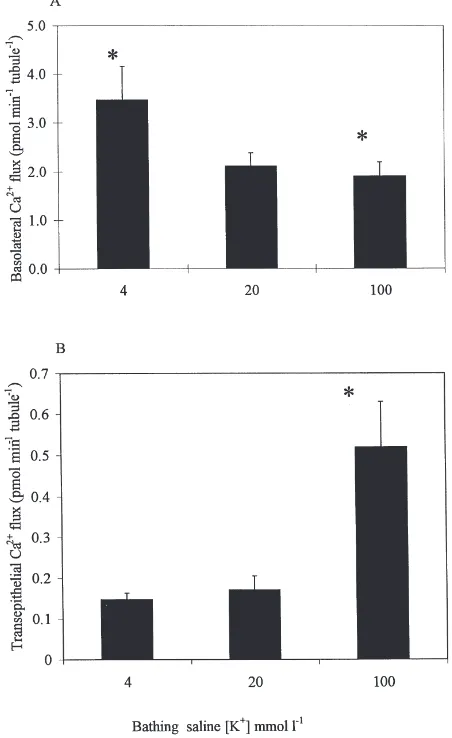
3.5. Effects of changes in bathing saline K+ concentration
Changes in bathing saline K+ concentration alter the basolateral membrane potential of Drosophila MTs (O’Donnell et al., 1996) and might be expected, there-fore, to produce corresponding changes in Ca2+ influx into MTs through voltage-dependent Ca2+ channels. When basolateral calcium fluxes were compared for tubules bathed in AARS containing 4, 20 and 100 mmol21 K+, a 25-fold reduction in saline K+ concen-tration was associated with a significant increase in baso-lateral Ca2+ flux (Fig. 5A).
However, there was no effect on secreted fluid cal-cium concentration or transepithelial calcal-cium flux in response to a 5-fold decrease in bathing saline K+ con-centration, from 20 to 4 mmol l21 (Fig. 5B), although secretion rates of isolated MTs decreased significantly (p,0.05) by 30% (data not shown). Secretion rates and transepithelial calcium flux of isolated MTs increased significantly by.150% in response to a 5-fold increase in K+concentration, but secreted fluid calcium concen-tration was unchanged (data not shown). The increased calcium flux in 100 mmol l21 K+ was therefore a response to the increase in fluid secretion rate.
3.6. Effects of changes in bathing saline Mg2+ concentration on transepithelial calcium transport
Mg2+is known to compete with Ca2+for transporters such as Ca2+ channels (Hagiwara, 1983). However, changes in bathing saline Mg2+ concentration had no effect on transepithelial Ca2+ flux. Transepithelial cal-cium fluxes of unstimulated or cAMP-stimulated whole anterior or posterior MTs in control AARS containing
Fig. 5. Effects of bathing saline K+concentration on basolateral and transepithelial calcium transport. (A) Basolateral calcium flux of iso-lated whole posterior MTs bathed for 40 min in 4 mmol l21 Ca2+
AARS containing 4, 20 or 100 mmol l21K+. Values are expressed as
mean±SEM. n=8 MT pairs. Bars marked with asterisks differ signifi-cantly (p,0.03) from each other. (B) Transepithelial calcium flux for isolated whole posterior MTs bathed for 40 min in 4 mmol l21 Ca2+
AARS containing 4, 20 or 100 mmol l21K+. The asterisk indicates a
significant (p,0.05) difference in flux of MTs isolated in 100 mmol l21 K+ compared with 20 mmol l21 K+. Values are expressed as
mean±SEM. n=5–10 MT pairs.
8.5 mmol l21Mg2+and 0.2 mmol l21Ca2+did not differ significantly from those of unstimulated or stimulated tubules bathed in Mg2+-free AARS or in AARS contain-ing 17 mmol l21 Mg2+ (n
$8 tubules for each experiment).
3.7. Effects of changes in bathing saline HCO32
and/or PO432 concentration
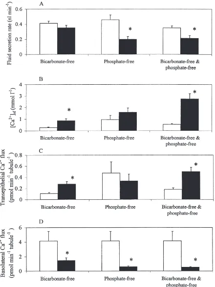
saline bicarbonate and phosphate concentration on basol-ateral and transepithelial Ca2+ flux in isolated tubules. There was no difference in fluid secretion rate for MTs isolated in nominally HCO32-free AARS in comparison to MTs isolated in standard AARS containing 10.2 mmol l21 HCO
32 (Fig. 6A). However, secreted fluid calcium concentration and transepithelial Ca2+ flux increased 221% and 156%, respectively, in HCO32-free AARS (Fig. 6B,C). Moreover, this increase occurred even though there was a 64% decrease in the calcium flux across the basolateral membrane of isolated MTs in nominally HCO32-free AARS compared to standard AARS (Fig. 6D). Increases in secreted fluid calcium con-centration were associated with decreases in secreted fluid pH. The pH of fluid secreted by MTs isolated for 60 min in nominally HCO32-free AARS was 6.99±0.13 (n=9), significantly more acid than the pH of 7.54±0.14 (n=8) measured in fluid secreted by MTs isolated in stan-dard AARS (p,0.001).
In PO432-free AARS, secretion rates were signifi-cantly lower than in control AARS containing 4.3 mmol l21NaH
2PO4(Fig. 6A). Secreted fluid calcium concen-tration increased in some, but not all, tubules; the mean value was not significantly different from that of the con-trol tubules. There was no change in transepithelial Ca2+ flux (Fig. 6B,C). In contrast, calcium flux across the basolateral membrane was significantly reduced in the absence of PO432(Fig. 6D). However, a 2-fold increase in PO432concentration from 4.3 to 8.6 mmol l21did not alter Ca2+flux (n=12 tubules).
In nominally HCO32-free and PO432-free AARS, secretion rates of isolated MTs decreased within 30 min (Fig. 6A), and there was a 5-fold increase in calcium concentration in the secreted fluid compared to isolated MTs in standard AARS (Fig. 6B). Transepithelial Ca2+ flux in HCO32-free and PO432-free AARS was signifi-cantly higher than in control AARS (Fig. 6C), in spite of the reduction in secretion rate. In contrast, basolateral Ca2+ flux was dramatically reduced in HCO
32-free and PO432-free AARS, relative to controls (Fig. 6D).
4. Discussion
4.1. Sequestration and secretion of Ca2+
The results demonstrate that all segments of both anterior and posterior Malpighian tubules have substan-tial capacity to transport Ca2+and to play a role, there-fore, in calcium homeostasis and elimination of excess dietary Ca2+. The role of deposition of Ca2+-rich con-cretions in the distal segment of the anterior MTs has been well established, primarily through studies of the larvae of the related species Drosophila hydei (Wessing and Zierold, 1992; Wessing et al., 1992). The distal seg-ment does not contribute to fluid secretion (Dow et al.,
1994; O’Donnell and Maddrell, 1995), and is used as a storage segment, storing calcium and magnesium in its lumen (Wessing et al., 1992). Our data show that the posterior MTs and the main and lower segments of the anterior MTs are also important in this regard.
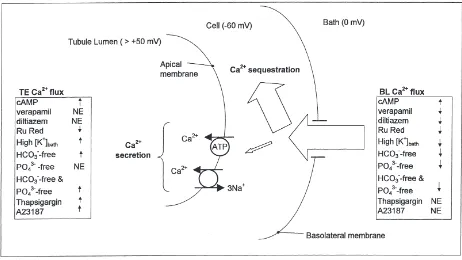
The implication of large basolateral calcium flux and much smaller transepithelial calcium flux is that most of the calcium which enters remains within the cell (Fig. 7). Calcium sequestration within the cells may involve mitochondria, endoplasmic reticulum, calcium-binding proteins and calcium-containing concretions.
The relative significance of sequestration versus secretion of Ca2+ can be appreciated by calculation of Ca2+turnover times. Basolateral Ca2+fluxes in unstimu-lated and cAMP-stimuunstimu-lated posterior MTs were 2.1 and 4.5 pmol min21 tubule21, respectively. The net rate of Ca2+ sequestration, after subtraction of transepithelial Ca2+ fluxes, is 1.8 and 3.6 pmol min21 tubule21 for unstimulated and cAMP-stimulated posterior MTs, respectively. For the pair of posterior tubules, therefore, these figures are equivalent to fluxes of 216 and 432 pmol h21, respectively. Adult females contain 3460 pmol Ca2+per fly (Dube et al., 2000), so the data above indi-cate that the Ca2+
content of the whole fly could be trans-ported into unstimulated posterior MTs in 16 h. This figure drops to 8 h for cAMP-stimulated tubules. Corre-sponding calculations show that the non-secretory distal segments of the pair of anterior MTs could sequester the amount of Ca2+ within the entire animal in
,2 h, and that basolateral Ca2+ flux for all segments of all four unstimulated tubules is sufficient to account for turnover of the Ca2+content of the whole fly in
|100 min. Secretion of Ca2+-rich fluids also contributes to elim-ination of significant quantities of calcium. Fluid secretion by all four unstimulated MTs could remove the entire Ca2+content of the whole fly in
|48 h. This value drops to |14 h for MTs stimulated with thapsigargin or cAMP. For MTs stimulated maximally with both cAMP and the peptide leucokinin 1 (O’Donnell and Maddrell, 1995), the transepithelial flux of 1.6 pmol min21 tubule21
is sufficient to remove the whole animal Ca2+ content in |9 h.
Fig. 6. Effects of nominally bicarbonate-free, phosphate-free or bicarbonate-free and phosphate-free AARS on fluid secretion rate (A), secreted fluid calcium concentration (B), transepithelial calcium flux (C) and basolateral calcium flux (D) for isolated whole posterior MTs. All salines contained 4 mmol l21Ca2+. Values (mean+SEM; n=7–14 tubules) in control and experimental saline indicated by open and closed bars,
Fig. 7. Schematic diagram summarizing secretion and sequestration of Ca2+by posterior Malpighian tubules of adult Drosophila melanogaster.
The width of the open arrows denotes the approximate magnitude of the indicated pathway. Most of the basolateral Ca2+flux which enters the
tubule (basolateral Ca2+flux) is sequestered and remains in the tubule, but some is secreted into the tubule lumen in soluble form and constitutes
transepithelial Ca2+flux. Increases (↑), decreases (↓) or no effect (NE) of particular experimental agents on TE and BL Ca2+flux are summarized
in the text boxes. The lumen positive transepithelial electrical potential precludes accumulation of Ca2+in the lumen by paracellular transport, and
it is assumed that all Ca2+ transport involves transcellular pathways. The significance of apical Ca2+influx (i.e. from lumen to cell) has not
been assessed.
levels of carbonate and/or phosphate for formation of intracellular concretions.
4.2. Why is there a need for high rates of MT calcium transport?
Adult D. melanogaster ingest 0.1 µl of food per hour (Schofield et al., 1997), equivalent to 9.6 nmol per day on food containing 4 mmol l21
Ca2+
. An adult fly thus ingests each day an amount of calcium equivalent to nearly three times the whole body calcium content. Cal-cium homeostasis in the face of such high dietary intake can be accomplished by gut absorption of only a low percentage of the calcium available in the diet, or by absorption of a high percentage followed by excretion of almost all of the calcium absorbed. Studies of other flies favour the latter explanation. Calcium absorption by the midgut of the blowfly Calliphora vicina is an active and rapid process; the absorption rate for flies fed on solutions containing 4–12.4 mmol l21Ca2+is
|120 nmol h21, sufficient to replace the total body calcium content (|650 nmol) in|6 h. Regulation of calcium by blowflies in response to variations in dietary calcium content is accomplished not by varying the rate of absorption
across the midgut, but by excretion of excess calcium by the MTs (Taylor, 1985).
Midgut absorption of Ca2+ followed by sequestration may also be a consequence of mechanisms which miti-gate the effects of exposure to toxic divalent cations. Concretions in the distal segment of D. hydei larval MTs concentrate Sr2+ and Ba2+ added to the diet, and an increase in one component in the diet results in a decrease in other elements in the concretions (Wessing and Zierold, 1992). Strontium, for example, is accumu-lated at the expense of calcium. High Ca2+transport rates by MTs and other dipterans may also permit survival in carbonate- or phosphate-rich environments. CaCO3 con-cretions in the MTs of the alkali fly Ephydra hyans may permit habitation of alkaline lakes containing up to 500 mmol l21 carbonate and bicarbonate (Herbst and Brad-ley, 1989).
4.3. Differential control of sequestration and secretion of Ca2+
Our results show that basolateral and transepithelial Ca2+
flux exceed transepithelial Ca2+ flux approximately 7-fold, but basolateral and transepithelial Ca2+ fluxes respond differently to changes in bathing saline K+, addition of Ca2+ channel blockers, increases in intra-cellular Ca2+levels, or to removal of bicarbonate and/or phosphate. These findings show that there is little coup-ling between basolateral and apical Ca2+transport mech-anisms; an increase in basolateral flux is not necessarily accompanied by a corresponding increase in transepi-thelial calcium flux, and vice versa.
The absence of coupling between basolateral and transepithelial Ca2+ fluxes is in striking contrast to the equivalence of basolateral and transepithelial fluxes for monovalent cations such as K+. Basolateral and apical fluxes across the lower MT of Rhodnius prolixus are identical, indicating that there is no significant seques-tration of K+within the tubule cells (Collier and O’Don-nell, 1997).
Both transepithelial and basolateral Ca2+ fluxes are stimulated by cyclic AMP in posterior MTs and in main and lower segments of anterior MTs. The large differ-ences in the magnitudes of basolateral and transepithelial Ca2+ fluxes suggest that cAMP enhances Ca2+ seques-tration in these tubules. By contrast, Ca2+
sequestration by the distal segments of the anterior MTs decreases in response to cAMP. Modulation of Ca2+sequestration by an intracellular second messenger such as cAMP raises the possibility that the process may be under the control of circulating first messengers (e.g. neuropeptides) in vivo.
4.4. Secretion of 45Ca2+by isolated MTs and estimation of exchangeable Ca2+
Any sequestration of calcium within the tubule cells will result in a decline in the percentage of exchangeable Ca2+. Importantly, good agreement between two inde-pendent measurements of exchangeable Ca2+within the MT justifies our calculation of transepithelial Ca2+ flux from the product of secreted fluid calcium concentration, measured by Ca2+
-selective microelectrodes, and secretion rate. For the average basolateral calcium flux of 4 pmol min21 tubule21, the corresponding average transepithelial calcium transport was 0.25 pmol min21 tubule21 (Fig. 2), indicating that 94% of the calcium which crosses the basolateral membrane remains within the tubule and only 6% is transported into the lumen (i.e. is exchangeable).
The latter figure was based on Ca2+ flux calculated as the product of secretion rate and secreted fluid Ca2+ concentration. A second and independent estimate of the percentage of exchangeable Ca2+ can be obtained by measuring transepithelial flux of 45Ca2+. The exchange-able calcium (ECa2+) within the MTs cells was calculated as follows:
SF is the calcium concentration in the secreted fluid (measured with a Ca2+-selective microelectrode), cpmSFis the cpm in the secreted fluid, SAMT is the specific activity of 45Ca2+ within the Mal-pighian tubule cell and cpmMTis the cpm in the Malpigh-ian tubule after 20 min in 45Ca2+-labelled saline.
By rearrangement, we avoid the need to use the term SAMT,
ECa2+5 [Ca2+]
SF×cpmMT
cpmSF
The average calcium concentration in secreted fluid from unstimulated MTs was 0.6 mmol l21. After subtrac-tion of the background counts (20 cpm), the mean con-tent of45 ured in a secreted droplet over 20 min was 41 cpm.
Therefore:
ECa2+5
(0.6 mmol l−1)×(383 cpm)
(41 cpm) 55.6 mmol l
−1
The volume of a pair of MTs, estimated using the tubule dimensions in Dow et al. (1994), is 2.94×1029 l. Therefore, the exchangeable Ca2+content of whole MTs was 0.01646 nmol (=5.6 mmol l21×2.94×1029 l), which is 5.5% of the total content of 0.297 nmol determined by atomic absorption spectrophotometry (Dube et al., in press). This is in close agreement with the value of 6% estimated above using flux calculated as the product of secreted fluid calcium concentration and fluid secretion rate. The good agreement between the two figures indi-cates that all of the Ca2+
in the secreted fluid is in dis-sociated form (i.e. it is not in the form of insoluble concretions).
4.5. Ca2+ channels and basolateral calcium transport
Sensitivity of basolateral Ca2+ flux to verapamil and diltiazem is consistent with the presence of L-type volt-age-dependent calcium channels (Hille, 1992) in the basolateral membrane of posterior MTs (Fig. 7). Reduction in basolateral Ca2+ in response to ruthenium red suggests that these channels may also be sensitive to ruthenium red (Tapia and Velasco, 1997). The increase in calcium flux across the basolateral membrane in response to a decrease in K+concentration in the bath-ing medium provides further evidence of voltage-depen-dent Ca2+
con-centration hyperpolarize the basolateral membrane (O’Donnell et al., 1996). The change in potential may provide a larger electrical gradient favouring Ca2+ move-ment from bath to cell through putative Ca2+ channels in the basolateral membrane, or it may activate quiescent channels. Increases or decreases in bathing saline Mg2+ concentration did not alter basolateral Ca2+ transport, indicating effective discrimination between the two divalent cations.
4.6. The nature of apical calcium transport
Movement of Ca2+from cell to lumen will be opposed by the large lumen-positive apical membrane potential (O’Donnell et al., 1996). Some form of active transport such as a Ca2+-ATPase is thus required, since Ca2+also moves against opposing chemical (i.e. concentration) gradients (Fig. 7). A Ca2+-ATPase has been found in the apical membrane of Lymantria dispar MTs using immu-nocytochemical techniques (Pannabecker et al., 1995). Alternatively, a 3Na+/Ca2+ exchanger could utilize a favourable electrochemical gradient for Na+ movement from lumen to cell to drive Ca2+from cell to lumen (Fig. 7). Na+ concentration in secreted fluid (|30 mmol l21; O’Donnell and Maddrell, 1995) exceeds typical intra-cellular levels cell (|7 mmol l21; Zierold and Wessing, 1990), and there is a large electrical gradient (i.e.|100 mV, lumen positive) which could in principle drive an electrogenic 3Na+/Ca2+exchange process across the api-cal membrane.
Our evidence for a Ca2+-ATPase is based on the sensi-tivity of transepithelial calcium transport to ruthenium red (Watson et al., 1971; Barkai and Williams, 1983). Ruthenium red may inhibit Ca2+-ATPase activity in the plasma membrane or in the endoplasmic reticulum or mitochondrial membranes (Tapia and Velasco, 1997). Cyclic AMP increases the lumen positive TEP (O’Donnell et al., 1996), but also increases transepi-thelial calcium secretion. Given the increase in the opposing electrical gradient across the apical membrane, cAMP may stimulate an apical Ca2+
transporter, such as an ATPase (e.g. Carafoli, 1991). It is worth noting that this study has not addressed the possibility of apical Ca2+ influx (i.e. from lumen to cell) and that our measure-ments of transepithelial Ca2+flux reveal net Ca2+ trans-port into the tubule lumen. Analysis of apical influx would require perfusion of the tubule lumen with sol-utions of known specific activity.
Given that both A23187 and thapsigargin increase transepithelial calcium flux but not basolateral calcium flux, it appears that both drugs must either alter calcium transport across the apical membrane or affect the down-stream movement or dissolution of lumenal concretions. Both drugs tend to enhance intracellular Ca2+ levels, thereby tending to increase the transepithelial Cl2
per-meability (O’Donnell et al., 1996). Thapsigargin is a
cium mobilizing agent which inhibits the uptake of cal-cium by the Ca2+-ATPase in the endoplasmic reticulum The result is a slow increase in cytosolic calcium con-centration in stellate cells only (Rosay et al., 1997). Ca2+ secretion into the lumen may thus be enhanced in response to thapsigargin because of the decline in the opposing lumen-positive apical membrane potential that results from the increase in transepithelial Cl2
per-meability (O’Donnell et al., 1996).
A23187 increased both fluid secretion rates and secreted fluid calcium concentration, and has been shown in other studies to depolarize transepithelial volt-age and decrease transepithelial resistance in MTs of
Aedes, consistent with a Ca2+-dependent enhancement of the transepithelial Cl2shunt (Clark et al., 1998;
O’Don-nell and Spring, 2000). In general, the effects of A23187 reflect an increase in the membrane permeability of cal-cium (Alberts et al., 1994). In this context, our finding that A23187 did not alter basolateral Ca2+ flux was entirely unexpected. This result may reflect the very high influx of Ca2+across the basolateral cell surface. In other words, permeability of the basolateral cell membrane to Ca2+may be sufficiently high at rest that the addition of a Ca2+
ionophore has relatively little effect. Alternatively, A23187 may act primarily on stellate cells, enhancing transepithelial Cl2 transport and increasing
transepi-thelial Ca2+transport by reducing the opposing lumen-positive transepithelial potential. Under this scenario, the basolateral membrane of the principal cells may be refractory to the effects of A23187. The latter compound is an organic anion, and its effective concentration may be reduced by transport into the lumen by the organic anion transport systems present in Drosophila tubules (Riegel et al., 1999).
References
Alberts, B., Bray, D., Lewis, J., Raff, M., Roberts, K., Watson, J.D., 1994. Molecular Biology of the Cell, 3rd edn. Garland Publishing, New York.
Ammann, D., 1986. Ion-Selective Microelectrodes. Principles, Design and Application. Springer-Verlag, Berlin.
Barkai, A.I., Williams, R.W., 1983. The exchange of calcium in larvae of the mosquito Aedes aegypti. Journal of Experimental Biology 104, 139–148.
Beyenbach, K.W., 1995. Mechanism and regulation of electrolyte transport in Malpighian tubules. Journal of Insect Physiology 41, 197–207.
Busto, M., Iyengar, B., Campos, A.R., 1999. Genetic dissection of behavior: modulation of locomotion by light in the Drosophila
mel-anogaster larva requires genetically distinct visual system
func-tions. Journal of Neuroscience 199, 3337–3344.
Carafoli, E., 1991. Calcium pump of the plasma membrane. Physio-logical Reviews 71, 129–153.
Charuk, J.H., Pirraglia, C.A., Reithmeier, R.A., 1990. Interaction of ruthenium red with Ca2+-binding proteins. Analytical Biochemistry
188, 123–131.
concentration-dependence for CRF-like diuretic peptide: mech-anisms of action. Journal of Experimental Biology 201, 1753–1762. Collier, K.A., O’Donnell, M.J., 1997. Analysis of epithelial transport by measurement of K+, Cl2 and pH gradients in extracellular unstirred layers: ion secretion and reabsorption by Malpighian tubules of Rhodnius prolixus. Journal of Experimental Biology 200, 1627–1638.
Dow, J.A.T., Maddrell, S.H.P., Go¨rtz, A., Skaer, N.J.V., Brogan, S., Kaiser, K., 1994. The Malpighian tubules of Drosophila
mel-anogaster: a novel phenotype for studies of fluid secretion and its
control. Journal of Experimental Biology 197, 421–428. Dow, J.A.T., Davies, S.A., Sozen, M.A., 1998. Fluid secretion by the
Drosophila Malpighian tubule. American Zoologist 38, 450–460.
Dube, K., McDonald, D.G., O’Donnell, M.J. 2000. Calcium homeo-stasis in larval and adult Drosophila melanogaster. Archives of Insect Biochemistry and Physiology 44, 27–39.
Hagiwara, S., 1983. Membrane Potential-Dependent Ion Channels in Cell Membrane: Phylogenetic and Developmental Approaches. Raven Press, New York.
Hanai, H., Kameyama, M., Kaneko, E., Fujita, M., 1991. Properties of two calcium transport systems of isolated rat ileal epithelial cells: effects of Ca2+channel modulators and membrane potential
examined with fluorescent dye, fura-2. Pflugers Archives 419, 184–189.
Herbst, D.B., Bradley, T.J., 1989. A Malpighian tubule lime gland in an insect inhabiting alkaline salt lakes. Journal of Experimental Biology 145, 63–78.
Hille, B., 1992. Ionic Channels of Excitable Membranes. Sinauer, Sun-derland, MA, 607pp.
Maddrell, S.H.P., Whittembury, G., Mooney, R.L., Harrison, J.B., Overton, J.A., Rodriguez, B., 1991. The fate of calcium in the diet of Rhodnius prolixus; storage in concretion bodies in the Malpigh-ian tubules. Journal of Experimental Biology 157, 483–502. Maddrell, S.H.P., O’Donnell, M.J., Caffrey, R., 1993. The regulation
of haemolymph potassium activity during initiation and mainte-nance of diuresis in fed Rhodnius prolixus. Journal of Experimental Biology 177, 273–285.
O’Donnell, M.J., Maddrell, S.H.P., 1995. Fluid reabsorption and ion transport by the lower Malpighian tubules of adult female
Droso-phila. Journal of Experimental Biology 198, 1647–1653.
O’Donnell, M.J., Spring, J.H., 2000. Modes of control of insect Mal-pighian tubules: synergism, antagonism, cooperation and auton-omous regulation. Journal of Insect Physiology 46, 107–117. O’Donnell, M.J., Dow, J.A., Huesmann, G.R., Tublitz, N.J., Maddrell,
S.H.P., 1996. Separate control of anion and cation transport in Mal-pighian tubules of Drosophila melanogaster. Journal of Experi-mental Biology 199, 1163–1175.
Pannabecker, T.L., Smith, C.A., Beyenbach, K.W., Wasserman, R.H., 1995. Immunocytochemical localization of plasma membrane
cal-cium pump in the insect (Lymantria dispar) Malpighian tubule. Journal of Insect Physiology 41, 1105–1112.
Peterson, M.W., Gruenhaupt, D., 1990. A23187 increases permeability of MDCK monolayers independent of phospholipase activation. American Journal of Physiology 259, C69–C76.
Riegel, J.A., Farndale, R.W., Maddrell, S.H.P., 1999. Fluid secretion by isolated Malpighian tubules of Drosophila melanogaster Meig.: effects of organic anions, quinacrine and a diuretic factor found in the secreted fluid. Journal of Experimental Biology 202, 2339– 2348.
Rosay, P., Davies, S.A., Yu, Y., So¨zen, M.A., Kaiser, K., Dow, J.A., 1997. Cell-type specific calcium signalling in a Drosophila epi-thelium. Journal of Cell Science 110, 1683–1692.
Saunders, K.D., Cates, J.A., Abedin, M.Z., Kleinman, R., Roslyn, J.J., 1990. Ca2+ channel blockade inhibits gallbladder ion transport.
Journal of Surgical Research 49, 306–310.
Schofield, R.M.S., Postlethwait, J.H., Lefevre, H.W., 1997. MeV-ion microprobe analyses of whole Drosophila suggest that zinc and copper accumulation is regulated storage not deposit excretion. Journal of Experimental Biology 200, 3235–3243.
Tapia, R., Velasco, I., 1997. Ruthenium red as a tool to study calcium channels, neuronal death and the function of neural pathways. Neu-rochemistry International 30, 137–147.
Taylor, C.W., 1985. Calcium regulation in blowflies: absence of a role for midgut. American Journal of Physiology 249, R209–R213. Taylor, C.W., 1986. Calcium regulation in insects. Advances in Insect
Physiology 19, 155–186.
Thastrup, O., Cullen, P.J., Drøbak, B.K., Hanley, M.R., Dawson, A.P., 1990. Thapsigargin, a tumor promoter, discharges intracellular cium stores by specific inhibition of the endoplasmic reticulum cal-cium ATPase. Proceedings of the National Academy of Science USA 87, 2466–2470.
Watson, E.L., Vincenzi, F.F., Davis, P.W., 1971. Ca2+-activated
mem-brane ATPase: selective inhibition by ruthenium red. Biochimica Biophysica Acta 249, 606–610.
Wessing, A., Zierold, K., 1992. Metal-salt feeding causes alternations in concretions in Drosophila larval Malpighian tubules as revealed by X-ray microanalysis. Journal of Insect Physiology 38, 623–632. Wessing, A., Zierold, K., Hervert, F., 1992. Two types of concretions in Drosophila Malpighian tubules as revealed by X-ray microanal-ysis: a study on urine formation. Journal of Insect Physiology 38, 543–554.
Zhuang, Z., Ahearn, G.A., 1996. Ca2+transport processes of lobster
hepatopancreatic brush-border membrane vesicles. Journal of Experimental Biology 199, 1195–1208.