KEYWORDS
ABSTRACT
Cytotoxicity and Antioxidant Activity of
Zingiber officinale
and
6-Gingerol on HepG2 cells
Harliansyah1,2, Noor Azian Murad3,Wan Zurinah Wan Ngah1, Yasmin Anum Mohd Yusof1
1Department of Biochemistry, Faculty of Medicine, Universiti Kebangsaan Malaysia, Jalan Raja Muda Abdul Aziz, 50300 Kuala Lumpur, Malaysia
2Department of Biochemistry, YARSI University, School of Medicine, Jakarta Indonesia
3Centre of Lipids and Engineering and Applied Research, Universiti Teknologi Malaysia, Jalan Semarak, 50300 Kuala Lumpur, Malaysia
antioxidant; antiproliferation; [6]-Gingerol; HepG2; Zingiber officinale
The present study was designed to compare the effects of ethanolic extract of ginger (Zingiber officinale) and its phenolic component [6]-Gingerol on viability, antiproliferation and apoptotic levels of human hepatoma cell lines (HepG2) and its antioxidant activity. HepG2 cells were cultured in Eagle’s minimum essential medium (EMEM) and the percentage of cell cytotoxicity was evaluated by tetrazolium salt (MTS) assay. Antiproliferation and apoptotic levels were measured by 5’Bromo-2’deoxyuridine (BrdU) colorimetry. Antioxidant capacity was studied by 1.1-diphenyl-2-picryl-hydrazyl radical (DPPH) using spectrophotometry. We found that cytotoxicity and antiproliferative effect of ginger extract and [6]-Gingerol could be associated with induction of apoptosis. The ginger ethanol extract and [6]-Gingerol also showed remarkable antioxidant activities in comparison with ascorbic acid and N-acetyl-L-cysteine.
Oxidative damage caused by free radicals is known to participate in the pathogenesis of several diseases such as cardiovascular, aging, rheumatoid arthritis and cancer. Extensive research in the last few years has revealed that regular consumption of certain fruits, vegetables and spices can reduce the risk of acquiring specific cancers (Aggarwal and Shishodia, 2006). Phytochemicals derived from such fruits and vegetables, referred to as chemopreventive agents among other are curcumin, capsaicin, [6]-Gingerol. Because these agents have been shown to suppress cancer cell proliferation, inhibit growth factor signaling pathways, induce apoptosis, suppress the expression of anti apoptotic proteins, inhibits cyclooxygenase, they may have untapped therapeutic value (Taraphdar et al., 2001; Surh, 2002). Zingiber officinale Roscoe (ginger) is widely used all over the world as spice and condiments in daily cooking. Ginger has also been used in tra-ditional oriental medicines to ameliorate inflamma-tion, rheumatic disorder, and gastrointestinal discomforts (Geiger, 2005). Crude ginger contains up to 5-8% oleoresin of which 25% of the oleoresin, consists mainly gingerol (Chrubasik et al., 2005). [6]-Gingerol has been associated with analgesic, anti-inflammatory, sedative, antipyretic and antibacterial effects in both in vitro and in vivo studies (Bhattacharjee, 2000; Young et al., 2005).
Hepatocellular carcinoma (hepatoma) is one of the most common cancers in the world, with an annual incidence of approximately 1 million deaths, mainly in underdeveloped and developing countries (Pang et al., 2006). An imbalance between prolifera-tion and apoptosis is strongly linked to the cause of most cancers including liver cancer (Farinati et al., 2001). The search for chemopreventive agents found in natural products or foods is gaining a lot of interest in cancer research (Gosslau and Chen, 2004).
In the present study we compare the effects of ginger extract (Zingiber officinale) with its phenolic component [6]-Gingerol (component of ginger) in antioxidant activities, inhibiting proliferation and inducing apoptosis of hepatoma cell lines (HepG2).
MATERIALS AND METHODS
Materials
Ginger (Zingiber officinale) extract was obtained by ethanol extraction as provided by Dr Noor Azian Murad from Center for Lipids Engineering Applied Research (CLEAR), Universiti Teknologi Malaysia. [6]-Gingerol was purchased from WAKO, Japan.
Correspondence:
002 HARLIANSYAH, NOOR AZIAN MURAD, WAN ZURINAH WAN NGAH, YASMIN ANUM MOHD YUSOF
Methods Cell cultures
HepG2 (ATCC.HB 8065, Rockville, MD, USA) were maintained in Eagle’s minimum essential
medium (EMEM) supplemented with 10% heat
inactivated foetal bovine serum and 1% penicillin-streptomycin. The cells were cultured as a monolayer in plastic 75 cm2 tissue culture flash and grown at 370C in humidified atmosphere of 5% CO2. Cell’s viability, proliferation and apoptosis were performed when the cells reached 70-80% confluence. Ginger extract and [6]-Gingerol were added to cells after 24 hours incubation.
Sample analysis
Antioxidant activity in cell free system
The free radical-scavenging capacity of ethanolic extract from Zingiber officinale and [6] Gingerol were tested by its ability to bleach the stable 1,1-diphenyl-2-picryl-hydrazyl radical (DPPH). The reaction mixture contained one milliliter of different concentrations of ginger extract (from 10 to 1000
μg/ml) or [6]-Gingerol (100, 200, 500, 1000 μg/ml)
and 1 ml of freshly prepared 1 mM DPPH ethanolic solution. The resulting solution were left to stand for 30 min at room temperature, prior to being spectrophotometrically detected at 517 nm (Ito et al., 2005; Lee et al., 2004).
MTS assay for cell viability
HepG2 cells at a density of 2x10⁴ cells/ml were plated in 96 well microtiter plates. After 24 h of incubation to allow for cell attachment, the cells were treated with 100 l of varying concentrations of ginger extract and [6]-Gingerol (5, 10, 50, 100, 200, 500 and 1000 g/ml) in complex medium and incubated again for 24 h at 370C under 5% CO2. Three hours after the addition of MTS solution, the amount of formazan formed was measured spectrophoto-metrically at 490 nm with microplate reader Versamax-Molecular, Devices B-02865. Fifty percent inhibitory concentration (IC50) of ginger extract and [6]-Gingerol in HepG2 cells were calculated from triplicate wells.
Cell proliferation
Cellular proliferation of HepG2 cells were measured using BrdU kit (Roche Diagnostics, Germany). HepG2 cells were seeded into 96 well plates at a concentration of 2x10⁴ cells/ml in EMEM. Cells were incubated with various dilutions of ginger extract and [6]-Gingerol in a 96-well plates at a final volume of 100 l /well for 24 h in a humidified
atmosphere at 370C. 10 l of BrdU labeling solution were added in cells and incubated for another 24h at 370 C. 100 l/well anti-BrdU-POD working solution was added and incubated for 90 min at 250 C. After final rinsing, 100 l/well substrate solution was added and incubated at 250 C until color develop-ment was sufficient for photometric detection using ELISA reader (Versamax-Molecular, Devices.B-02865)at 450 nm (reference wavelength; 690 nm).
Analysis of DNA fragmentation for apoptosis HepG2 cells grown at density of 2 x 10⁶ cells/ 10ml were exposed to ginger extract and [6]-Gingerol (5, 10, 50, 100, 200, 500 and 1000 µg/ml) were added after 24 h incubation. Cellular DNA fragmentation was performed as per instruction in the ELISA kit (Roche Diagnostic, Germany). The absorbance of the samples was measured with ELISA reader (Versamax-Molecular, Devices.B-02865) at 450 nm (reference wavelength; 690 nm).
Analysis of data
Statistically significant differences were
assessed using the Student’s t test.
RESULTS
As shown in Figure 1, both ginger extract and [6]-Gingerol revealed potent antioxidant activities. Percent scavenging activity of [6]-Gingerol was higher compared to the ethanolic extract of ginger at lower concentration (< 100 µg/ml) but at higher concentration (500 µg/ml) ginger and [6]-Gingerol exhibited up to 92.68 ± 5.47% and 74.19 ± 5.36% respectively, of DPPH radical scavenging activity. The results showed that the order of potency of antioxidant activities as shown by DPPH radical scavenging capacity at concentration of 500 µg/ml is: diethyl dithiocarbamic (DDC) > ginger extract > buthyl hydroxyl toluene (BHT) > [6]-Gingerol > N-acetyl L-cysteine (NAc) > ascorbic acid. The isolation of bioactive compounds in the Zingiber officinale extracts in the future would help to ascertain the individual potency of the isolated compounds.
Ginger extract and [6]-Gingerol reduced viability of HepG2 cells significantly (p<0.01), after 24 h treatment with varying concentrations between 5 to 1000 µg/ml with an IC50 of 358.71 ± 17.12 and 431.70 ± 10.44 respectively (Table 1).
CELLS
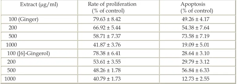
mentation assay assessed cells undergoing apoptosis. Ginger extract and [6]-Gingerol showed a dose dependent inhibition on the proliferation of HepG2 cells with a corresponding induction of apoptosis (Table 2). Ginger extract showed a higher percentage of apoptosis compared to its phenolic component
[6]-Gingerol at all concentrations which corresponds with its lower IC50. Both ginger extract and [6]-Gingerol exhibited maximal induction of apoptosis at 500 µg/ml.
0 20 40 60 80 100 120 140
0 10 20 50 100 200 500 1000
conce ntration (ug/m l)
ra
d
ic
al
s
ca
ve
n
g
in
g
a
ct
iv
it
y
(%
)
ginger N-acetyl L-cysteine
ascorbic acid buthylhydroxy toluene
diethyl dithiocarbamic gingerol
Fig.1. Free radical scavenging activity of ginger extract. Results are mean ± SD of three independent experiments.
Table 1. Cytotoxic activity of the ginger extract and [6]-Gingerol on HepG2 cells
Treatment ( g/ml) Ginger extract (% of control)
[6]-Gingerol (% of control)
0 100 100
100 59.22. ± 1.86 57.11 ± 2.07
200 44.76 ± 3.34 53.74 ± 2.97
500 31.81 ± 2.05 51.35 ± 2.25
1000 29.39 ± 3.81 11.65 ± 2.52
IC50 358.71 ± 17.12 431.70 ± 10.44
Data were presented as mean ± SD (n=3).
Table 2. Effect of ginger extract and [6]-Gingerol on proliferation and apoptosis on HepG2 cells
Extract ( g/ml) Rate of proliferation
(% of control)
Apoptosis (% of control)
100 (Ginger) 79.63 ± 8.42 49.26 ± 4.17
200 66.92 ± 5.44 54.38 ± 7.64
500 58.71 ± 7.37 73.58 ± 7.19
1000 41.87 ± 3.76 19.09 ± 5.01
100 ([6]-Gingerol) 78.38 ± 6.41 28.64 ± 3.10
200 53.61 ± 3.55 29.79 ± 3.12
500 48.26 ± 1.78 56.84 ± 6.33
1000 40.79 ± 1.73 12.73 ± 2.55
004 HARLIANSYAH, NOOR AZIAN MURAD, WAN ZURINAH WAN NGAH, YASMIN ANUM MOHD YUSOF
DISCUSSION
Free radicals are known to be a major factor in biological damages, and DPPH has been used to evaluate the free radical-scavenging activity of natural antioxidants. DPPH which is a radical itself with a purple color, change into a stable compound with a yellow color by reacting with an antioxidant, and the extent of the reaction dependents on the hydrogen donating ability of the antioxidant (Lee et al., 2004; Ito et al., 2005). Antioxidants are compounds that can delay or inhibit the oxidation of lipids or other molecules by inhibiting the initiation or propagation of oxidative chain reactions. Some spices or herbs contain bioactive phenolic substances with potent antioxidative and chemopreventive properties (Surh et al., 1998). The antioxidant activity of phenolic compounds is mainly due to their redox properties, which can play an important role in absorbing and neutralizing free radicals, quenching singlet and triplet oxygen or decomposing peroxides. Phenols are very important plant constituents because of their scavenging ability due to their hydroxyl groups. The phenolic compounds may contribute directly to antioxidative action. It is suggested that polyphenolic compounds have inhibitory effects on mutagenesis and carcinogenesis in humans, when up to 1.0 g daily ingested from a diet rich in fruits and vegetables
(Gűlcin et al., 2002).
Tumors are disease with proliferation disorder and apoptosis obstacle. The inhibition of proliferation and induction apoptosis are regulated by a network of signaling pathways and transcription factors, which are possible targets for a rational tumor therapy (Liu et al., 2004). Apoptosis is now recognized as an important mode of cell death in response to cytotoxic treatments. It has been well documented that the administration of many natural compounds with anti-tumor activities triggers the apoptotic death of cancer cells.
Table 1 showed that ginger extract and [6]-Gingerol at concentration of 100 µg/ml and above, significantly affected the viability of HepG2 cells, suggesting that the observed growth inhibition was caused by cytotoxic rather than a cytostatic effect of ginger and [6]-Gingerol. Cell viability can be reflected by the integrity of the mitochondria. A mitochondrial enzyme in living cells, succinate-dehydrogenase cleaves the tetrazolium salt, converting the MTS to an insoluble purple formazan. Therefore, the amount of formazan produced is directly proportional to the number of viable cells (Lee et al., 2004). The results presented here showed a
concentration dependent decrease in the percentage of cell viability and at a concentration of 200-1000 µg/ml of ginger extract and [6]-Gingerol were sufficient to effectively inhibit the cell proliferation.
We further investigated whether the cytotoxic effect was mediated via an apoptotic mechanism (Table 2). However, the mechanisms involved in these observations are largely unclear. Furthermore, it is likely that there are differences in the apoptotic process in some cells, which can be prevented by inhibitors of protein synthesis or inhibition of macromolecular synthesis and actually induce apoptosis in specific cells. Perhaps the ability of ginger or [6]-Gingerol to uncouple oxidative phosphorylation or increase production of free reactive oxygen species plays a role in this context. Ability in inhibiting or in enhancing apoptosis by plant extracts depends on several factors such as; extract concentration, concerted action of multiple micronutrients, cell type and redox status (Palozza et al., 2004). HepG2 cells are capable of undergoing apoptosis through the basic common signaling pathway p53 and c-Myc play an important role in the apoptosis signaling pathway in HepG2 cells treated with a number of apoptosis inducing compounds (Liu et al., 2002). The percentage of apoptotic cells was increased in a dose dependent manner by treatment with ginger extract and [6]-Gingerol at concentration ranging from 100-500 µg/ml, but decreased at 1000 µg/ml. It has been reported that the proto-oncogene Bcl-2 prevent apoptosis in multiple contexts. The induction of apoptosis by chemopreventive agent was inhibited by Bcl-2 family in in vitro models. HepG2 cells expressed a substantial amount of Bcl-2 but HuH-7 cells totally lacked Bcl-2 (Yamamoto et al., 2001). Overexpression of Bcl-2 or Bcl-x can protect against chemotherapy induced release of mitochondrial cytochrome c, caspase activation and DNA fragmentation (Tong et al., 2004). Apoptosis is a mechanistically driven form of cell death that is either developmentally regulated, or activated in response to specific stimuli or various forms of cell injury. In cancer biology, it is now evident that many cancer cells circumvent the normal apoptotic mechanisms to prevent their self destruction. Therefore, it would be advantageous in cancer chemotherapy and prevention to tip the balance in favor of apoptosis over mitosis (Yoo et al., 2002).
ginger as herbal medicine, has its unique properties, including: (a), no known adverse effect; (b), no difficulty for oral consumption; (c), low cost; and (d), a long history of use by the human population, all of which are indicative of its potential application as an anti tumor agents (Chrubasik et al., 2005).
In conclusion, the results of this study indicate that Zingiber officinale extract has higher DPPH radical scavenging activity. The anticancer effect of Zingiber officinale extract was demonstrated by inhibiting cellular proliferation and inducing apoptosis of hepatoma cells which could be associated mainly with the action of its main phenolic component, [6]-Gingerol.
REFERENCES
Aggarwal BB and Shishodia S 2006. Molecular targets of dietary agents for prevention and theraphy of ancer. Biochem. Pharmacol., 71: 1397-1421.
Bhattacharjee SK 2000. Ginger (Zingiber officinale). Handbook of aromatic plants, Pointer Publishers, India, pp: 473-474. Chrubasik S, Pittler MH and Roufogalis BD 2005. Zingiberis
rhizome: a comprehensive review on the ginger effect and efficacy profiles. Phytomed., 12: 684-701.
Farinati F, Cardin R, Fiorentino M, D’Errico A, Grigioni W and Cecchetto A 2001. Imbalance between cytoproliferation and apoptosis in hepatitis C virus related chronic liver disease. J. Viral Hepatitis., 8: 34-40.
Geiger JL 2005. The essential oil of ginger, Zingiber officinale, and anaesthesia. The Int. J. Aromathe., 15: 7-14.
Gosslau A and Chen KY 2004. Nutraceuticals, apoptosis and disease prevention. Nutr., 20: 95-102.
Gűlcin I, Oktay M, Kűfrevioğlu OI and Aslan A 2002.
Determination of antioxidant activity of lichen Cetraria islandica (L) Ach. J of Ethnopharmacol., 79: 325-329.
Ito M, Murakami K and Yoshino M 2005. Antioxidant action of eugenol compounds: role of metal ion in the inhibition of lipid peroxidation. Food Chem. Toxicol.,43:461- 466.
Lee J-Y, Hwang W-I and Lim S-T 2004. Antioxidant and anticancer activities of organic extracts from Platycodon grandiflorum A.de Candolle roots. J. Ethnopharm., 93: 409-415.
Liu H, Jin L, Tao S and Liu R 2002. Effect of Grape extract on HepG2 cell proliferation and apoptosis. Chin Med J. 115(3): 523-527.
Liu J, Hu W-X, He L-F, Ye M and Li Y 2004. Effects of lycorine on HL-60 cells via arresting cell cycle and inducing apoptosis. FEBS Lettr. 578: 245-250.
Paloza P, Serini S, Di Nicuolo F and Calviello G 2004. Modulation of apoptotic signaling by carotenoids in cancer cells. Arch Biochim Biophys. 430: 104-109.
Pang R, Tse E and Poon RTP 2006. Molecular pathways in hepatocellular carcinoma. Cancer Lettr., 240: 157-169. Surh Y-J, Lee E and Lee JM 1998. Chemoprotective properties of
some pungent ingredients present in red pepper and ginger. Mutation Res. 402: 259-267.
Surh Y-J 2002. Anti-tumor promoting potential of selected spice ingredients with antioxidative and anti-inflammatory activities: a short review. Food and Chem. Toxicol., 40: 1091-1097.
Taraphdar AK, Roy M and Bhattacharya RK 2001. Natural products as inducers of apoptosis: Implication for cancer therapy and prevention. Current Sci., 80(11): 1387-1396. Tong X, Lin S, Fujii M and Hou D-X 2004. Molecular mechanisms
of echinocystic acid-induced apoptosis in HepG2. Biochem. Biophys Res Comm. 321: 539-546.
Yamamoto Y, Nakajima M, Yamazaki H and Yokoi T 2001. Cytotoxicity and apoptosis produced by troglitazone in human hepatoma cells. Life Sci. 70: 471-482.
Yoo S-M, Oh S-H, Lee S-J, Lee W, Ko W-G, Moon C-K and Lee B-H 2002. Inhibition of proliferation and induction of apoptosis by tetrandrine in HepG2. J of Ethnopharmacol. 81: 225-229. Young H-Y, Luo Y-L, Cheng H-Y, Hsieh W-C, Liao J-C and Peng