Abstract
Background: PV and ET have high predisposition to thrombosis and recurrence of thrombosis. We determined VEGF, D-dimer and coag-ulation activation markers in clinically stable patients and recurrence of thrombosis.
Methods: Thirty-five Indonesian patients diagnosed with PV and ET and under treatment for the disease were recruited. The following as-says were performed: VEGF, D-dimer, fibrinogen, TAT-complex, vWF, β-TG and JAK2 V617F mutation. Data between patients who were clinically stable (n=20) and those with recurrent thrombosis (n=15) at the time of study was analysed.
Results: The mean age for PV/ET was 51.7 ± 14.9 years. Thrombosis episode was recorded for 94.3% (33/35) patients. Twenty (57.1%) clinically stable and 15 (42.9%) patients had recurrence of thrombosis. D-dimer (P=<0.001), fibrinogen (P=0.005) were statistically significant and VEGF (P=0.06) were seen in recurrence of thrombosis compared to clinically stable patients who had normal D-dimer. Elevated D-dimer seen in recurrence thrombosis was significantly correlated with VEGF (P=0.002) levels. Elevated VEGF were seen in 45% of clinically stable patients and 73.3% in recurrence of thrombosis.
VEGF, D-dimer and Coagulation
Activation Markers in Indonesian
Patients with Polycythemia Vera
and Essential Thrombocythemia
and Their Relation with Recurrence
of Thrombosis
ORIGINAL
SrySuryani Widjaja1, Karmel L Tambunan2, Yahwardiah Siregar1, Rahajuningsih Dharma3, Stephen CL Koh4
1 Biochemistry department, Medical Faculty, University Sumatera Utara, Medan, Indonesia
2 Hemato-Oncology Department, Medical Faculty, University Indonesia, Jakarta, Indonesia
3 Clinical Pathology Department, Medical faculty, University Indonesia, Jakarta,Indonesia
4 Clinical Pathology Department, Faculty of Medicine, University Sumatera Utara, Medan, Indonesia
Contact information:
Dr Widjaja Sry Suryani
[email protected]Keywords
Introduction
Polycythemia vera (PV) and essential thrombo-cythemia (ET) are Philadelphia negative myelopro-liferative neoplasms (MPNs) with cardinal features of an increased red cell mass in PV and high platelet count in ET (1, 2). They have high predisposition to thrombosis with a rate of major thrombosis as high as about 50% (3, 4). It is associated with JAK2V617F somatic mutation which carries an enhanced risk of thrombosis (5) but their role in thrombotic com-plications is not entirely clear. Clinical factors (age, previous history of thrombotic events, JAK2 V617F somatic mutation (6) as well as leukocytosis, eryth-rocytosis and thrombocytosis is associated with en-hanced risk of thrombosis (7). Blood hyperviscosity causes high shear stress of vessel wall and accounts for chronic endothelial dysfunction, platelet and leu-kocyte activation (8). Blood cell activation effects on the endothelium and plasma clotting factors are most likely the cause of thrombin formation in MPN have been reported (9). The mechanisms leading to thrombosis remain largely speculative, but are likely to be complex and multifactorial (10). Vascular endothelial growth factor (VEGF) a potent regulator of vascular permeability and angiogenesis is also an indirect procoagulant released by platelets (11, 12). Increased angiogenic activity has been reported in PV and ET (13, 14) and platelets are the major phys-iological transporter of VEGF in blood (15). VEGF induces platelet activation and elevated levels stim-ulate endothelial activation, enhance tissue factor
synthesis and induce thrombin formation (16,17). In-creased levels might be responsible for endothelium activation and for occurrence of a thrombotic event (18). The role of D-dimer assays in the diagnosis of patients with suspected deep vein thrombosis (DVT) has been extensively studied (19). It is a very useful diagnostic tool in the management of patients with suspected DVT and pulmonary embolism (PE) (20) and has been shown to have a high sensitivity and a high negative predictive value for DVT exclusion (21). It is a marker for hypercoagulability and links with venous as well as arterial thrombotic events and has been used to determine the hypercoagu-lable state leading to thrombosis in PV and ET (22, 23). The main strategy of management in PV and ET is to prevent thrombosis besides treating the dis-ease (24).
The aim of the study is to determine VEGF, D-di-mer and coagulation activation markers in Indone-sian patients with polycythemia vera and essential thrombocythemia and their relation to recurrence of thrombosis.
Methods
Subjects
The study received ethical approval from the Health Research Ethical Committee (197/KOMET/ FK/USU2010), Medical School, University of North Sumatra, Indonesia. Only patients diagnosed with PV and ET as defined by the WHO Protocol 2008
(25) and who gave signed informed consent were admitted to the study. Patients were recruited be-tween August 2010 and January 2011 from the hospitals in Medan, Indonesia: Adam Malik Hos-pital, Dr Pirngadi HosHos-pital, Herna HosHos-pital, Imelda and Elizabeth Hospital. Recruitment of patients with either PV/ET on their visit to various hospitals and clinics was referred to the study. This include four-teen patients who were diagnosed with recurrence
of thrombosis at admission to the study and one patient who developed recurrence of thrombosis three days later.
A total of 45 patients were recruited and only 35 patients (PV n=24 females n=3; ET n=11 females n=4) who fulfilled the inclusion criteria was admit-ted into the study. Their age ranged between 14 years and 79 years (mean 52.0 ± 15.1 years). Their characteristics are shown in Tables 1 and 2
Table 1. Characteristics of patients with PV and ET
PV ET PV/ET
N 24 11 35
Age mean (SD) years 54.6 (12.9) 45.3 (17.4) 51.7 (14.9)
Sex: Male/Female 21/3 7/4 28/7
History of thrombosis
Arterial 7 2 9
Venous 5 3 8
Arterial + venous 2 0 2
Microvascular 10 4 14
No history 0 2 2
At time of Study
Clinically stable 12 8 20
Clinical symptoms 12 3 15
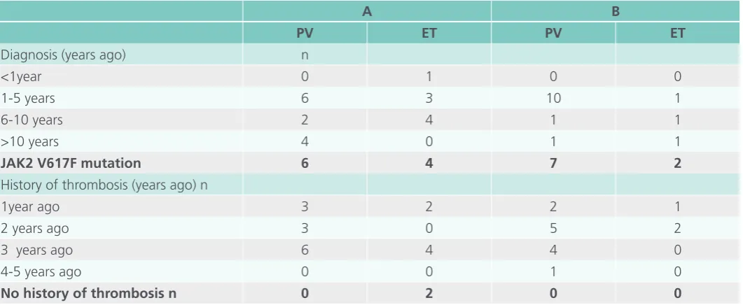
Table 2. Duration of diagnosis, JAK2 V617F mutation and history of thrombosis in clinically stable patients (A) and those who developed recurrent thrombosis (B).
A B
PV ET PV ET
Diagnosis (years ago) n
<1year 0 1 0 0
1-5 years 6 3 10 1
6-10 years 2 4 1 1
>10 years 4 0 1 1
JAK2 V617F mutation 6 4 7 2
History of thrombosis (years ago) n
1year ago 3 2 2 1
2 years ago 3 0 5 2
3 years ago 6 4 4 0
4-5 years ago 0 0 1 0
Blood collection
Blood sampling was performed from a clean venepuncture using the Vacutainer system (Beck-ton Dickenson, New Jersey, USA). About 8 mL of blood was collected and 3 mL gently mixed into citrate tubes (sodium citrate 0.109M) and the re-mainder blood into plain tubes for serum collec-tion. The citrated blood samples were double spun for 15 min each time at 2000g within an hour of blood collection. Serum and plasma samples were aliquoted and kept at -70° C until assayed. Blood sampling was performed only at admission to the study; in recurrence of thrombosis blood sampling was performed post-diagnosis at between 1 to 5 days (n=8), 10-20 days (n=5), and 30 days (n=1) except for one blood sampling was performed 3 days before diagnosis. Most of the patients were on aspirin treatment regime.
Laboratory assays
The following assays were performed: JAK2 V617F mutation (IPSOGEN JAK2 MutantQuant Kit) was performed at the Molecular Diagnostic Centre, National University Health System (NUHS), Yong Loo Lin School of Medicine, National University of Sin-gapore. Serum VEGF Immunoassay (R&D systems, Minneapolis, USA) and beta Thromboglobulin (βTG) (Asserachrome Kit, Stago, France). Von Willebrand Factor (VWF), in-house modified Elisa method us-ing International Standard from National Biological Standards and Control (NIBSC), Potters Bar, Hert-fordshire, UK, Fibrinogen (Clauss method) using in-ternational standard from NIBSC, plasma D-dimer (Zymu D-dimer Kit, France). The above tests were performed at the Coagulation Laboratory, Depart-ment of Obstetrics & Gynaecology, NUHS.
Statistical Analysis
The Statistical Package for Social Sciences (SPSS17; SPSS Inc, Chicago, Il, USA) was used to perform the statistical analysis. Non-parametric Mann Whitney U test was performed together with independent
t-test to evaluate patients under treatment who were clinically stable and those who developed clinical symptoms of recurrence of thrombosis at the time of admission to the study. Pearsons corre-lation analysis was used to determine for correcorre-lation between elevated D-dimer against VEGF seen in re-currence of thrombosis. A P value of less than 0.05 was considered statistically significant.
Results
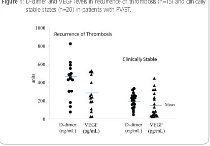
In total, 35 patients who fulfilled the WHO de-fined criteria for PV/ET was studied. Their charac-teristics with history of thrombotic episodes are shown in Tables 1 and 2, VEGF and haemostatic parameters in Table 3. The statistics of evaluating PV/ET patients who were clinically stable and those who developed clinical symptoms of recurrence of thrombosis are shown in Table 4. The scatter plots of VEGF and D-dimer levels between clinically sta-ble and recurrent thrombosis patients are shown in Figure 1 and the correlation between elevated D-dimer and VEGF levels in patients who developed recurrence of thrombosis in Figure 2.
Characteristics of patients studied
Table 3. VEGF and haemostatic parameters in PV/ET patients (n=35)
Mean (SD) Range Normal Reference
VEGF pg/mL 202.4(164.1) 24.4 – 529.8 <115.0
D-dimer ng/mL 295.6 (179.4) 49.0 - 825.0 <400.0
TAT-complex µg/L 2.80 (1.74) 1.01 - 9.13 <2.0
vWF IU/mL 1.20 (0.43) 0.49 – 2.41 <1.5
Fibrinogen g/L 3.47 (1.71) 1.00 – 8.00 2.00 – 4.00
β-TG IU/mL 799.3 (883.8) 105.2 – 3987.0 <40.0
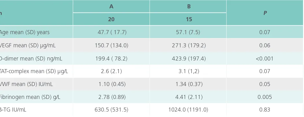
Table 4. Statistics of parameters studied in PV/ET patients who are clinically stable (A) compared to those who developed recurrent thrombosis (B).
n
A B
P
20 15
Age mean (SD) years 47.7 ( 17.7) 57.1 (7.5) 0.07
VEGF mean (SD) µg/mL 150.7 (134.0) 271.3 (179.2) 0.06
D-dimer mean (SD) ng/mL 199.4 ( 78.2) 423.9 (197.4) <0.001
TAT-complex mean (SD) µg/L 2.6 (2.1) 3.1 (1,2) 0.07
VWF mean (SD) IU/mL 1.10 (0.45) 1.34 (0.37) 0.05
Fibrinogen mean (SD) g/L 2.78 (0.89) 4.41 (2.11) 0.005
β-TG IU/mL 630.5 (531.5) 1024.0 (1191.0) 0.83
(B): MVD 5; DVT 3; CHD 2; stroke 4; PE 1
disease (MVD) 5, Deep Vein thrombosis (DVT) 3, Coronary heart disease (CHD) 2, stroke 4 and Pul-monary Embolism (PE) 1. In clinically stable patients 16 received aspirin 100 mg and one Clopidogrel 75mg with phlebotomy treatment in 11 patients and 15 patients received both phlebotomy and as-pirin at the time of blood sampling. In patients who developed recurrence of thrombosis (n=15) seven received aspirin and one Clopidogrel while eleven
had phlebotomy treatment. In 3 patients diagnosed with DVT (PV n=2, ET n=1) low molecular weight heparin was given in addition to aspirin (n=2) and clopidogrel (n=1).
VEGF and haemostatic parameters
anal-Figure 1: D-dimer and VEGF levels in recurrence of thrombosis (n=15) and clinically stable states (n=20) in patients with PV/ET.
ysis. Elevated levels of VEGF (mean 202.4±164.1 pg/mL), TAT-complex (mean 2.80±1.74ug/mL) and β-TG (mean 799.3± 883.8 IU/mL) were evident in PV/ET patients compared to normal reference. Al-though D-dimer, VWF and fibrinogen mean levels were within normal reference range their ranges were widely variable (Table 3). Elevated VEGF levels were seen in 20/35 (57.1%) patients, D-dimer 10/35 (28.6%), Fibrinogen 9/35 (25.7%) and VWF 8/35 (23.6%) patients. The age of patients at 65 years old and above was 20% (7/35).The reference ranges quoted was from the manufacturer’s normal refer-ence and therefore, no age-matched to the study population was done (Table 3).
Statistics of parameters studied in PV/ET patients who were clinically stable (n=20) were compared to those who had developed clinical symptoms of recurrence of thrombosis (n=15) at admission to the study (Table 4). Recurrence of thrombosis oc-curred in 13.3% (2/15) of patients at 65 years old and above. The mean age was higher for patients who experienced a recurrence of thrombosis (mean 57.1 ±7.5 years vs mean 47.7 ±17.7 years) relative to those that were clinically stable but this observa-tion did not reach statistical significance (P=0.07). Statistically significant elevated D-dimer (P= <0.001) and fibrinogen (P=0.005) were seen in patients with recurrence of thrombosis compared to patients who were clinically stable. Higher mean trends were seen for TAT-complex (P=0.07) and VWF (P=0.05) in recurrence of thrombosis but they did not reach statistical significance. No significant differences were also seen for VEGF and Beta-TG levels even though the levels were elevated above the normal reference in both groups (Table 4). Elevated VEGF levels were seen in 45.0% (9/20) of clinically stable patients and 73.3% (11/15) in patients with recur-rence of thrombosis. Clinically stable patients had no elevated D-dimer levels whilst in the recurrence of thrombosis group 66.7% (10/15) patients had el-evated levels (>400ng/mL).The scatter plot for VEGF and D-dimer in both groups is shown in Fig. 1.
Correlation between VEGF and D-dimer levels
Statistically significant correlation between elevat-ed D-dimer (10/15) and VEGF levels in recurrence of thrombosis group were seen (r=0.8540, P=0.002) (Fig. 2). D-dimer was not elevated in clinically sta-ble patients even though 45% (9/20) had elevated VEGF levels (>115 pg/mL).
JAK2 V617F mutation
JAK2 V617F mutation was confirmed in 54.2% (13/24) patients with PV and in 54.5% (6/11) pa-tients with ET. The overall mutation seen in PV/ET was 54.3%. No statistically significant differenc-es in the parameters studied were seen between JAK2 V617F positive and negative patients except for VWF (P=0.04) in negative patients (mean 1.32 ± 0.35 IU/mL vs mean 1.10 ±0.47 IU/mL) in JAK2 V617F positive mutation.
Discussion
between year one to twelve from diagnosis, the incidence for PV/ET was 94.3% which was much higher than reported (3, 4). At admission to the study, 15 patients (42.9%), PV n=12, ET n=3 were reported to have developed recurrence of bosis between one and five years from last throm-botic episode whilst the remaining 20 patients were clinically stable including two (ET) patients with no history of thrombosis. This recurrence of thrombosis was higher than reported (26, 8). A hypercoagulable state for this population was evident from elevat-ed D dimer and fibrinogen levels seen in patients with recurrence of thrombosis, which is in agree-ment with the study of Wautier and co-workers (9). In support of this observation, D-dimer levels were not elevated in clinically stable patients. VEGF levels were elevated in 45.0% of patients who were clinically stable and 73.3% in patients who experi-enced recurrence of thrombosis. The elevated levels are of concern as they are known as a pro-coagu-lant, inducing endothelial activation and thrombin formation (16, 17). Markedly increased D-dimer was found in PV/ET and no differences of coagulation activation markers was found in relation to JAK2-V617F mutational status (29). JAK2 JAK2-V617F mutation was confirmed in 54.2% PV and 54.5% ET patients. We did not find any significant differences in the plasma levels of D-dimer and coagulation activation markers between JAK2 V617F positive and nega-tive patients even though JAK2 V617F mutation associated with thrombosis (16, 17). Considerable variation of JAK2 V617F mutation frequency have been reported with PV of between 65- 97% and ET 23-57% (30) and PV 95%, ET 50-60% (31). The frequency of the mutation in PV patients (54.2%) in our study is rather low, whilst it was comparable to the published literature in ET patients (54.5%). The published reports for JAK2 V617F frequency are for Caucasian population and the frequency in Asians especially with PV need to be further investi-gated. Possible variations in mutation discrepancies
observed in PV include assay sensitivity, diagnostic accuracy and treatment effect (32) have been sug-gested.
Conclusion
In conclusion, plasma VEGF levels were elevated (57.1%) in PV/ET patients relative to controls, al-though the difference was not significant. Similarly, plasma D-dimer and fibrinogen levels were signifi-cantly higher in patients with recurrence of throm-bosis relative to clinically stable patients, whereas plasma VEGF levels were non-significantly elevated. Elevated plasma D-dimer seen in patients with re-currence of thrombosis was found to correlate sig-nificantly with elevated plasma VEGF levels. VEGF and D-dimer measurements have been used in clin-ical practice to determine the risk of PV/ET patients with vascular complications.
Acknowledgements
We wish to express our sincere gratitude to the following hospitals for allowing us access to their patients for the study: Adam Malik Hospital, Hos-pital Dr Pirngadi, Herna HosHos-pital, Imelda and Eliza-beth Hospital and National University Health System (NUHS), Yong Loo Lin School of Medicine, National University of Singapore for access to their research laboratories’ facilities at Molecular Diagnostic Cen-tre and the Coagulation Laboratory, Department of Obstetrics and Gynaecology. We wish to thank Pro-fessor Sorimuda Sarumpaet for statistical assistance.
Conflict of interest
References
1. Vannucchi MA, Guglielmelli P, Tefferi A. Advances in understanding and management of myeloproliferative neoplasms. A Cancer J for Clinicians 2009; 59:171-91.
2. Campbell PJ, Green AR. Mechanisms of diseases: the myeloproliferative disorders. N Engl J Med 2006; 355:2452-66.
3. Elliot MA, Tefferi A. Thrombosis and haemorrhage in polycythemiavera and essential thrombocythemia. Br J Haematol 2005; 128:275-90.
4. Cortelazzo S, Viero P, Finazzi G, D’EmilioA, Rodeghiero F, Barbui T. Incidence and risk factors for thrombotic complications in a historical cohort of 100 patients with essential thrombocythemia. J ClinOncol 1990; 8:556-62.
5. Tefferi A, Vainchecker W. Myeloproliferative neoplasms in molecular pathophysiology, essential clinical understanding and treatment strategies. J Clin Oncol 2011; 29:573-82.
6. Robertson B, Urquhart C, Ford I, Townend J, Watson GH, Vickers MA et al. Platelet and coagulation markers in myeloproliferative diseases:relationships with JAK2V617F status, clonality, and antiphospholipid antibodies. Journal of Thrombosis and Haemostasis 2007; 5:1679-85.
7. Vianello F, Battisti A, Cella G, Marchetti M, Falanga A. Defining the thrombotic risks in patients with myeloproliferative neoplasms. The Scientific World J 2011; 11:1131–37.
8. Landolfi R, Di Gennaro L. Pathophysiology of thrombosis in myeloproliferative neoplasms. Haematologica 2011; 96(2):183-6.
9. Wautier MP, El Nemer W, Gane P, Rain JD, Cartron JP, Colin Y, Le Van Kim C,Wautier JL. Increased adhesion to endothelial cells of erythrocytes from patients with polycythemiavera is mediated by laminin alpha5 chain and Lu/BCAM. Blood 2007; 110:894-901.
10. Casini A, Fontana P, Lecompte TP. Thrombotic complications of myeloproliferative neoplasms: risk assessment and risk-guided management. JThrombHaemost 2013; 11:1215-27.
11. Connolly DT. Vascular permeability factor: a unique regulator of blood vessel function. J Cell Biochem 1991; 47:219-23.
12. Mohle R, Green D, Moore MAS, Nachman RL, Rafii S. Constitutive production of thrombin-induced release of vascular endothelial growth factor by human megakaryocytes and platelets. ProcNatlAcadSci 1997; 94:663-8.
13. Raimondo DF, Giuseppe PA, Stefano M, Rosario G. Angiogenesis in chronic myeloproliferative diseases. ActaHaematologica 2001; 106:177-83.
14. Stamatia T, Timoleon V, Sofia V, Vassilios P, Konstantinos T, Athanassions V et al. Elevated levels of serum vascular
endothelial growth factor in patients with polycythemiavera. ActaHaematologica 2003; 110:16-9.
15. Verheul HM, Hoekman K, Luykx-de Bakker S et al. Platelet transporter of vascular endothelial growth factor. Clinical Cancer Research 1997; 3:2187-90.
16. Dvorak HF, Senger RD, Dvorak AM, Harvey VS, McDonagh J. Regulation of extravascular coagulation by microvascular permeability. Science (Washington DC) 1985; 227:1059-61.
17. Zucker S, Mirza H, Conner CE. Vascular endothelial growth factor induces tissue factor and matrix metalloproteinase production in endothelial cells: conversion of prothrombin to thrombin results in progelatinase-A activation and cell proliferation. Int J Cancer 1998; 75:780-6.
18. Cacciola RR, Di Francesco E, Glustolisi R, Cacciola E. Elevated serum vascular endothelial growth factor levels in patients with polycythemiavera and thrombotic complications. Haematologica 2002; 87:774-5.
19. Goodacre S, Sampson FC, Sutton AJ, Mason S, Morris F. Variation in the diagnostic performance of D-dimer for suspected deep vein thrombosis. Q J Med 2005; 98:513-27.
20. Kovacs MJ, MacKinnon C, Ginsberg J, Wells PS. A comparison of three rapid D-dimer methods for the diagnosis of venous thromboembolism. Br J Haematol 2001; 115:140-4.
21. Prisco D, Grifori E. The role of D-dimer testing in patients with suspected venous thromboembolism. SeminThrombHemost 2009; 35:50-9.
22. Kleinegris MC, Ten Cate H, Ten Cate HAJ., 2013. D-dimer as a marker for cardiovascular and arterial thrombotic events in patients with peripheral arterial disease. A systematic review. ThrombHaemost 2013; 110(2):233-43.
23. Gomez K, Tuddenham EGD, McVey JH., 2011. Normal Haemostasis. Post Graduate Haematology 2011; 6thed:747-71. Wiley Blackwell.
24. Landolfi R and Marchioli R et al. Efficacy and safety of low dose aspirin in polycythemia vera .The New England Journal of Medicine 2004;350:114-24.
25. Tefferi A, VardimanJw. Classification and diagnosis of myeloproliferative neoplasms: the 2008 World Health Organization criteria and point of care diagnostic alogrithms. Leukemia 2008; 22:14-22.
26. Marchioli R, Finazzi G, Landolfi R, Kutti J, Gisslinger H, Patrono C et al. Vascular and neoplastic risk in a large cohort of patients with polycythemiavera. J ClinOncol 2005; 23:2224-32.
28. De Stefano V, Tommaso Z, Rossi E, Vannucchi AM, Ruggeri M, Elli E, et al. Recurrent thrombosis in patients with polycythemiavera and essential thrombocythemia: incidence, risk factors, and effects of treatments. Haematologica 2008; 93:372-80.
29, Trelinski J, Wierzbowska A, KrawczynskaA,Sakowicz A, Pietrucha T, Smolewski P, Robak T, Chojnowski K. Circulating endothelial cells in essential thrombocythemia and polycythemiavera: correlation with JAK2-V617F mutational status, angiogenic factors and coagulation activation markers. Int J Hematol 2010; 91:792-8.
30. Nelson ME, Steensma DP. JAk2 V617 in myeloid disorders: what do we know now, and where ae we headed? Leuk Lymphoma 2006; 47:177-94.
31. Cross NC. Genetic and epigenetic complexity in myeloproliferative neoplasms. Hematology Am SocHematolEduc Program 2011; 2011:208-14.
32. Jones AV, Silver RT, Waghorn K, Cutis C KreilS, Zoi K et al. Minimal molecular response in polycythemiavera patients treated with imatinib or interferon alpha. Blood 2006; 107:3339-41.
Where Doctors exchange clinical experiences, review their cases and share clinical knowledge. You can also access lots of medical publications for free. Join Now!
http://medicalia.org/
Comment on this article:
International Archives of Medicine is an open access journal publishing articles encompassing all aspects of medical scien-ce and clinical practiscien-ce. IAM is considered a megajournal with independent sections on all areas of medicine. IAM is a really international journal with authors and board members from all around the world. The journal is widely indexed and classified Q1 in category Medicine.
Publish with iMedPub