Brain Research 886 (2000) 113–164
www.elsevier.com / locate / bres
Interactive report
1
Cerebral hemisphere regulation of motivated behavior
*
Larry W. Swanson
The Neuroscience Program, Hedco Neuroscience Building, Rm. 428, University of Southern California, 3614 Watt Way, Los Angeles, CA90089-2520, USA
Accepted 10 September 2000
Abstract
The goals of this article are to suggest a basic wiring diagram for the motor neural network that controls motivated behavior, and to provide a model for the organization of cerebral hemisphere inputs to this network. Cerebral projections mediate voluntary regulation of a behavior control column in the ventromedial upper brainstem that includes (from rostral to caudal) the medial preoptic, anterior hypothalamic, descending paraventricular, ventromedial, and premammillary nuclei, the mammillary body, and finally the substantia nigra and ventral tegmental area. The rostral segment of this column is involved in controlling ingestive (eating and drinking) and social (defensive and reproductive) behaviors, whereas the caudal segment is involved in controlling general exploratory or foraging behaviors (with locomotor and orienting components) that are required for obtaining any particular goal object. Virtually all parts of the cerebral hemispheres contribute to a triple descending projection — with cortical excitatory, striatal inhibitory, and pallidal disinhibitory components — to specific parts of the behavior control column. The functional dynamics of this circuitry remain to be established.
2000 Elsevier Science B.V. All rights reserved. Theme: Neural basis of behavior
Topic: Motivation and emotion
Keywords: Amygdala; Basal ganglia; Cerebral cortex; Hypothalamus; Motor behavior; Striatum
1. Introduction the brain, and its paired cerebral hemispheres. Instead,
there is not even a consensus list of parts for the brain, let Consciousness — thinking and feeling, the rational and alone a global scheme for classifying the parts and emotional sides of our mental life — is, the clinical and describing the basic plan of their interconnections or experimental evidence would suggest, a product of activity wiring diagram. In short, there remains an unfortunate lack in neural networks of the cerebral hemispheres. As a of fundamental models describing how the brain works as matter of fact, the notion that conscious or voluntary a system [285].
control of behavior is mediated by cerebral influences The reason for this lack of understanding is simple: descending onto the paired sensorimotor nerves of the sheer complexity. Until the late 19th century, when the brainstem and spinal cord has evolved over a very long cornerstones of brain systems analysis — the neuron period of time. Threads of its history can be traced back doctrine and theory of functional polarity [33,249] — were several thousand years to Greco–Roman antiquity, espe- elaborated and became widely accepted, only a few parts cially in the work of Galen [48,156]. of the brain were distinguished, and it was relatively easy The basic structural plan and functional organization of to propose global models of brain structure–function (e.g. most organs in the body are taken for granted by now — [77,173,332]). However, the introduction of the Golgi the kidney, heart, and stomach are good examples. But this method, and of experimental degeneration methods for does not apply to far and away the most important organ, pathway tracing, in the latter half of the 19th century yielded orders of magnitude more information about brain
´
1 structural organization. The situation was clear to Ramon y
Published on the World Wide Web on 2 November 2000.
Cajal, who contributed far more than any other single *Tel.11-213-740-5892; fax:11-213-741-0561.
E-mail address: [email protected] (L.W. Swanson). investigator to our understanding of the vertebrate nervous 0006-8993 / 00 / $ – see front matter 2000 Elsevier Science B.V. All rights reserved.
Behavior &
system. In 1909 he predicted that ‘to extend our under- unambiguously. A crippling handicap of systems neuro-standing of neural function to the most complex human science today is the confused, ambiguous state of the physiological and psychological activities, it is essential nomenclature used to describe brain structure, much of that we first generate a clear and accurate view of the which is unavoidably based on historical accident rather structure of the relevant centers, and of the human brain than contemporary knowledge [285].
itself, so that the basic plan — the overview — can be
grasped in the blink of an eye.’ [33] 1.1. Motor system and the tripartite control of behavior The latest neuroanatomy revolution started around 1970,
and was based on a combination of axonal transport Before examining the structural organization and pos-pathway tracing methods and immunohistochemistry (sup- sible functional significance of what will be referred to as plemented with hybridization histochemistry in the 1980s). the behavior control column, and then the organization of It produced another avalanche of data on previously cerebral inputs to the column, it is useful to outline a high unknown neural connections — this time including neuro- level scheme for the nervous system control of behavior in transmitters and their receptors — and a natural preoccupa- general [292]. As a starting point, we assume that behavior tion with subjecting individual circuit elements to detailed is the product of, or is driven by, activity in the motor analysis. Fortunately, these methods have clarified for the system — behavior is a function of activity in the motor first time the basic connections of certain long obscure system (Fig. 1). It seems incontrovertible that the behavior regions of the forebrain, including the hippocampus, we observe in others is the product of somatic muscle amygdala, septum, and hypothalamus, so that the time may contractions that in turn are controlled directly by activity be opportune to reexamine from a synthetic perspective the in somatic motoneuron pools of the brainstem and spinal overall organization of brain regions that control behavior cord. In this Section we shall consider three key features of — looking for simplifying principles instead of ever more the motor system: (1) its neural inputs fall into three broad
subdivision and detail. functional classes (sensory, cognitive, and behavioral
Eventually, a consensus description of nervous system state), (2) it is organized hierarchically, and (3) it has three organization will emerge, as happened long ago (basically divisions — somatic, autonomic, and neuroendocrine. in the 18th century) for the skeletal, muscular, circulatory, Turning first to the organization of neural inputs to the and other systems. The following synthesis is presented in motor system, Cajal pointed out long ago that sensory the spirit of providing the crude outlines of at least one systems generally have a dual projection within the central basic plan or model to rekindle discussion of this topic — nervous system. One branch goes directly to the motor and to stimulate the formulation of even better models. The system, and the other goes (more or less directly) to the analysis is based on four converging lines of evidence: cerebral cortex, where sensations and perceptions are development, gene expression patterns, circuit connectivi- elaborated, and voluntary motor impulses are generated. ty, and function, no one of which by itself is necessarily For example, dorsal root ganglion cell axons branch convincing. The ultimate model will be internally con- within the spinal cord, with some collaterals innervating sistent yet comprehensive in terms of system components, components of the intraspinal motor system and others and each component will be defined clearly and named innervating neurons that project to the thalamus and then
L.W. Swanson / Brain Research 886 (2000) 113 –164 115 cortex; and retinal ganglion cell axons branch to innervate intrinsic activity [327] that controls behavioral state — the the lateral geniculate-cortical projection and the superior sleep / wake cycle and levels of arousal within a particular colliculus-reticulospinal projection. Cajal illustrated this state. Obviously, behavior is quite different when one is principle in a series of brilliant diagrams (see Fig. 2 for asleep or awake, and when awake there is a certain basic one of them), and established two fundamental classes of level of arousal or spontaneous activity that is independent inputs to motoneurons, primary mediators of the behavior of, though modulated by, sensory inputs.
we observe in others: direct sensory inputs, which are In summary, there are three major classes of inputs to responsible for the reflex initiation of behavior, and the motor system: (a) sensory, which mediate reflex cortical inputs, which are responsible for the voluntary or behavior; (b) cortical, which mediate cognition and vol-conscious initiation of behavior and are informed by untary behavior; and (c) behavioral state control (Fig. 1).
sensory and other information. Turning now to the hierarchical organization of the
A third fundamental class of inputs to motoneurons motor system itself, the clinical observations and theories gradually became recognized in the 20th century, and can of John Hughlings Jackson over a century ago [301] no longer be ignored. This is the class of inputs arising pioneered the now generally held view that the motor from the still rather poorly understood brain system with system is organized hierarchically [28,306,328]. Neverthe-less, after accepting by definition that motoneuron pools constitute the lowest level of the hierarchy, there is little or no consensus today about organizing principles and nomenclature for the higher levels. The basic idea is illustrated nicely, however, by what is known in a general way about the organization of the neural system that mediates and controls locomotor behavior [192,328,330]. The lowest or first level of the locomotor system is formed of course by a subset of motoneuron pools in the spinal cord ventral horn that innervates the limb muscles respon-sible for locomotor behavior (Fig. 1). The second major level is referred to as the locomotor pattern generator, which lies entirely within the spinal cord, near the motoneuron pools that it regulates. In fact, it is itself a hierarchy of increasingly complex motor pattern generators that coordinate and time muscle contractions across in-dividual joints, then across multiple joints within a par-ticular limb, and finally amongst all four limbs. A third major level is represented, at least in part, by an ill-defined region of the dorsal tegmentum known as the mesence-phalic locomotor region, and rostroventral to this is a fourth major level in an ill-defined region of the caudal hypothalamus / rostral midbrain — the so-called subtha-lamic or hypothasubtha-lamic locomotor region.
A crude though nevertheless useful understanding of functional differentiation between these major levels of the locomotor hierarchy has been gained by transecting the neuraxis at different rostrocaudal levels. The existence of a spinal locomotor pattern generator is demonstrated by the Fig. 2. One of Cajal’s early circuit diagrams based on the Golgi method.
fact that whereas a spinal animal displays no spontaneous It shows how sensory information is directed toward both the motor
system and the cognitive system — and how there are two main inputs to locomotor activity, coordinated limb movements charac-the motor system. The peripheral, dendritic process (d) of a dorsal root teristic of locomotion may be elicited when the limbs of ganglion cell (D) ends in the skin (D9), where sensory stimuli are such an animal are placed on a moving treadmill, thus detected. Impulses generated by the stimulus travel up the peripheral
providing somatic sensory input to the pattern generator process (arrows) and then the central, axonal process (c), which bifurcates
[192,330]. Furthermore, undisturbed midbrain animals also in the spinal cord (B). The descending branch generates collaterals in the
spinal gray matter, which end on interneurons (not shown) that in turn show no spontaneous locomotor activity [88], although it innervate ventral horn motoneurons (shown for convenience on contrala- can be elicited either by certain sensory stimuli (for teral side near (b) that innervate muscle fibers (C). The other bifurcation example, auditory or nociceptive), or by experimental branch of the central process ascends to another neuron (f) whose axon
stimulation of the mesencephalic locomotor region (g) in turn relays sensory impulses to the cerebral cortex (A).
Psychomo-[192,330], which apparently sends direct and indirect tor neurons in the cortex send via their axons (a) a second major class of
generator. Viewed in this way, the mesencephalic system consists of a series of primary, transversely ar-locomotor region can be thought of as a ar-locomotor pattern ranged ‘segments’ that, from rostral to caudal, include the
initiator. In contrast, undisturbed chronic hypothalamic forebrain, midbrain, hindbrain, and spinal cord.The other
animals do present spontaneous locomotor behavior, which structural model dates back even further to Vesalius in the by definition is not influenced or directed by cognitive 16th century and consists of three parts: a trunk or core inputs from the telencephalon because it has been removed that generates a series of paired cranial and spinal nerves or disconnected [88]. In the sense of providing a certain (from an essentially continuous brainstem and spinal cord, level of endogenous activity (perhaps some form of ‘set- respectively), and suprasegmental cerebrum and cerebel-point’), the hypothalamic locomotor region can be thought lum.
of as a locomotor pattern controller, which generates Present unresolved ambiguity centers around how to spontaneous inputs (in an unknown way), ultimately, to the define the brainstem and cerebrum in terms of forebrain, spinal locomotor pattern generator. midbrain, and hindbrain divisions. From the functional Although less understood, there is good evidence for schematic point of view (Fig. 1), the thalamus relays conceptually similar central pattern generators for other sensory information to the cognitive system in the cerebral complex motor behaviors, related for example to coordi- hemispheres, and the hypothalamus is a key part of the nated eye movements (centered in the midbrain), orofacial neuroendocrine and autonomic motor systems, so these behaviors (centered in the dorsolateral hindbrain), and two forebrain areas can reasonably be included within the orientation of the head via the neck musculature (centered brainstem. On the other hand, there is a strong bias in the in the lower medulla and upper cervical spinal cord). It literature to treat the forebrain as a structural unit. Realisti-seems reasonable to postulate that these pattern generators cally, the extent to which these structural distinctions are are also influenced by higher levels of the motor hierarchy, artificial or inaccurate remains to be determined; there is namely pattern initiators and controllers. no compelling evidence to support or reject either model at In closing this section, we should point out that there are this time. For the sake of convenience, then, we shall now three divisions of the motor system. Thus far we have present a working hypothesis of basic subdivisions in the considered the well-known somatomotor system, which mammalian forebrain because they appear to play such regulates the contraction of skeletal, striate, or voluntary critical roles in the expression of both voluntary and reflex muscle. The motoneuron pools for this system extend from behaviors.
the midbrain rostrally to the caudal end of the spinal cord. The gradual progression from simple to complex mor-The second is the autonomic visceromotor system, where phology revealed during embryological development pro-the first stage motoneurons form pools of preganglionic vides a time-honored way to appreciate the basic organiz-sympathetic and paraorganiz-sympathetic neurons; together they ing principles of forebrain architecture (for reviews of the also stretch from the midbrain to the caudal end of the approach adopted here, see Refs. [7,9]). At early stages spinal cord, with a few gaps here and there. For the most when neurulation occurs — when the neural plate fuses part they innervate smooth muscle, cardiac muscle, and dorsally to form the neural tube — one can identify glands. The third is the neuroendocrine secretomotor primary forebrain, midbrain, and hindbrain vesicles, with
system; its motoneuron pools are centered in and around tiny paired optic vesicles evaginating from the presumptive
the periventricular zone of the hypothalamus and they hypothalamic region of the forebrain vesicle. The next exert their influence throughout the body via the pituitary major event in forebrain vesicle differentiation involves the gland (Section 1.3). It seems reasonable to hypothesize formation rostrodorsally of an external groove, the hemis-that the second and third systems are hierarchically orga- pheric sulcus, which at least initially is complemented by nized along the lines outlined for the somatomotor system. an internal bulge, the torus hemisphericus. The hemis-And as we shall see, the hypothalamus appears to contain pheric sulcus unambiguously divides the forebrain vesicle mechanisms for coordinating appropriate responses in all into three secondary vesicles — paired telencephalic
three motor systems. vesicles (endbrain vesicles, with their incipient lateral
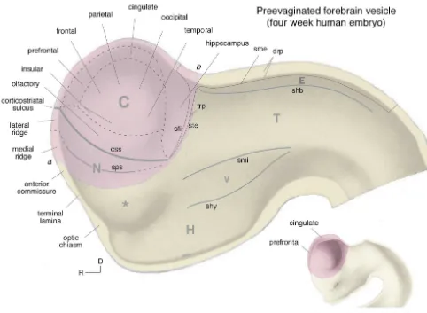
ventricles) and diencephalon (interbrain, with its incipient 1.2. Basic organization of the forebrain third ventricle). This stage, which occurs in the four week human and eleven day rat embryo, is very instructive for The fundamental plan of nervous system organization understanding later development because the entire wall of just presented — a motor system controlled by reflex, the forebrain vesicle can be observed in a midsagittal view voluntary, and behavioral state inputs — is obviously a (Fig. 3) — the telencephalon has not yet begun its functional schema. On the structural side of the coin two massive, complex, and still not fully understood process of distinct though not necessarily mutually exclusive plans for evagination (Fig. 4).
L.W. Swanson / Brain Research 886 (2000) 113 –164 117
Fig. 3. Major forebrain subdivisions. A fate map of the major forebrain subdivisions projected onto the forebrain vesicle of a four week human embryo, before the telencephalic vesicle (pink) has evaginated to the extent it has a week later (inset, lower right). At the four week stage it is easy to envision qualitatively where the major regions of the cerebral cortex (C) will normally differentiate at later stages, as will the major ‘longitudinal’ divisions of the interbrain or diencephalon: the hypothalamus (H), ventral thalamus (V), dorsal thalamus (T), and epithalamus (E). The asterisk indicates the presumptive preoptic region, rostral (R) and dorsal (D) to the optic sulcus (gray streak extending from the optic chiasm). a–b indicates the junction between telencephalon and diencephalon (yellow), the presumptive interventricular foramen of Monro. Unless stated otherwise, all nomenclature in this article follows Refs. [9,282]. Other abbreviations: css, corticostriatal sulcus; drp, diencephalic roof plate; N, cerebral nuclei / basal ganglia; sfi, fimbrial sulcus; shb, habenular sulcus; shy, hypothalamic sulcus; sme, sulcus medullaris; smi, middle diencephalic sulcus; sps, striatopallidal sulcus; ste, sulcus terminalis; trp, telencephalic roof plate. Adapted with permission from Refs. [9,113].
diencephalon become distinguishable, two longitudinal presumptive nuclear or noncortical division of the telen-grooves appear in the inner wall of the right and left halves cephalon, which starts before neurogenesis in the cortical of the diencephalic vesicle — the middle diencephalic and region. Around this time a third longitudinal sulcus hypothalamic sulci. They define, in between, the ventral (habenular) also appears in the diencephalon, just ventral thalamus and a rostroventral extension of it that lies just to the roof plate; it separates presumptive epithalamus caudal to the optic sulcus, where the first neurogenesis in from presumptive dorsal thalamus.
ganglia / cerebral nuclei region of the telencephalic vesicle, along with at least part of the amygdala. We shall examine this problem further in Section 3, where information about connections and neurotransmitter expression will be added to the developmental evidence.
Overall, the simplest interpretation of a vast literature on forebrain embryology is that the telencephalic neuro-epithelium differentiates into a topologically dorsal cere-bral cortex and ventral cerecere-bral nuclei (basal ganglia), which in turn differentiate into a dorsolateral striatum and ventromedial pallidum. On the other hand, the diencephalic neuroepithelium differentiates, to a first order of approxi-mation, into a stack of four more or less longitudinal bands — hypothalamus, ventral thalamus, dorsal thalamus, and epithalamus. By and large, this view is based on more than a century’s worth of morphological embryology. An understanding of the genomic program that assembles the neural plate and tube will undoubtedly lead to major new insights, although analysis of certain homeobox gene expression patterns already tends to confirm and clarify the location of certain fundamental borders, such as that between telencephalon and diencephalon (Fig. 6) and between cortex and basal nuclei (see [211]).
1.3. Basic organization of the hypothalamus
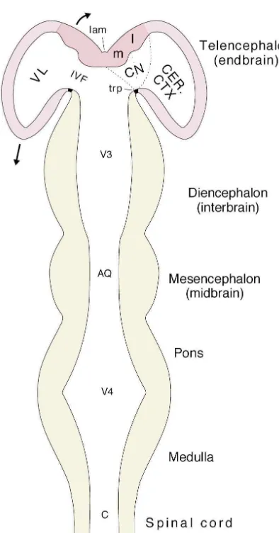
The majority of connections within the adult forebrain are accounted for by a familiar qualitative scheme: the dorsal thalamus projects topographically to the entire ‘neocortex’ (see [267]), which in turn projects topographi-cally to the ‘basal ganglia,’ especially the striatum (see Fig. 4. The major divisions of the central nervous system in a horizontal
[84]). In contrast, whereas the hypothalamus is exception-section of a schematic neural tube with a straightened longitudinal axis.
ally important from the functional perspective — it is The cerebral hemispheres (pink) are rapidly evaginating (arrows), and
essential not only for survival of the individual but for the their two divisions — cerebral cortex (CER. CTX), and cerebral nuclei /
basal ganglia (CN), with their two differentiations, the medial (m) and species as well — reasonably accurate data about its neural lateral (l) ventricular ridges — are indicated. The major divisions of the connections only began appearing with the neuroanatomy ventricular system associated with major tissue divisions are also shown:
revolution of the 1970s, and the vague outlines of its basic central canal (C), fourth ventricle (V4), cerebral aqueduct (AQ), third
structural organization — its place in the forebrain as a ventricle (V3) and its rostromedial border the lamina terminalis (lam),
whole — are just now beginning to emerge [283]. and lateral ventricles (VL) with the associated interventricular foramina
of Monro (IVF). Other abbreviation: trp, telencephalic roof plate (see The origins of deep interest in the hypothalamus can be Fig. 3). Adapted with permission from Ref. [278]. traced to 1901 and Frolich’s detailed account of the¨ adiposogenital syndrome — excessive truncal obesity The situation in the telencephalic vesicle at this stage is combined with genital atrophy, which was thought to be
neces-a
b
c
a
b
c
A.
B.
L.W. Swanson / Brain Research 886 (2000) 113 –164 119
Fig. 5. Embryonic cortex and ventricular ridges. (A) Transverse section through the telencephalic vesicles of a 19 mm human embryo (about 8 weeks) showing the pallium or cortex (between a and the sulcus ventralis) and the two ventricular ridges — a dorsal or lateral ridge between a and b, and a ventral or medial ridge between b and c. The neuroepithelium is a black layer of varying thickness lining the ventricular cavity; a mantle layer of young neurons can be seen superficial to the neuroepithelium of the ventricular ridges, and in the most ventrolateral cortex just dorsal to a. (B) Transverse section of the left telencephalic vesicle at a later stage of development to show the further precocious growth of the mantle layer associated with the ventricular ridges — which begin bulging dorsally and partly obliterating the originally relatively huge lateral ventricle (from an embryonic day 16 hamster embryo). Abbreviations: a, corticostriatal sulcus; Ang. ven., ventral angle; b, striatopallidal sulcus; c, ventral angle; Cor. str. lat., lateral corpus striatum (striatum); Cor. str. med., medial corpus striatum (pallidum); CPC, caudoputamen; Fas. den., fascia dentata (dentate gyrus); Fis. hip., hippocampal fissure; Hip, hippocampus; Nuc. med. sept., medial septal nucleus; Sul. vent., ventral sulcus; SVZ, subventricular zone; VZ, ventricular zone (neuroepithelium). For clarity, the abbreviations a, b, and c have been added to the original figures. Part A adapted from Ref. [113], Part B adapted with permission from Ref. [302].
sary for survival of the individual and of the species controllers for ingestive, defensive, and reproductive
be-(reviewed in Ref. [275]). haviors (as well as controllers for thermoregulatory
tropic hormones into a portal system for transport to the anterior pituitary, where they act on five classic endocrine cell types (the parvicellular neurosecretory system). The neuroendocrine motor zone is centered in the ventromedial diencephalon, in three contiguous differentiations of the docrine neurons that migrate away from the periventricular zone during development; and the gonadotropin releasing hormone (GnRH) motoneurons, which are unique insofar as they are generated during development from the olfac-tory epithelium instead of from the third ventricular neuroepithelium, and come to lie scattered in the adult septal and preoptic regions.
The hypothalamic medial nuclei form a column of very distinct cell groups that, arranged from rostral to caudal, include the medial preoptic nucleus, anterior hypothalamic nucleus, descending division of the paraventricular nu-cleus, ventromedial nucleus (and adjacent tuberal nucleus), Fig. 6. Homeobox gene expression distinguishes telencephalon from
dorsal and ventral premammillary nuclei, and mammillary diencephalon. This is a transverse section through the forebrain vesicle of
body. They form the greater part of the behavior control an embryonic day 13 rat embryo. Brain-1 (Brn-1) is a POU-III homeobox
column discussed in Section 2. gene, and an autoradiogram of its mRNA expression pattern is shown on
the right side of the figure. The white arrows indicate the boundary The hypothalamic lateral zone remains poorly under-between telencephalic and diencephalic vesicles, and it is clear that Brn-1 stood, and in the widest sense may be involved in the expression in the telencephalic vesicle at this stage of development stops
regulation of behavioral state and arousal mechanisms at this boundary, although patches of expression are also seen in the
[275]. It would appear that the projections of the lateral presumptive hypothalamic paraventricular nucleus (PVH) and
epibranchi-preoptic area are distinct from more caudal regions of this al placodes (ebp). Structural features seen at this level in an adjacent
Nissl-stained section are drawn on the left, with the telencephalic vesicle zone (Ref. [272] and R.F. Thompson, L.W. Swanson, indicated in pink and the diencephalic vesicle in yellow. Other abbrevia- unpublished observations with PHAL), and recently it has tions: css, corticostriatal sulcus; CTXl,m, cortex, lateral, medial regions;
become clear that the tuberal level of the lateral hypo-hf, hippocampal fissure; HIP, hippocampus; HY, hypothalamus; let,
thalamic area is distinguished by separate populations of epithelial lamina; ME, median eminence; mtl, mantle layer; ppa,
neurons that express melanin-concentrating hormone and parahypophysial arch (adjacent to presumptive subfornical organ); PR,
pallidal (medial ventricular) ridge; pts, pallidothalamic sulcus; sfi, fimbrial hypocretin / orexin (see [25,130]), as well as corticotropin sulcus; she, hemispheric sulcus; shy, hypothalamic sulcus; sps, striatopal- releasing hormone in response to dehydration [234,323]. lidal sulcus; SR, striatal (lateral ventricular) ridge; TEM, thalamic
This would suggest that the supraoptic or anterior, and the eminence. Adapted with permission from Ref. [7].
mammillary levels of the lateral zone may also have distinct features that remain to be characterized.
A.
B.
L.W. Swanson / Brain Research 886 (2000) 113 –164 121
Fig. 7. Hypothalamic obesity. (A) Twenty-year-old female patient on the right suffered from a tumor confined to ventral and medial regions of the hypothalamus and displayed excessive hunger, thirst, and rage — and had lost her menstrual cycle — whereas obesity in the rat shown on the left was produced by an experimental lesion in the same general region of the hypothalamus. (B) Levels of central nervous system transection where animals can (a, b) and cannot (c) survive independently and display spontaneous behavior, including eating, drinking, and locomotion. Part A is reproduced with permission from Refs. [215,268], and Part B is reproduced with permission from Ref. [114].
control column, and projects to the neuroendocrine motor medial nuclei form the rostral segment of a behavior zone and preautonomic cell groups. In addition, it contains control column extending through the ventromedial mid-central rhythm generators such as the suprachiasmatic brain, and that as a whole this column receives a
topo-nucleus. graphically organized input from virtually the entire
cere-bral hemisphere.
1.4. Perspective This analysis of forebrain organization relies heavily on
a model of overall nervous system organization postulating Voluntary behavior is controlled directly by projections that behavior is equivalent to activity in the motor system, from the cerebral cortex to the somatic motor system. So it which in turn is modulated by three classes of inputs — seems reasonable to focus an analysis of neural systems voluntary or cognitive, reflex or sensory, and behavioral mediating this class of behavior on the reasonably well- state (Fig. 1). In this scheme, the hypothalamic medial known topographic map of the cerebral cortex, and on the nuclei are part of the behavior control column, and thus lie less clear higher levels of the somatic motor system at the apex of the motor system hierarchy (Fig. 9), based hierarchy. The overall organization of projections from the on similarities with the hierarchical control of locomotor various thalamic nuclei to the entire cortical mantle is also behavior (Fig. 1). Thus, it would be expected that the firmly established, along with the organizing principles of hypothalamic medial nuclei receive three classes of inputs: outputs from the whole isocortex (neocortex) to the cognitive, sensory, and behavioral state [226], although ‘classical’ basal ganglia (cerebral nuclei). this review focuses only on the first class — inputs from
The rest of this paper deals with two major aspects of the cerebral hemispheres. forebrain organization that remain problematic. First, how
do certain long enigmatic regions including the
hippocam-pus, amygdala, and septum (usually included within the 2. The behavior control column
‘limbic system’) fit into the grand scheme of cerebral
hemisphere organization? As a simple working hypothesis It is convenient first to discuss a new concept, the based on developmental, connectional, and neurotrans- ‘behavior control column,’ and then go on in Sections 3 mitter utilization criteria it is proposed that all parts of the and 4 to analyze how cerebral hemisphere inputs map onto cerebral hemispheres belong to either the cerebral cortex or it in a topographically organized way.
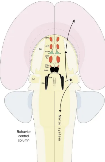
Hypothalamic
L.W. Swanson / Brain Research 886 (2000) 113 –164 123 (3) Overall, the evidence (Sections 1.3, 2.1–2.3) indi-cates that the ventromedial column of nuclei shown in Fig. 10 forms at least the core of a behavioral control column at the top of the motor system hierarchy (as defined in Figs. 1 and 9), and that this column may be divided into rostral and caudal segments. The rostral segment, from the medial preoptic to premammillary nuclei (the preoptic-premammillary segment) plays a critical role in circuits regulating the three basic classes of goal-oriented be-havior common to all animals: ingestive, reproductive, and defensive; whereas the caudal segment (the mammillary-nigral segment; black in Fig. 10) plays a critical role in circuits underlying the expression of exploratory or forag-ing behavior in general. The rostral segment can be divided further into two major parts, one dealing with the social behaviors just mentioned, and another, the descend-ing division of the paraventricular nucleus (PVHd; green in Fig. 10), dealing with ingestive (eating and drinking) behaviors, which we shall now consider in more detail.
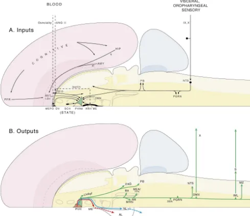
2.1. Thirst as a model system (ingestive behavior)
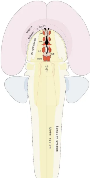
The behavior control column is formed by a longitudinal Fig. 9. A model of hypothalamic controllers at the top of the motor
array of cell groups whose functional significance is system hierarchy, with a trio of sensory, behavioral state, and cerebral
hemisphere inputs (see Fig. 1). Although not shown for the sake of known at least in a general way. However, these cell clarity, all three classes of inputs can go to all levels of the motor system groups are not ‘centers’ in some isolated, naive sense hierarchy. Abbreviations: r, reflex; v, voluntary.
[180]. Rather, they are nodes in circuits, systems, or networks that mediate particular classes of behavior cleus (shown in red in Fig. 10) — all generate a dual [275,293] — nodes that appear to act as controllers (or projection, with a primary branch descending to the parts of controller networks) providing set-points or some brainstem motor system and a secondary branch ascending baseline level of endogenous activity (Sections 1.1, 1.3). to the thalamus [226]. This is, in principle, just like the Furthermore, whereas the evidence strongly indicates that projections of the caudally adjacent medial and lateral they are essential for the control of these behaviors, it is mammillary nuclei, which form the caudal medial zone of certainly not known at the present time whether or not the hypothalamus. It has long been known from develop- there are nearby cell groups in the hypothalamus and mental studies that individual neurons in the mammillary midbrain that are also integral parts of the behavior control nuclei send a descending axon to the brainstem tegmen- column. In short, while the behavior control column as tum, and a collateral of this axon to the anterior thalamic defined in a preliminary way here appears to form an nuclei [33,75,311]. It is also similar in principle to the essential core of the associated circuitry, it may well caudally adjacent reticular part of the substantia nigra include additional components.
[39,226], which sends a branched projection to the brain- For purposes of description and analysis, motivated stem motor system (including the deeper layers of the behavior in general can be divided into three sequential superior colliculus and reticular formation) and to the phases — initiation, procurement, and consummatory thalamus [16,266]; and it is similar to the adjacent ventral [293]. In terms of defining underlying neural circuits, thirst tegmental area, which sends projections to the brainstem has provided an unusually good model, in large part motor system and thalamus, in addition to other sites (see because so much is known about the physiology of body
[17,273]). water regulation, and water intake in animals is so easy to
(2) The rostral medial zone nuclei just listed (medial measure and to manipulate with well-defined physiological preoptic, anterior hypothalamic, ventromedial and tuberal, stimuli [228,334]. Even more specifically, perhaps the best and premammillary; red in Fig. 10) are interconnected in a understood motivated behavior of all in terms of neural massive, highly differentiated way, and other connectional circuitry is drinking associated with a particular stimulus, and functional evidence indicates that they form critical hypovolemia, because at least one mechanism and site of parts of circuitry underlying the expression of reproductive initiation is known with certainty — high circulating levels and defensive behaviors, that is, social behaviors (involv- of the peptide hormone angiotensin II acting on neuronal ing interactions between animals) critical for survival of receptors in the subfornical organ [73,257,305].
Fig. 10. An overview of the behavior control column, with the rostral segment in red and green, and the caudal segment in black. Almost all of the nuclei in this column generate a dual, typically branched projection — descending to the motor system on one hand and ascending to thalamocortical loops on the other. Abbreviations: AHN, anterior hypothalamic nucleus; MAM, mammillary body; MPN, medial preoptic nucleus (lateral part in particular); PMd,v, premammillary nuclei, dorsal, ventral; PVHd, descending division of paraventricular nucleus; SC, superior colliculus, deeper layers; SNr, reticular substantia nigra; TH, dorsal thalamus; TU, tuberal nucleus; VMH, ventromedial nucleus; VTA, ventral tegmental area.
L.W. Swanson / Brain Research 886 (2000) 113 –164 125
Fig. 11. Outline of circuitry involved in controlling thirst and drinking behavior. The scheme focuses on one member of the behavior control column, the descending paraventricular nucleus (PVHd), and its major inputs (A) and outputs (B). As discussed in the text, various classes of input play a key role in the initiation phase of the behavior, whereas outputs of the PVH are involved in the procurement and consummatory phases of the behavior, as well as in coordinating the appropriate visceral (neuroendocrine and autonomic) responses to maintain homeostasis during these latter phases, before enough water is located and ingested. The PVHd is perhaps the best established component of the hypothalamic thirst control network, although there are almost certainly others, which remain obscure at this point. Interestingly, the PVHd is a critical component of the hypothalamic hunger control network as well. Other abbreviations: AL, anterior lobe pituitary; AMY, amygdala; ANG II, angiotensin II; ARH, arcuate nucleus; BST, bed nuclei stria terminalis; DMH, dorsomedial nucleus; DMX, dorsal motor nucleus vagus; fi, fimbria; HIP, hippocampus; IML, intermediolateral preganglionic column; IX, glossopharynge-al nerve; LHApf, perifornicglossopharynge-al laterglossopharynge-al hypothglossopharynge-alamic area (tuberglossopharynge-al level); LSv, ventrglossopharynge-al laterglossopharynge-al septglossopharynge-al nucleus; ME, median eminence; MEA, midbrain extrapyramidal area; MEPO, median preoptic nucleus; MRN, mesencephalic reticular nucleus; MZ, marginal zone; NL, neural (posterior) lobe pituitary; NTS, nucleus of the solitary tract; OV, vascular organ lamina terminalis; PAG, periaqueductal gray; PB, parabrachial nucleus; PFR, prefrontal region;
´
PGRN, paragigantocellular reticular nucleus (ventrolateral medulla); PPN, pedunculopontine nucleus; RA, raphe nuclei; RR, retrorubral area; S, sympathetic ganglia; SCH, suprachiasmatic nucleus; SFO, subfornical organ; SSN, superior salivatory nucleus; st, stria terminalis; X, vagus nerve.
regions. The best evidence for this comes from lesions of (and hunger), there is, however, a great deal of evidence the periaqueductal gray region (but not the medulla), that it is at least an integral part of the control mechanism which attenuate the primary polydipsia and subsequent or network [145].
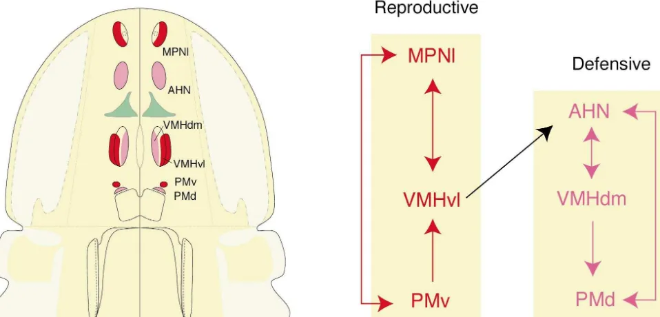
car-diovascular volume receptors, hepatic osmoreceptors, and 2.2. Social behavior network (reproductive and defensive a ‘dry mouth’). This information is relayed, at least in part, behaviors)
by direct connections from the nucleus of the solitary tract
to the PVHd, and by less direct inputs relayed from the Pathway tracing methods demonstrate that there are two nucleus of the solitary tract via the ventrolateral medulla highly interconnected sets of nuclei in the rostral behavior (the lateral paragigantocellular reticular nucleus), projec- control column (Fig. 12). One set [36,37,226,256] includes tions to the PVHd that in part use norepinephrine, epineph- the lateral part of the medial preoptic nucleus, ventrolateral rine, galanin, and neuropeptide Y as neurotransmitters part of the ventromedial nucleus, and ventral premammil-[148,239,240]. All four substances induce primary poly- lary nucleus. There is abundant evidence that these three dipsia followed by hyperphagia when injected into the cell groups form a core part of the sexually dimorphic PVH (see [145]). Humoral sensory information (angioten- circuit mediating reproductive behavior (see [254]), and sin II levels) is detected in the subfornical organ, which each of them expresses abundant levels of estrogen re-also projects directly to the PVHd, a pathway that, ceptor mRNA [255]. For example, the medial preoptic interestingly, also uses angiotensin II as a neurotransmitter nucleus appears to be involved selectively in the expres-[149]. In fact, the total output of the subfornical organ is of sion of masculine sexual behavior whereas the ventrolater-considerable interest because it innervates directly neuro- al ventromedial nucleus is important for the expression of nal cell groups participating in all three classes of motor feminine sexual behavior (in particular, the lordosis reflex). responses to hypovolemia — behavioral, autonomic, and In contrast, the other set includes the anterior hypo-neuroendocrine [275–277,291]. One of these cell groups thalamic nucleus, dorsomedial part of the ventromedial surrounds the rostral end of the third ventricle, in and nucleus, and dorsal premammillary nucleus [37,39,222]. perhaps around the median preoptic nucleus and vascular Abundant evidence reviewed elsewhere indicates that the organ of the lamina terminalis. Injections of angiotensin II circuitry established by these three cell groups plays a into this general region elicit drinking [248,296], and the critical role in the expression of defensive behaviors, projection to it from the subfornical organ also contains especially with respect to predators [34,51,225,226], and angiotensin II [149]. Furthermore, this region, like the they all express abundant levels of androgen receptor subfornical organ [253], is known to be osmoreceptive mRNA [255].
[24] (another humoral stimulus to thirst) and to project to The only major direct connection between these two sets the PVHd [97]. It should also be mentioned that there is a of nuclei [37] is formed by a projection from the ventrola-neuropeptide Y-containing projection from the hypo- teral part of the ventromedial nucleus (part of the re-thalamic arcuate nucleus to the PVH, and it is possible that productive behavior network) and the anterior hypo-circulating leptin entering through the nearby median thalamic nucleus (part of the defensive behavior network). eminence acts on this pathway to influence ingestive There are no known major direct connections between behavior responses associated with the PVHd (see [69]). these six nuclei and the PVHd (part of the ingestive
Not surprisingly, there are also presumed cognitive / behavior network). voluntary and behavioral state inputs to the PVHd. Current
evidence suggests that the former are relayed to the PVHd 2.3. Exploration segment of the column ( foraging by the bed nuclei of the stria terminalis and ventral lateral behavior)
septal nucleus, which in turn receive inputs from the
L.W. Swanson / Brain Research 886 (2000) 113 –164 127
Fig. 12. Left. Cell groups of the rostral behavior control column. The descending paraventricular nucleus, which is involved in the control of eating and drinking (ingestive behavior, see Fig. 11) is shown in green. The rest of the cell groups play a major role in controlling two classes of social behaviors, that is, behaviors involving interactions between animals — reproductive (red) and defensive (magenta). Right. The organization of major direct connections between components of the rostral behavior control column. See text for details.
when the animal’s head is pointed in a certain direction tional organization of a motivated behavior control column within the environment. Interestingly, whereas neurons that can be thought of as lying at the apex of the motor with this basic neurophysiological profile were discovered system hierarchy in the upper brainstem, we shall now go in the subicular complex of the hippocampal formation, on to consider how cerebral influences map onto the lesions there do not alter dramatically the physiological column. This will be facilitated by introducing a con-properties of head direction cells in the anterior thalamic ceptually simple model of cerebral hemisphere organiza-nuclei, whereas lesions in this part of the diencephalon tion, and later (in Section 6) comparing it briefly with abolish head direction responses in the subicular complex. other current ways of dealing with this problem. In Preliminary evidence suggests that perhaps vestibular essence, the model postulates that the cerebral hemispheres information about head orientation is relayed via the dorsal have only three parts — cortex, striatum, and pallidum — tegmental nucleus to the mammillary body (and / or anterior which generate a triple descending projection to the motor thalamic nuclei), and then on to the subicular complex system — excitatory, inhibitory, and disinhibitory, respec-[260,300]. It has been suggested that the mammil- tively. The model is based on a combination of em-lothalamic-cortical system containing head direction neu- bryological, gene expression, connectional, and functional rons is critically involved in elaborating a sense of arguments.
direction [300].
Thus, a case can be made for the caudal behavior control
column being involved critically in two basic aspects of 3.1. Development and fast neurotransmitters exploratory or foraging behavior in general — locomotion
and orientation of the eyes, head, and neck, with the As discussed in Section 1.2, there is currently rather reticular substantia nigra more involved in orienting move- broad general agreement among neuroembryologists that ments and the mammillary body in orientation direction. the mammalian telencephalon (endbrain, cerebral hemi-By way of contrast, the rostral behavior control column spheres, and cerebrum are considered synonyms here) appears to play a critical role in establishing particular consists of two basic parts, a cortex dorsally and a nuclear goals, such as food or water, a mate, or escape from a mass ventrally (Figs. 3–6). As an extension of this view,
predator. most of the major human neuroanatomy textbooks of the
last quarter century [42,186,195,331] have referred to the noncortical part of the adult cerebral hemisphere as the
3. A model of cerebral hemisphere organization basal ganglia or basal nuclei (cerebral nuclei is used as a
hemisphere as cortical or nuclear, or the problem is simply use glutamate as an excitatory neurotransmitter
ignored (see Section 6). [206,226,295]. This is simply an application of the
poly-A second, independent argument for a basic dichotomy transmitter hypothesis that all neurons (at least at some between cerebral cortex and nuclei comes from extensive stage of the life cycle) use either an excitatory amino acid evidence [84] that most, if not all, cortical projection or GABA as a neurotransmitter, along with various neurons (pyramidal cells) use glutamate as a fast, excitat- combinations of other peptides and molecules [281]. ory neurotransmitter, whereas in contrast the descending In the mammalian cerebral cortex, glutamate appears to projections of two classic parts of the basal ganglia / be used as a neurotransmitter by (all) pyramidal neurons cerebral nuclei (the caudate nucleus / putamen and the whereas GABA is used by (many) interneurons but not by globus pallidus) use GABA as a fast, inhibitory neuro- pyramidal cells. In a series of experiments with fundamen-transmitter (Fig. 13). One postulate of the model outlined tal theoretical implications, Rubenstein and colleagues here is that in general descending projections of the basal [11,12], and now others [143,202], have recently presented ganglia / cerebral nuclei use GABA as an inhibitory neuro- evidence that at least most GABAergic interneurons of the transmitter, and descending projections of cerebral cortex adult cerebral cortex are actually generated by the neuro-epithelium of the ventricular ridges (which generate the basal ganglia / cerebral nuclei) at early stages of develop-ment and then migrate dorsally to the pallium (cortex) along tangential routes. These results imply another fun-damental difference between the cerebral cortex and nuclei — this time with respect to developmental gene expression patterns, related to whether neurons use glutamate (a default neurotransmitter [281]) or GABA as a neuro-transmitter. Perhaps during normal mantle layer formation in mammals, only the ventricular ridges generate neurons that retain the capacity to express glutamic acid decarbox-ylase (GAD), and thus synthesize GABA from glutamate, throughout life.
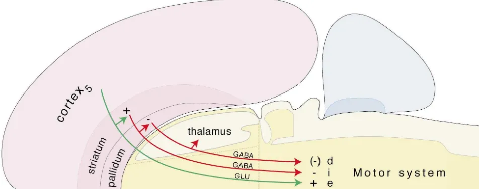
3.2. Triple projection to the motor system
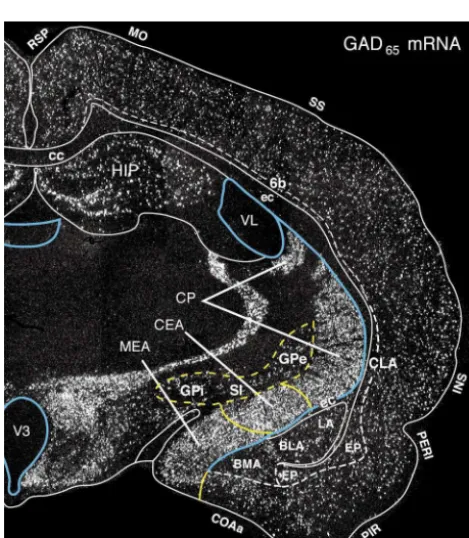
Another postulate of our model is that the cerebral hemispheres generate a fundamental triple descending projection to the motor system, based on the ‘classical’ isocortical-striatal-pallidal model (see Sections 4 and 5 for references), and that the projections from structurally and functionally differentiated regions of the cerebrum are variations on this arrangement. The adult ‘minimal or prototypical circuit element’ (Fig. 14) consists of: (1) an excitatory (glutamatergic) projection from cortex to the Fig. 13. The distribution of neurons expressing GAD65 mRNA in a
brainstem and spinal cord motor system, with an excitatory transverse histological section through the rat forebrain. GAD65 (along
with GAD67) is the enzyme that converts the default neurotransmitter collateral [146] to the striatum; (2) an inhibitory glutamate to the neurotransmitter GABA. In the cerebral cortex, GAD is (GABAergic) projection from the striatum to the brainstem expressed in interneurons, whereas in the cerebral nuclei / basal ganglia it motor system, with an inhibitory collateral [197] to the is expressed in descending projection neurons. Various cerebral nuclei
pallidum; and (3) an inhibitory (GABAergic) projection regions as interpreted here are indicated with yellow; the lateral and third
from the pallidum to the brainstem motor system (with an ventricular ependyma is shown in blue, along with the obliterated
(ventral) part of the lateral ventricle (see Figs. 15, 17 and 18). This inhibitory collateral [197] to the thalamus). Functionally, section corresponds approximately to the one shown in Fig. 17B. the pallidal projection is disinhibitory [227] because it is Abbreviations: BLA, basolateral amygdalar nucleus; BMA, basomedial inhibited by the striatal input, which in turn is excited by amygdalar nucleus; cc, corpus callosum; CEA, central amygdalar nucleus;
the cortical input. The descending projection to the brain-CLA, claustrum proper; COAa, anterior cortical amygdalar nucleus; CP,
stem / spinal cord motor system from the isocortex caudoputamen; ec, external capsule; EP, endopiriform nucleus; GPe,i,
globus pallidus, external, internal segments; HIP, hippocampus; INS, (synonymous with neocortex, a term better left unused in insular cortex; LA, lateral amygdalar nucleus; MEA, medial amygdalar light of unfounded evolutionary implications; see Section nucleus; MO, motor cortex; PERI, perirhinal area; PIR, piriform area; 6) arises primarily from layer 5, whereas the isocortical RSP, retrosplenial cortex; SI, substantia innominata; SS, somatosensory
projection to thalamus arises predominantly from layer 6; cortex; VL, lateral ventricle; V3, third ventricle; 6b, cortical layer 6b or 7
cortical associational / commissural projections arise pref-(subplate). From an in situ hybridization autoradiogram. Adapted with
L.W. Swanson / Brain Research 886 (2000) 113 –164 129
Fig. 14. Triple cascading projection from the cerebral hemispheres to the brainstem motor system. This minimal or prototypical circuit element consists of a glutamatergic (GLU) projection from layer 5 pyramidal neurons of the isocortex (or equivalent pyramidal neurons in allocortex), with a glutamatergic collateral to the striatum. This dual projection appears to be excitatory (e,1, green). The striatum then generates a GABAergic projection to the motor system, with a GABAergic collateral to the pallidum. This dual striatal projection appears to be inhibitory (i,2, red). Finally, the pallidum generates a GABAergic projection to the brainstem motor system, with a GABAergic collateral to the dorsal thalamus. This dual pallidal projection can be viewed as disinhibitory [d, (2)] because it is inhibited by the striatal input.
[123,147]). Recall that the brainstem part of the motor of the cortical mantle into areas or fields. As an obvious system, as defined here, includes the hypothalamus (e.g. example, there is a clear difference in lamination between
Fig. 9). olfactory and somatic sensorimotor cortical areas (Fig. 15).
Although alternative schemes are available [129], the one 3.3. A basic taxonomy of parts used here is derived from the classical work of Brodmann [78]. We have developed a graphical way to show the Thus far we have suggested that embryological, neuro- cortical areal map in a topographically accurate way transmitter, and connectional evidence all converges to (where the surface area of particular cortical fields is indicate that the cerebral hemispheres present two basic maintained along with correct borders between fields) for divisions, cortex and nuclei, and that the latter have two the rat [8,278,282] (Fig. 16) and human [280], and subdivisions, striatum and pallidum. This brings us to the references to the primary literature for the various cortical crux of the problem — how might all of the various parts areas may be found in Ref. [282]. The flatmap approach is of the cerebral hemispheres be classified as either cortical, based on embryology, and in principle it is a fate map of striatal, or pallidal, and how does the triple descending the neural plate, which topologically is a flat sheet, one cell projection to the motor system apply to this classification? thick. At early stages of the neural tube, it is easy to The answers to these two questions are, of course, entirely appreciate how the presumptive cortical protomap could be interrelated. For the sake of clarity, the classification visualized on a flat sheet, before the telencephalic vesicles scheme for parts we have arrived at thus far will be evaginate (Fig. 3).
discussed now, followed in the next Section by an analysis There are only a few points about regions included in of the connectional evidence. Here there are two concerns: the cerebral cortex that merit comment here, and they what are the major divisions of the cerebral cortex, and revolve around the admittedly unusual olfactory region, what are the major divisions of the cerebral nuclei / basal which is unique because it is the only part of the cerebral
ganglia? hemispheres to receive direct input from a (sensory)
capsule (Figs. 16A and 17D), is derived embryologically from the cortical subplate, and thus amounts to a layer variously called 6b or 7. Ventral to the claustrum proper lies the endopiriform ‘nucleus,’ which more often than not has been regarded as a ventral division of the claustrum, deep to the three classical layers of the piriform area and just superficial to the rostroventral end of the extreme capsule (Fig. 17A). Dorsal to the claustrum proper, recent work has identified in rodents a very distinct though thin layer 6b or 7 that stretches all the way dorsomedially into the cingulate gyrus and may well be derived from the cortical subplate (see [63,282,310,312,315]).
The suggestion here is that a cortical subplate region, which becomes progressively thinner from ventral to dorsal (Fig. 17A), consists of the endopiriform nucleus, claustrum proper, and layer 6b / 7, respectively. The final component is the most speculative, but follows Meynert’s original suggestion in 1867 [170–172] that the basolateral complex of the amygdala is a thick, caudoventral extension of the claustrum. Recent Golgi analyses have emphasized the pyramidal cell-like morphology of projection neurons in this complex (e.g. [161]), which probably use glutamate rather than GABA as a neurotransmitter (see [54,161]), and it is possible to arrange the various parts of the Fig. 15. Cerebral cortex versus basal ganglia / cerebral nuclei. This is a basolateral complex in positions deep to various olfactory transverse Nissl-stained histological section through the adult rat
telen-(amygdalar) and temporal cortical areas [294], just superfi-cephalon to show the disposition of cerebral cortex (pink) versus cerebral
cial to the caudoventral end of the external capsule (Fig. nuclei / basal ganglia (yellow). Notice how differentiated cortical
lamina-17B, C). tion patterns can be; for example, compare somatic sensorimotor cortex
with olfactory cortex. Correspondingly, note how differentiated various To summarize, our working hypothesis suggests that the regions of striatum can be; for example, compare caudoputamen (CP) claustral complex (basolateral amygdalar nuclei, endo-with olfactory tubercle (OT). The claustral division of cerebral cortex is
piriform nucleus, claustrum proper, and isocortical layer shown in darker pink, deep to the traditional cortical plate, and the lateral
6b / 7) is derived embryologically from the cortical sub-ventricular ependyma with its obliterated ventral extension are shown in
plate region deep to the cortical plate, and that its blue. This section corresponds to level A in Fig. 17. Other abbreviations:
a, corticostriatal sulcus (obliterated, see Fig. 5); ACB, nucleus accum- projection neurons use glutamate as a neurotransmitter. bens; LS, lateral septal nucleus; MS, medial septal nucleus; NDB, nucleus
of the diagonal band; SI, substantia innominata. Photomicrograph from 3.3.2. The cerebral nuclei (striatum and pallidum) Ref. [282].
According to our simple model of the cerebral hemi-spheres, everything that is not cortical (as defined in the of the amygdala on the surface of the hemisphere as flatmap of Fig. 16) is either striatal or pallidal. In a seminal components of the (olfactory) cortex [294]. They include paper, Heimer and Wilson [104] stressed the utility of the well-known cortical ‘nucleus’ of the amygdala and distinguishing between dorsal and ventral regions of the ‘nucleus’ of the lateral olfactory tract, along with the more basal ganglia with very similar connectional patterns (Fig. obscure postpiriform transition area and piriform-amygda- 19). According to this now widely accepted view, the lar area — all of which appear to contain classical caudate nucleus and putamen form the dorsal striatum, pyramidal neurons that use glutamate as a neurotrans- which projects to the globus pallidus or dorsal pallidum; mitter. These four parts of the amygdala, along with the whereas in contrast the nucleus accumbens, striatal fundus, piriform area, anterior olfactory ‘nucleus,’ and tenia tecta and olfactory tubercle form the ventral striatum, which might be thought of as the secondary olfactory region of projects to the substantia innominata or ventral pallidum
cortex. broadly conceived. More recently, we have expanded this
L.W. Swanson / Brain Research 886 (2000) 113 –164 131
Fig. 16. A flatmap of the rat cerebral cortex. In this projection, surface areas and boundary conditions are accurate (at least qualitatively), so that shapes and distances are inevitably distorted. In principle, this is a topological transformation of the cerebral cortex as observed in the unevaginated telencephalic vesicle, early in embryonic development (see Fig. 3). Abbreviations: FRP, frontal pole; OCP, occipital pole; TEP, temporal pole. Adapted from Ref. [282].
the striatum for hippocampal cortex. It is now known that of the pallidum (Fig. 19, Section 4.2, and Ref. [295]) there is a topographically organized projection from Am- related to amygdalar parts of the caudal striatum.
mon’s horn cortex and subiculum proper to the lateral
septal nucleus (Section 4), that the medium spiny neurons 3.3.3. Overview of cerebral regional anatomy
there are GABAergic (as in the dorsal and ventral The regional anatomy of the adult cerebral hemispheres striatum), and that there is a projection from the lateral is exceptionally difficult to appreciate. However, its gener-septal nucleus to the medial gener-septal / nucleus of the diagonal al organization is much simpler to understand if one begins band complex. It seems obvious that the latter, with its early in development with the unevaginated telencephalic mixture of GABAergic and cholinergic neurons is a medial vesicle and its presumptive cortical region and two ven-differentiation of the substantia innominata that is special- tricular ridges (Fig. 5) — and then assumes that the former ized with respect to connections with the hippocampal generates the cerebral cortex proper (the cortical plate) and
formation [124,169,232]. claustral complex (the subplate), whereas the latter
Fig. 17. Adult cortical plate versus subplate. The relationship between cerebral cortex proper (the cortical plate, light pink) and the claustral complex (the cortical subplate, darker pink), as seen in three transverse sections through the adult rat forebrain. The approximate location of the sections, which are arranged from rostral (A) to caudal (C), is indicated in the schematic sagittal outline at the upper left, which also shows corresponding atlas levels in Ref. [282]. As in Figs. 13, 15 and 18, the ventricular ependyma, and the obliterated lateral ventricle, are indicated in blue. A Nissl-stained section for level A is shown in Fig. 15; and the distribution of GAD-expressing neurons at about level B is shown in Fig. 13. Abbreviations: a, obliterated corticostriatal sulcus; ec, external capsule; H, hypothalamus; hf, hippocampal fissure; hippo., hippocampus; PAL, pallidum; rf, rhinal fissure; STR, striatum; T, thalamus. Drawings adapted from Ref. [282].
latter indicates the border between cerebral cortex and dorsal striatum to dorsal pallidum projection system as a cerebral nuclei / basal ganglia as defined in this article. prototype. A topographically organized projection from most of the isocortex to the entire caudoputamen was first demonstrated in 1961 by Webster [325], and since that
4. Cerebral inputs to the behavior control column time it has become clear [59,166] that major contributors include the visual, auditory, somatosensory, and gustatory Based on the general classification of cerebral parts just areas, the somatomotor areas, the orbital cortex and outlined we can see how the triple descending projection cingulate region, and posterior parietal and temporal (Fig. 14) is organized topographically with respect to the association areas (Fig. 20A). Pyramidal cells in this broad various components of the behavior control column (Fig. region of cortex that project to the brainstem and spinal 10). In this Section the defining [269], primary, or major cord motor system are concentrated in layer 5, use projection from a cortical area to the striatum, or from a glutamate as a neurotransmitter, and generate a gluta-region of the striatum to the pallidum, will be described to matergic collateral to the dorsal striatum (see emphasize organizing principles. A more detailed account [84,123,146,147]). In terms of basal ganglia / cerebral of cerebral connections, which recognizes that a particular nuclei inputs to the behavior control column, there is a cortical area may project to multiple sites within the topographically organized GABAergic projection from the striatum (and pallidum), and that a particular striatal region caudoputamen to the substantia nigra (see [59]), and most may project to multiple sites within the pallidum, will be of these axons generate a GABAergic collateral in the
presented in Section 5. globus pallidus (see [197]) — and then there is a dense
a-CP ACB FS OT LSC MEA CEA AAA IA
GPe GPi SI MA MS/DBB BST
Dorsal Ventral Medial Caudorostral
STRIATUM
PALLIDUM
L.W. Swanson / Brain Research 886 (2000) 113 –164 133
Fig. 18. Development of mature lateral ventricle shape. This sequence of cartoons illustrates how a great deal of the lateral ventricle’s (VL) wall (in blue) becomes obliterated during embryogenesis, as the dorsal ventricular ridge of the cerebral nuclei / basal ganglia (CN, in yellow) grows dorsally with the massive thickening of its mantle layer (m). Ventricle obliteration begins ventrally at the corticostriatal sulcus (a; see Figs. 3, 5, 6, 13, 15 and 17) and gradually extends dorsally (B–D). Axonal projections from the olfactory cortex and immediately adjacent regions that differentiate near the cerebral nuclei / basal ganglia, descend in the medial forebrain bundle (mfb), ventral to the obliterated lateral ventricle (B–D). Descending projections from other, more dorsal regions of cortex pass through the earlier obliterated lateral ventricle in the internal capsule (int), fornix system (fx), and stria terminalis (st). As descending, association, and commissural projections form, they construct a deep fiber system consisting of the external (ec) and extreme (ee) capsules, just superficial to the ventricular layer of the neural tube (v). It would appear that the claustral complex (CLA), the remnant of the cortical subplate, differentiates within this deep fiber system, and that it becomes progressively thinner from ventral to dorsal (D). Adapted with permission from Ref. [294].
Fig. 19. A simple scheme for topographic regionalization of the basal ganglia / cerebral nuclei. Abbreviations: AAA, anterior amygdalar area; ACB, nucleus accumbens; BST, bed nuclei stria terminalis; CEA, central amygdalar nucleus; CP, caudoputamen; FS, striatal fundus; GPe,i, globus pallidus, external, internal segments; IA, intercalated amygdalar nuclei; LSC, lateral septal complex; MA, magnocellular (preoptic) nucleus; MEA, medial amygdalar nucleus; MS / DBB, medial septal / nucleus of the diagonal band complex; OT, olfactory tubercle; SI, substantia innominata.
course through the internal capsule / lateral forebrain bun-granular insular regions of cortex also send a glutamatergic
dle fiber system [282] (Fig. 20A). Other brainstem projec-projection directly to the substantia nigra (see [137,183]).
tions of the caudoputamen and globus pallidus are dis-This is the basic triple descending projection from the
cussed in Section 5.1. cerebrum to the motor system (including the behavior
Let us now compare this arrangement with defining control column) discussed in Section 3.2, and it tends to
L.W. Swanson / Brain Research 886 (2000) 113 –164 135
cortical inputs to the ventral striatum, and in particular tral premammillary, and medial mammillary nuclei; this with defining inputs to its major component, the nucleus projection arises mostly from the ventral half of the accumbens (Fig. 20B). These inputs arise predominantly in subiculum (see [40,132,286]). The lateral septal complex the entorhinal area of the hippocampal formation [308] and in turn sends a descending, presumably GABAergic, adjacent perirhinal area of the inferior temporal association projection to the medial septal / diagonal band complex region [168], and in the medial prefrontal region and (medial pallidum) and to most components of the rostral caudally adjacent agranular insular region [19]. Then, the behavior control column (see [225,289]). Because of the nucleus accumbens sends dense projections to the sub- highly topographic nature of projections from the hip-stantia innominata and to the ventral tegmental area pocampus to the lateral septal complex, and from the latter component of the behavior control column to the behavior control column, it would appear that [104,105,188,287], projections that are GABAergic (see different dorsoventral zones within the hippocampus (and [126]). And finally, the substantia innominata (ventral in particular field CA1) influence different functional pallidum in the sense of Fig. 19) sends a presumably components of the behavior control column [223,225] GABAergic projection to the ventral tegmental area, and (Fig. 21).
other brainstem regions (see [92]). Fibers associated with Next we come to cortical areas sending their most dense this component of the triple descending projection from the striatal inputs to the central and medial amygdalar nuclei cerebrum tend to course through the medial forebrain — the caudal striatum (Fig. 20E; also see Fig. 24 below). bundle system [282] rather than the lateral forebrain By far the most dense striatal projection of the accessory
bundle system (Fig. 20A, B). olfactory bulb (primary pheromonal cortex) is to the
Thus far we have examined the major triple descending medial amygdalar nucleus [242], which also receives cerebral input to the substantia nigra and ventral tegmental massive inputs from areas of cortex associated with the area. Defining inputs to the third component of the caudal amygdala (cortical ‘nucleus’ and postpiriform transition segment of the behavior control column, the mammillary and piriform-amygdalar areas) (see [162,165]) — all of body, are associated with yet another region of cortex, the which in turn receive massive inputs from the main or subicular complex of the hippocampal formation (Fig. accessory olfactory bulb [242]. Perhaps the most dense 20C). Interestingly, it is this region and not Ammon’s horn striatal projections from the visceral area and adjacent (the hippocampus proper) that projects through the post- posterior agranular insular area are to the central nucleus, commissural fornix in a topographically organized way to which also gets heavy inputs from the amygdala-related the mammillary body (see [132,286,288,316,317]). The cortical regions just mentioned as projecting massively to subicular complex has a very light and limited projection the medial amygdalar nucleus (see [162,165]). The medial to the striatum [316,317]. In particular, it appears to and central nuclei send a dense GABAergic projection to innervate a narrow vertical band of the caudoputamen the bed nuclei of the stria terminalis or rostral pallidum adjacent to the lateral wall of the lateral ventricle (J.M. (see [38,141,206,247]), and the medial nucleus also pro-Wyss, personal communication). The exact projections of jects to all components of the rostral behavior control this dorsal striatal zone to the nuclei of the caudal behavior column except the descending paraventricular nucleus and column zone, and to the pallidum, remain to be de- dorsal premammillary nucleus (see [38]). Finally, the bed
termined. nuclei of the stria terminalis project to much of the
As discussed in Section 2.3, the ventral tegmental area behavior control column (see [289]), although the details plays a role in controlling locomotor behavior, the sub- remain to be analyzed.
stantia nigra in orienting movements of the eyes, head, and And last, there is the most idiosyncratic part of the neck, and the mammillary body in the orientation direction cerebrum, associated with the main olfactory bulb (Fig. of the head — all important components of exploratory or 20F). The main olfactory bulb, and three principal cortical
foraging behavior. recipients of its output, the anterior olfactory nucleus, tenia