DAFTAR PUSTAKA
1. WHO | Dengue and severe dengue.
http://www.who.int/mediacentre/factsheets/fs117/en/#.VoY-RO_-qqU.mendeley. Accessed January 1, 2016.
2. Treatment, Prevention and Control Global Strategy for Dengue Prevention and Control 2. World Health Organization
3. Mustafa MS, Rasotgi V, Jain S, Gupta V. Discovery of fifth serotype of dengue virus (DENV-5): A new public health dilemma in dengue control.
Med J Armed Forces India. 2015;71(1):67-70.
doi:10.1016/j.mjafi.2014.09.011.
4. Guzman MG, Halstead SB, Artsob H, et al. Europe PMC Funders Group Dengue : a continuing global threat Europe PMC Funders Author
Manuscripts. 2015;8(12 0):1-26. doi:10.1038/nrmicro2460.Dengue.
5. Yohan B, Kendarsari RI, Mutia K, Bowolaksono A, Harahap AR, Sasmono RT. Growth characteristics and cytokine/chemokine induction profiles of dengue viruses in various cell lines. Acta Virol. 2014;58(1):20-27. http://www.ncbi.nlm.nih.gov/pubmed/24717025.
6. Jessie K, Fong MY, Devi S, Lam SK, Wong KT. Localization of dengue virus in naturally infected human tissues, by immunohistochemistry and in situ hybridization. J Infect Dis. 2004;189(8):1411-18. doi:10.1086/383043.
7. Fink J, Gu F, Ling L, et al. Host gene expression profiling of dengue virus infection in cell lines and patients. PLoS Negl Trop Dis. 2007;1(2):e86. doi:10.1371/journal.pntd.0000086.
Epidemiology, Virus Evolution, Antiviral Drugs, and Vaccine Development. Curr Infect Dis Rep. 2010;12(3):157-164. doi:10.1007/s11908-010-0102-7.
9. Wang W-K, Chao D-Y, Kao C-L, et al. High Levels of Plasma Dengue Viral Load during Defervescence in Patients with Dengue Hemorrhagic Fever: Implications for Pathogenesis. Virology. 2003;305(2):330-338. doi:10.1006/viro.2002.1704.
10. Murray NE, Quam MB, Wilder-Smith A. Epidemiology of dengue: past, present and future prospects. Clin Epidemiol. 2013;5:299-309.
doi:10.2147/CLEP.S34440.
11. Anand P, Kunnumakkara AB, Newman RA, Aggarwal BB. Bioavailability of Curcumin: Problems and Promises. Mol Pharm. 2007;4(6):807-818. doi:10.1021/mp700113r.
12. Sharma R a., Gescher a. J, Steward WP. Curcumin: The story so far. Eur J Cancer. 2005;41(13):1955-1968. doi:10.1016/j.ejca.2005.05.009.
13. Singh RK, Rai D, Yadav D, Bhargava A, Balzarini J, De Clercq E.
Synthesis, antibacterial and antiviral properties of curcumin bioconjugates bearing dipeptide, fatty acids and folic acid. Eur J Med Chem.
2010;45(3):1078-1086. doi:10.1016/j.ejmech.2009.12.002.
14. Chen T, Chen D, Wen H, et al. Inhibition of Enveloped Viruses Infectivity by Curcumin. PLoS One. 2013;8(5):1-11.
doi:10.1371/journal.pone.0062482.
15. Anggakusuma, Colpitts CC, Schang LM, et al. Turmeric curcumin inhibits entry of all hepatitis C virus genotypes into human liver cells. Gut.
2014;63(7):1137-49. doi:10.1136/gutjnl-2012-304299.
vitro. Arch Virol. 2014;159(3):573-579. doi:10.1007/s00705-013-1849-6.
17. Kaushik G, Kaushik T, Yadav SK, Sharma SK, Ranawat P. Curcumin sensitizes lung adenocarcinoma cells to apoptosis via intracellular redox status mediated pathway. 2012;50(December):853-861.
18. Haryanto S, Hayati RF, Yohan B, et al. The molecular and clinical features of dengue during outbreak in Jambi, Indonesia in 2015. Pathog Glob Health. May 2016:1-11. doi:10.1080/20477724.2016.1184864.
19. Ni H, Barrett AD. Molecular differences between wild-type Japanese encephalitis virus strains of high and low mouse neuroinvasiveness. J Gen Virol. 1996;77 ( Pt 7):1449-55. doi:10.1099/0022-1317-77-7-1449.
20. Marbawati D, Rahmah S. Effects of Curcumin and Pentagamavunon-0
Against Dengue-2 Virus Infection In Vero Cells ; an In Vitro Study.
Procedia Environ Sci. 2015;23(Ictcred 2014):215-221. doi:10.1016/j.proenv.2015.01.033.
21. Mukhopadhyay S, Kuhn RJ, Rossmann MG. A structural perspective of the flavivirus life cycle. Nat Rev Microbiol. 2005;3(1):13-22.
doi:10.1038/nrmicro1067.
22. Kuno G, Chang G, Tsuchiya K, Karabatsos N, Cropp C. Phylogeny of the genus Flavivirus. J Virol. 1998;72(1):73-83.
23. Rodenhuis-Zybert IA, Wilschut J, Smit JM. Dengue virus life cycle: viral and host factors modulating infectivity. Cell Mol Life Sci.
2010;67(16):2773-2786. doi:10.1007/s00018-010-0357-z.
24. Zybert I a., van der Ende-Metselaar H, Wilschut J, Smit JM. Functional importance of dengue virus maturation: Infectious properties of immature virions. J Gen Virol. 2008;89(12):3047-3051.
25. Noisakran S, Onlamoon N, Songprakhon P, Hsiao H-M, Chokephaibulkit K, Perng GC. Cells in Dengue Virus Infection In Vivo. Adv Virol.
2010;2010:1-15. doi:10.1155/2010/164878.
26. Sydow FF, Santiago M a, Neves-Souza PC, et al. Comparison of dengue infection in human mononuclear leukocytes with mosquito C6/36 and mammalian Vero cells using flow cytometry to detect virus antigen. Mem Inst Oswaldo Cruz. 2000;95(4):483-489.
doi:10.1590/S0074-02762000000400007.
27. Chu JJH, Ng ML. Infectious Entry of West Nile Virus Occurs through a Clathrin-Mediated Endocytic Pathway Infectious Entry of West Nile Virus
Occurs through a Clathrin-Mediated Endocytic Pathway. J Virol. 2004;78(19):10543-10555. doi:10.1128/JVI.78.19.10543.
28. Fischl W, Bartenschlager R. Exploitation of cellular pathways by Dengue virus. Curr Opin Microbiol. 2011;14(4):470-475.
doi:10.1016/j.mib.2011.07.012.
29. Clyde K, Kyle JL, Harris E. Recent Advances in Deciphering Viral and Host Determinants of Dengue Virus Replication and Pathogenesis. J Virol. 2006;80(23):11418-11431. doi:10.1128/JVI.01257-06.
30. Modis Y, Ogata S, Clements D, Harrison SC. Structure of the dengue virus envelope protein after membrane fusion. Nature. 2004;427(6972):313-319.
doi:10.1038/nature02165.
31. Krishnan MN, Ng A, Sukumaran B, et al. RNA interference screen for human genes associated with West Nile virus infection. Nature. 2008;455(7210):242-245. doi:10.1038/nature07207.
33. Viswanathan K, Fruh K, DeFilippis V. Viral hijacking of the host ubiquitin system to evade interferon responses. Curr Opin Microbiol.
2010;13(4):517-523. doi:10.1016/j.mib.2010.05.012.
34. Milly CMJ, Gubler DJ. Investigation of the role of the ubiquitin proteasome pathway in dengue virus life cycle. PhD Progr Integr Biol Med. 2015
35. COOPER PD. The plaque assay of animal viruses. Adv Virus Res. 1961;8:319-378.
36. Baer A, Kehn-Hall K. Viral Concentration Determination Through Plaque Assays: Using Traditional and Novel Overlay Systems. J Vis Exp.
2015;(93). doi:10.3791/52065.Viral.
37. Medina F, Medina JF, Colon C, Vergne E, Santiago GA, Munoz-Jordan JL. Dengue virus: isolation, propagation, quantification, and storage. Curr Protoc Microbiol. 2012;Chapter 15:Unit 15D.2.
doi:10.1002/9780471729259.mc15d02s27.
38. Edelman DC, Barletta J. Real-time PCR provides improved detection and titer determination of bacteriophage. 2003;35(2).
39. Huang MT, Ma W, Lu YP, et al. Effects of curcumin, demethoxycurcumin, bisdemethoxycurcumin and tetrahydrocurcumin on
12-O-tetradecanoylphorbol-13-acetate-induced tumor promotion.
Carcinogenesis. 1995;16(10):2493-97.
40. Dutta K, Ghosh D, Basu A. Curcumin protects neuronal cells from
Japanese encephalitis virus-mediated cell death and also inhibits infective viral particle formation by dysregulation of ubiquitin-proteasome system. J Neuroimmune Pharmacol. 2009;4(3):328-337. doi:10.1007/s11481-009-9158-2.
inhibits hepatitis C virus replication via suppressing the Akt-SREBP-1 pathway. FEBS Lett. 2010;584(4):707-712.
doi:10.1016/j.febslet.2009.12.019.
42. Si X, Wang Y, Wong J, Zhang J, McManus BM, Luo H. Dysregulation of the ubiquitin-proteasome system by curcumin suppresses coxsackievirus B3 replication. J Virol. 2007;81(7):3142-3150. doi:10.1128/JVI.02028-06.
43. Chen DY, Shien JH, Tiley L, et al. Curcumin inhibits influenza virus infection and haemagglutination activity. Food Chem. 2010;119(4):1346-1351. doi:10.1016/j.foodchem.2009.09.011.
44. Ingolfsson HI, Koeppe 2nd RE, Andersen OS. Curcumin is a modulator of bilayer material properties. Biochemistry. 2007;46(36):10384-91.
doi:10.1021/bi701013n.
45. Mahy BWJ, Kangro HO. Virology Methods Manual. London: Academic Press; 1996.
46. Kang JH, Kang HS, Kim IK, et al. Curcumin sensitizes human lung cancer cells to apoptosis and metastasis synergistically combined with carboplatin.
Exp Biol Med (Maywood). 2015;240(11):1416-25. doi:10.1177/1535370215571881.
47. Wang YJ, Pan MH, Cheng AL, et al. Stability of curcumin in buffer
solutions and characterization of its degradation products. J Pharm Biomed Anal. 1997;15(12):1867-76.
48. Na HS, Cha MH, Oh D-R, Cho C-W, Rhee JH, Kim YR. Protective
mechanism of curcumin against Vibrio vulnificus infection. FEMS Immunol Med Microbiol. 2011;63(3):355-362. doi:10.1111/j.1574-695X.2011.00855.x.
49. Tan GKX, Ng JKW, Lim AHY, Yeo KP, Angeli V, Alonso S.
Strain Induces Systemic Vascular Leakage in AG129 Mice. 1997:523-532.
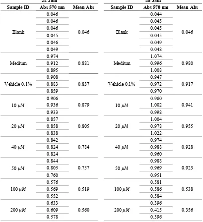
LAMPIRAN 3. Data Penelitian
Tabel Absorbansi 570nm Cell Toxicity Assay
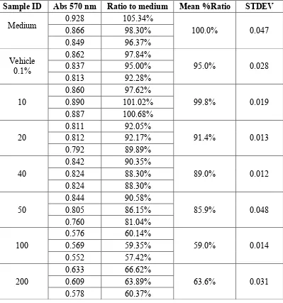
Tabel Analisis Hasil Curcumin A549 Cell Toxicity Assay 24 Jam
Sample ID Abs 570 nm Ratio to medium Mean %Ratio STDEV
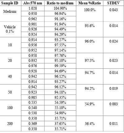
Tabel Analisis Hasil Curcumin A549 Cell Toxicity Assay 48 Jam
Sample ID Abs 570 nm Ratio to medium Mean %Ratio STDEV
* = outliers
LAMPIRAN 4. Uji Analisis statistik
Uji Normalitas Data Cell Toxicity Assay 24 dan 48 Jam
Tests of Normality
Group
Kolmogorov-Smirnova Shapiro-Wilk Statistic df Sig. Statistic df Sig.
24 Jam Medium .308 3 . .902 3 .393
Vehicle .176 3 . 1.000 3 .977
10 .353 3 . .824 3 .174
20 .369 3 . .787 3 .085
40 .385 3 . .750 3 .000
50 .186 3 . .998 3 .921
100 .273 3 . .945 3 .549
200 .200 3 . .995 3 .860
48 Jam Medium .333 3 . .862 3 .274
Vehicle .359 3 . .810 3 .138
10 .354 3 . .821 3 .165
20 .215 3 . .989 3 .800
40 .175 3 . 1.000 3 1.000
50 .177 3 . 1.000 3 .970
100 .219 3 . .987 3 .780
200 .385 3 . .750 3 .000
Analisis Regresi Linear Cell Toxicity Assay 24 jam
Best-fit values ± SE
Slope -0.1945 ± 0.06142
Y-intercept 95.06 ± 5.859
X-intercept 488.6
1/slope -5.14
95% Confidence Intervals
Slope -0.3651 to -0.02401
Y-intercept 78.79 to 111.3
X-intercept 288.7 to 3467
Goodness of Fit
R square 0.7149
Sy.x 9.75
Is slope significantly non-zero?
F 10.03
DFn, DFd 1, 4
P value 0.0339
Deviation from zero? Significant
Equation Y = -0.1945*X + 95.06
Data
Number of X values 6
Maximum number of Y replicates 1
Total number of values 8
Analisis Regresi Linear Cell Toxicity Assay 48 Jam
Best-fit values ± SE
Slope -0.3566 ± 0.05497
Y-intercept 103.9 ± 5.244
X-intercept 291.4
1/slope -2.804
95% Confidence Intervals
Slope -0.5092 to -0.204
Y-intercept 89.34 to 118.5
X-intercept 221 to 461
Goodness of Fit
R square 0.9132
Sy.x 8.726
Is slope significantly non-zero?
F 42.09
DFn, DFd 1, 4
P value 0.0029
Deviation from zero? Significant
Equation Y = -0.3566*X + 103.9
Data
Number of X values 6
Maximum number of Y replicates 1
Total number of values 8
Number of missing values 0
Uji Anova Cell Toxicity Assay 24 Jam
ANOVA
Abs24
Sum of
Squares df Mean Square F Sig. Between
Groups .403 7 .058 83.579 .000
Total .415 23
95% Confidence Interval Lower
200 .244333* .021442 .000 .17010 .31857
40 Medium -.097000
*
.021442 .006 -.17123 -.02277
Vehicle -.053333 .021442 .267 -.12757 .02090 10 -.095000* .021442 .008 -.16923 -.02077
20 -.021000 .021442 .971 -.09523 .05323
50 .027000 .021442 .901 -.04723 .10123
100 .264333* .021442 .000 .19010 .33857
200 .223333* .021442 .000 .14910 .29757
50 Medium -.124000* .021442 .001 -.19823 -.04977 Vehicle -.080333* .021442 .029 -.15457 -.00610 10 -.122000* .021442 .001 -.19623 -.04777
20 -.048000 .021442 .380 -.12223 .02623
40 -.027000 .021442 .901 -.10123 .04723
100 .237333* .021442 .000 .16310 .31157
200 .196333* .021442 .000 .12210 .27057
100 Medium -.361333* .021442 .000 -.43557 -.28710 Vehicle -.317667* .021442 .000 -.39190 -.24343 10 -.359333* .021442 .000 -.43357 -.28510 20 -.285333* .021442 .000 -.35957 -.21110 40 -.264333* .021442 .000 -.33857 -.19010 50 -.237333* .021442 .000 -.31157 -.16310
200 -.041000 .021442 .562 -.11523 .03323
200 Medium -.320333* .021442 .000 -.39457 -.24610 Vehicle -.276667* .021442 .000 -.35090 -.20243 10 -.318333* .021442 .000 -.39257 -.24410 20 -.244333* .021442 .000 -.31857 -.17010 40 -.223333* .021442 .000 -.29757 -.14910 50 -.196333* .021442 .000 -.27057 -.12210
100 .041000 .021442 .562 -.03323 .11523
200 Medium -.623667* .017437 .000 -.68404 -.56330 Vehicle -.560667* .017437 .000 -.62104 -.50030 10 -.584333* .017437 .000 -.64470 -.52396 20 -.599000* .017437 .000 -.65937 -.53863 40 -.571667* .017437 .000 -.63204 -.51130 50 -.567000* .017437 .000 -.62737 -.50663 100 -.181333* .017437 .000 -.24170 -.12096 *. The mean difference is significant at the 0.05 level.
Uji Normalitas Data After Entry
Tests of Normality
Group
Kolmogorov-Smirnova Shapiro-Wilk Statistic df Sig. Statistic df Sig. After_Entr
y
Medium .352 3 . .826 3 .178
Vehicle .343 3 . .842 3 .220
10 .337 3 . .855 3 .253
25 .355 3 . .819 3 .161
50 .345 3 . .840 3 .214
a. Lilliefors Significance Correction
Uji Normalitas Data Full Time
Tests of Normality
Group
Kolmogorov-Smirnova Shapiro-Wilk Statistic df Sig. Statistic df Sig. Full_Tim
e
Medium .310 3 . .899 3 .381
Vehicle .276 3 . .942 3 .537
10 .353 3 . .824 3 .174
25 .200 3 . .995 3 .860
50 .349 3 . .832 3 .194
Uji Anova MTT After Entry
Dependent Variable: After_Entry Tukey HSD
(I) Group (J) Group
Mean Difference
(I-J) Std. Error Sig.
95% Confidence Interval Lower
Uji Anova MTT Full Time
Dependent Variable: Full_Time Tukey HSD
(I) Group (J) Group
Mean Difference
(I-J) Std. Error Sig.
95% Confidence Interval Lower
Uji Normalitas Data Titer Virus After Entry
Tests of Normality
Group
Kolmogorov-Smirnova Shapiro-Wilk Statistic df Sig. Statistic df Sig.
a. Lilliefors Significance Correction
Uji Normalitas Data Titer Virus Full Time
Tests of Normality
Group
Kolmogorov-Smirnova Shapiro-Wilk Statistic df Sig. Statistic df Sig.
a. Lilliefors Significance Correction
Uji Anova After Entry
ANOVA Within Groups 5348958333
33.333 10
53489583333 .333
Total 7085725000
Multiple Comparisons
Dependent Variable: After_Entry Tukey HSD
(I) Group (J) Group
Mean Difference
(I-J) Std. Error Sig.
95% Confidence Interval
Uji one way anova Full Tme Within Groups 4081000000
00.000 10
Dependent Variable: Full_Time Tukey HSD
95% Confidence Interval
Lower Bound Upper Bound Medium Vehicle -191666.67 164944.43509 .772 -734512.3512 351179.0178
LAMPIRAN 6. Dokumentasi
Hasil Plaque Assay After Entry
Hasil Plaque Assay Full Time
LAMPIRAN 7. Biodata Mahasiswa
Identitas
Nama : Jonathan Alvin Nugraha Halim
NIM : 22010112130167
Tempat/tanggal lahir : Semarang, 14 Juli 1994 Jenis kelamin : Laki-laki
Alamat : Jl. Pekunden Timur V/14,Semarang Nomor Telepon : (024)8318819
Nomor HP : 081390601606
e-mail : [email protected]
Riwayat Pendidikan Formal
1. SD Xaverius 1 Jambi Lulus tahun : 2006 2. SMP Xaverius 1 Jambi Lulus tahun : 2009 3. SMA Kolese Loyola Semarang Lulus tahun : 2012 4. Fakultas Kedokteran Universitas Diponegoro Masuk tahun : 2012
Riwayat Organisasi
1. Ketua BK Basket HIMA KU Undip (2013-14)
2. Kordiv. Eksternal dan Olahraga Pelayanan Rohani Mahasiswa Katolik Fakultas Kedokteran UNDIP (2015)
Publikasi
1. Haryanto S, Hayati RF, Yohan B, Sijabat L, Sihite IF, Fahri S, Meutiawati F,
Halim Jonathan A.N, Halim SN, Soebandrio A, Sasmono RT. The molecular and clinical features of dengue during outbreak in Jambi, Indonesia in 2015.