Corresponding author: Nur Atteyya Natasha MZ, Department of Prosthodontics Faculty of Dentistry Universitas Padjadjaran Sekeloa Selatan No.1 Bandung, West Java-Indonesia, Ph./Fax.: +6222-2504985/2532805
Correlation between the distance of maxillary central incisors and
incisive papilla in diferent arch form
Nur Atteyya Natasha Mohd Zali *, Rasmi Rikmasari*, Hasna Dziab*
*Department of Prosthodontics Faculty Of Dentistry Universitas Padjadjaran, Indonesia
ABSTRACT
In edentulous treatment, relocation of anterior teeth in the preexisting natural position is the
utmost importance. It is necessary to refer to the signiicant anatomical landmarks, one of them is incisive papilla. To make it more eicient both functionally and biologically, the teeth were arranged in particular geometric manner known as a dental arch. The author has chosen to conducted the research among the Malay race represented by the Malay undergraduate students. The purpose of this study was to evaluate the correlation between the distance of maxillary central incisors and incisive papilla (CI-IP) in diferent arch form and gender. Maxillary impressions of 34 dentate individuals were taken, and the measurements were performed using a digital caliper. The results showed the CI-IP distance was ranging between 7.65 to 9.90 mm, with the average of 8.77 mm. There was no signiicant diference of the CI-IP distance between male and female regardless of their arch forms (p>0.05). Individuals with ovoid and tapered arch form, however, showed a signiicant diference of the CI-IP distance between male and female (p<0.05). Meanwhile, Individuals with square arch form showed no signiicant diference of the CI-IP distance between male and female (p>0.05). It can be concluded that gender factor was irrelevant towards the CI-IP distance regardless of the individual arch form. However, there was a correlation between the CI-IP distance in diferent arch forms in both male and female sample.
Keywords: Incisive papilla, arch form, maxillary central incisors
INTRODUCTION
The increasing level of dental awareness in
the community has decreased the total edentulism
case due to increasing number of edentulous patients seeking treatment. The reasons for having treatment may vary, but the predominant reason was appearance improvement.1 Also,
it is clear that the increasing demand for edentulous treatment also escalated the patient’s
expectations, therefore, required a high-qualiied skill of the dentists.2
Prosthodontists who treat a large number
of edentulous patients realize that some patients
cannot be satisied aesthetically, functionally or both. For these patients, even a more objective selection criteria will be unsuccessful. However, for the majority of edentulous patients, a simple objective technique involving anatomical measurements would provide at least a starting
point for tooth selection.3
Once the maxillary anterior teeth were chosen, the rest of the teeth can be selected, and the denture can be placed on their bases. The selection of anterior teeth formed the basis for selecting tooth position will, therefore, afect the
entire.4 In spite of various methods, determination
anterior teeth has not deined yet. Although more advances in techniques and materials were made
in prosthodontics, there is still no accurate method for selection and arrangement of the maxillary
anterior teeth available for dentists.5 The key is
the placement of the maxillary central incisors.
The correct position of these teeth will directly inluence the position of every denture.
Conirmation of anterior tooth position will be accomplished by referring to the anatomical landmarks such as the incisive papilla,
pre-extraction records such as radiographic image,
speech sounds, and patient’s feedback. Without patient’s dental record before the extraction
performed, the selection of maxillary anterior
teeth for the edentulous patient would be mostly subjective. To keep the premium aesthetical part, dentists should follow anatomic landmarks on
assisting the relocation the original tooth position.4
The incisive papilla is a stable landmark that remains unchanged following the extraction of the maxillary anterior teeth. This landmark is used for
assessing the position of maxillary incisors of the
patient’s denture, and as a biometric guideline in the placement of removable central incisors and
maxillary dentures in a comprehensive denture
therapy. The use of this biometric guideline is based on the need for the artiicial anterior teeth settlement as close as possible to positions the
edentulism and aligning the tooth arrangement in edentulism therapy thus improving the aesthetical aspect for the patient.6
Many factors such as hereditary factor,
the bone growth, tooth eruption and inclination, external inluences, function, and ethnic background could afect the size and shape of
the dental arches.7 The diferences in arch shape
and dimension are able to afect the clinical treatment. Also, people from diferent ethnic groups will also have diferent morphological
conditions, and clinicians should anticipate these
diferences rather than generalizing all cases to a
single treatment.8
A parallel research has performed recently
to determine the distance between maxillary central incisors with incisive papilla based on race and gender involving the students of Faculty of
Dentistry Universitas Sumatera Utara,9 between
Caucasians and Mongoloids male and female students, however, the researcher did not
mention about the incisive papilla and maxillary central incisors correlation with diferent arch forms based on ethnicity. In the present as well
as previous literature, the arch forms assessment
was performed by their geometrical description. Therefore, through this research, we intended to prove whether measurements of the
incisive papilla and maxillary central incisors
distance in dentate individuals would be able to
provide meaningful guidelines for the maxillary anterior teeth arrangement in prosthodontics
procedures for edentulous patients with similar dental arches. The research was conducted towards Malaysian Malay undergraduate students of Universitas Padjadjaran batch 2007 and 2008.
METHODS
This research was descriptive with
analytical survey methods amongst the Malaysian Malay undergraduate students of Universitas
Padjadjaran batch 2007 and 2008. The sample was taken with the random sampling method. The total number of the population was 255, with 77 male and 178 female. The tools and materials used in this research were as follows: Rinsing cup; Mixing spatula; Rubber bowl; Alginate powder; Dental plaster; Various sizes of dental impression tray; Mouth mirror; Dental explorer; Tweezers; Le crown dental; Wax knife; and Digital caliper with 0.05 mm scale. First of all, the average distance from the centre of incisive papilla to the labial incisal of the one-third of central incisors in both male and female subjects was obtained using the
mean and standard deviation formula. Then the average distance from the centre of incisive papilla
to the labial incisal of the one-third of central incisors according to the arch form (square, ovoid, and tapered) was obtained. The value taken was analysed using a Student’s t-distribution.
RESULTS
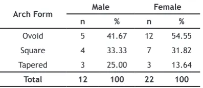
The study was conducted towards the total of 34 subjects selected using the random sampling method. The results obtained throughout this study were presented in Table 1 to Table 4. Table 1 showed that the ovoid arch form was the most
common type of arch found in male and female
respectively. The tapered arch form was the least common type of arch found in both genders, with the value of 25.00% in male and 13.64% in female, while the square arch form was found as much as 33.33% in male and 31.82% in female.
The mean and SD (standard deviation), of unpaired (independent) sample diferences, and also the t-value and p-value of the maxillary central incisors to incisive papilla (CI-IP) distance were described in Table 2 to Table 4. Table 2 showed the statistical analysis of data taken from male and female subjects. The CI-IP distance of male sample was ranged from 7.62-9.90 mm with the average of 9.02 mm and standard deviation of ±0.64. The CI-IP distance of female sample was ranged from 7.65-9.30 mm with the average of 8.68 mm and standard deviation of ±0.36. The average value of male samples was higher than the
average value of female samples. A low standard
deviation indicated that the data points tend to
be very close to the average value.
Table 3 and Table 4 showed the measurement of central incisor to incisive papilla (CI-IP) distance based on gender and arch form. The square arch form had the minimum CI-IP distance (ranged from 7.62-8.89 mm with the average of 8.49 in male, and ranged from 7.65-8.88 mm with the average of 8.42 mm in female). The tapered arch form had the maximum CI-IP distance (ranged from 9.80-9.90 mm with the average of 9.86 in male, and ranged from 8.88-9.30 mm with the average from 9.06 mm in female). The ovoid arch form had the CI-IP distance in the range of 8.85-9.22 mm with the average of 8.97 mm in male, and in the range of 8.25-9.16 mm with the average of 8.73
in female.
Table 1. Frequency distribution of diferent arch forms
Arch Form Male Female
n % n %
Ovoid 5 41.67 12 54.55
Square 4 33.33 7 31.82
Tapered 3 25.00 3 13.64
Total 12 100 22 100
Table 2. CI-IP distance based on gender
Male Female
Range Mean ± SD Range Mean ± SD t-value p-value
7.62-9.90 9.02 ± 0.64 7.65-9.30 8.68 ± 0.36 1.74 >0.05 Reference CI-IP = Maxillary central incisors-incisive papilla; SD Standard deviation
Table 3. CI-IP distance based on gender and arch form
Male Female
Arch form Range (mm) Mean±SD (mm) Range (mm) Mean±SD (mm)
Ovoid 8.85-9.22 8.97±0.16 8.25-9.16 8.73±0.24
Square 7.62-8.89 8.49±0.60 7.65-8.88 8.42±0.44
Tapered 9.80-9.90 9.86±0.05 8.88-9.30 9.06±0.22
Table 4. CI-IP distance based on arch of both gender and arch form
Male Female t-value p-value Mean ± SD (mm) Mean ± SD (mm)
DISCUSSION able to provide meaningful guidelines for maxillary anterior teeth arrangement while in prosthodontic procedures for patients with similar arch forms. The upper jaw impressions of as much as 34 dentate individuals with diferent arch form were taken. The measurements were performed using a digital caliper, and all the data has been
interpreted.
The result of this study showed that the ovoid arch form was the most common type of arch form found in both male and female (41.67% and 45.55%). The square arch forms were the second type found after the ovoid forms in both genders (33.33% in male and 31.82% in female). The tapered arch forms were found as much as 25.00% in male and 13.64% in female. Zia et al.3
conducted a study on 150 samples and also found that ovoid arch form was the most common type of arch form among Pakistani population (57.30% in male and 68.00% in female). However, Nakatsuka10
found that the most frequent arch form among Japanese was the round square. Kook11 had studied
the diference between Korean (Mongoloid race) and North American (Caucasian race) populations and found that in the Caucasian population, the tapered arch form was predominated and the square arch form was predominated in the Korean population, and the Korean arches tended to be larger and deeper than Caucasian. Gafni et al.12
stated that the most frequent arch form of the Israeli group was found to be the ovoid form, as opposed to the North American group with the
tapered form as the predominant form. The reason
for these results was because the North American population had statistically signiicant lower arch widths and higher arch depths compared to the
Israeli population.
The average CI-IP distance in both male and female subjects regardless of their arch forms were 9.02 mm and 8.68 mm respectively. The statistical analysis (unpaired sample t-test) result revealed that there was no signiicant
diference (p>0.05) in CI-IP distance in male and female subjects with ovoid, square and tapered arch forms (p>0.05). This result meant that gender factor was an irrelevant factor of the CI-IP distance. This study was consistent with the indings of an earlier study stated that there was no signiicant diference between the CI-IP distance of both male and female regardless of
their arch form.5,3,13,14 However, a study conducted
by Seok15 and more recent study by Simanungkalit9
were contradicted with this study, which found that there was a signiicant diference between the CI-IP distance of male and female subjects. In the research conducted by Simanungkalit discovered that in Mongoloid race, there was a signiicant diference of CI-IP distance between male and female (p=0.036). However, in Caucasia race, there was no signiicant diference between male and female (p=0.0226).
In the ovoid arch form subjects, the average CI-IP distance in male was diferent from female, which was 8.97 mm and 8.73 mm respectively. The statistical analysis (unpaired sample t-test) result revealed that there was a signiicant diference (p<0.05) in the CI-IP distance between male and female subjects with ovoid arch form. In subjects with square arch form, a slight diference in the average value was observed (8.49 mm in male and 8.42 in female). The statistical analysis result of the p-value resulted in p>0.05, meant that there was no signiicant diference in the CI-IP distance between male and female subjects with square arch form. In the tapered arch form subjects,
the statistical analysis result revealed that there
was a signiicant diference between male and female (p<0.05) with the average value of 9.86 mm and 9.06 mm respectively. These results were consistent with the research conducted by Zia et
al.3 In their research, they measured the distance
between the mesial edges of the maxillary central incisors to the posterior border of the incisive papilla in dentate individuals with diferent arch forms. They discovered that individuals with ovoid and tapered arch forms showed a signiicant diference in the CI-IP distance between males and females (p<0.05). In subjects with square forms, however, a slight diference in was found (no signiicant diference).
The distance from the center of the incisive
central incisor was ranged between 7.18-11.51 mm, with the average of 9.21 mm.16 In the
research conducted by Fu et al. in 2007 amongst young adults in Taiwan discovered that the mesio-labial incisal edge of the upper central incisor was 7.30 ± 0.64 mm anterior to the center of the
incisive papilla.17 Elfadil’s study in 2008 measured
the distances from the labial surface of the
central incisor to the center and posterior point of the incisive papilla.14 The data obtained suggested
that the average distance of 12.4 mm when the posterior point of the incisive papilla was used as the reference point and 8.93 mm when the center of the papilla was used as the reference point.
Ortman and Tsao stated that the distance of the anterior part of the maxillary central incisors
and the posterior of the incisive papilla was 12.45
mm.18 Also, Grave and Becker in 1987 have proved
this similar measurement, which was as much as 12-13 mm.19 Lassila et al. discovered that the
measurement of the anterior part of the maxillary central incisors and the posterior of the incisive
papilla was 12 mm. The measurement methods of all these studies were a two-dimensional
method.20 However, Park et al. in 2007 suggested
similar results (11.96 ± 1.37) mm with the three-dimensional measurement method, with a virtual
model of the maxillary anterior teeth and incisive
papilla. The results of these studies using both two and three-dimensional measuring methods showed that both methods were reliable.21
Chalsuthipan and Boonsiri investigated the relationship between the incisive papilla,
maxillary central incisors, and canines in the Thai
population. As much as 360 selected maxillary models were analyzed in their study.22 In this
study, we found that the vertical distance from
the most distal point of the incisive papilla to the
incisal edge of the central incisors was ranged from 6.94-7.23 mm with the average of 7.08 mm. Guldag et al. in 2008 discovered that the average vertical distance between the maxillary central
incisors and the midpoint of the incisive papilla on
the stone casts was 6.70 ± 0.81 mm. The vertical distance was ranged from 5.51-8.89 mm.23
The average value variation may caused by the reference point diferences on the incisive papilla because some researchers used the most posterior border of the incisive papilla. The most posterior part of incisive papilla was the farthest
from the occlusal plane and the maxillary central incisors.23
In 1993, Lau and Clark studied the
relationship of the incisive papilla to the maxillary central incisors and the canine in the Southern
Chinese population.24 The results showed that
there was a slight diference between the Southern Chinese population and other most ethnic groups. The guidelines used was the incisive papilla as a
reference for the setting of denture construction
recommended for Caucasians, and was able to applied to the Southern Chinese patients. Elfadil in 2008 also mentioned that in his study amongst Sudanese population showed insigniicant diferences compared to other ethnic groups.14
Overall, the distance from the center of
incisive papilla to the labio-incisal of the
one-third of central incisors among the Malaysian
Malay students was ranged between 7.65-9.90 mm, with the average of 8.77 mm. There was no signiicant diference in the CI-IP distance between male and female subjects regardless of their arch form. Individuals with ovoid and tapered arch forms showed a signiicant diference in the CI-IP distance between male and female (p<0.05). Meanwhile, individuals with square arch forms showed no signiicant diference in the CI-IP distance between male and female (p>0.05).
The guidelines used was the incisive
papilla as a reference for the setting of denture
construction recommended for Caucasians that was able to applied for Mongoloid patients.24 However,
the wax rims should be modiied intraorally to
incorporate individual characteristics, and the
anterior teeth should be arranged on the modiied wax rims.23 The bases carried the occlusion rims
should be rigid and stable. The upper rim was modiied to give the correct lip support. The incisive papilla provides a useful biometric guide to the prominence of the rim. Patients’ wishes,
or previous satisfactory dentures, may dictate
otherwise.
CONCLUSION
REFERENCES
1. Owen CP. Fundamentals of Removable Partial Dentures. 2nd ed. Cape Town: Juta Academic;
2000. p. 1.
2. McCord JF, Grant AA, Youngson CC, Watson RM,
Davis DM. Missing Teeth: A Guide to Treatment Options. London: Churchill Livingstone; 2003.
p. 1.
3. Zia M, Azad AA, Ahmed S. Comparison of distance between maxillary central incisors
and incisive papilla in dentate individuals
with diferent arch forms. J Ayub Med Coll Abbottabad. 2009 Oct-Dec;21(4):125-8. 4. West Virgina University Libraries [homepage
on internet]. Morgantown: Davis BA. 2005.
Anatomical measurements of orthodontic and
edentulous casts to determine the width of the maxillary anterior teeth. [cited 2011 Apr]; [about 1 screen]. Available from: http:// wvuscholar.wvu.edu:8881//exlibris/dtl/ d3_1/apache_media/L2V4bGlicmlz2R0b/ 5. Rajiv Gandhi University of Health Sciences
[homepage on internet]. Bangalore: Qazi SN. 2006. Nasal width, intercanine distance
and incisive papilla as guides for selection and arrangement of maxillary anterior
teeth. [cited 2011 Apr]; [about 2 screens]. Available from: http://shazana%20nasal%20 papilla&ei=57oToaiHMS3ren2IjOCg&usg=AF/. 6. International & American Dental Research
[homepage on internet]. Alexandria, VA: Kathree A, Wilson VJ, Solomons CS. 2006. Biometric relationship between the incisive papilla and maxillary central incisors. [cited 2011 Jun]; [about 2 screens]. Available from: http://iadr.confex.com/iadr/safdiv03/ liminaryprogram/abstract65559.htm/.
7. Al-Khateeb SN, Abu Alhaija ES. Tooth size
discrepancies and arch parameters among
diferent malocclusions in a Jordanian sample. Angle Orthod. 2006 May;76(3):459-65.
8. Burris BG, Harris EF. Maxillary arch size and shape in American blacks and whites. Angle Orthod. 2000 Aug;70(4):297-302.
9. Simanungkalit P. Perbedaan jarak antara gigi
insisivus sentralis rahang atas dengan papila
insisivum berdasarkan ras dan jenis kelamin pada mahasiswa FKG USU Angkatan 2007 dan 2008 [minor thesis]. Medan: USU. 2010.
10. Nakatsuka M, Iwai Y, Jue SS, Oh SH, Guo L, Tominaga Y, et al. A morphological study on the classiication of maxillary dental arches. Okajimas Folia Anat Jpn. 2004 May;81(1):5-13.
11. Kook YA, Nojima K, Moon HB, McLaughlin
RP, Sinclair PM. Comparison of arch forms between Korean and North American white
populations. Am J Orthod Dentofacial Orthop.
126:680–6.
12. Gafni Y, Tzur-Gadassi L, Nojima K, McLaughlin
RP, Abed Y, Redlich M. Comparison of arch forms between Israeli and North American white populations. Am J Orthod Dentofacial Orthop. 2004 Dec;126(6):680-6.
13. Amin WM, Taha ST, Al-Tarawneh SK, Saleh MW, Ghzawi A. The relationships of the maxillary
central incisors and canines to the incisive
papilla in Jordanians. J Contemp Dent Pract. 2008 Jul 1;9(5):42-51.
14. Idris RAE. The incisive papilla as a guide for anterior teeth arrangement [thesis]. Khartoum: University of Khartoum. 2002. 15. Seok HY, Wan SS. A study on the positioning
of the maxillary central incisor in Korean. J Korean Acad Prost. 1995;33:85-97.
16. Huang SJ, Chou TM, Lee HE, Wu YC, Yang YH, Ho CD, et al. Exploring the Distance between Upper Central Incisor Edge and Incisive Papilla in Taiwanese Population. Taiwan J Oral Med Health Sci. 2004;20:4-10.
17. Fu PS, Hung CC, Hong JM, Wang JC, Tsai CF, Wu YM. Three-dimensional relationship of the
maxillary anterior teeth to the incisive papilla
in young adults. Kaohsiung J Med Sci. 2007 Oct;23(10):519-25.
18. Ortman HR, Tsao DH. Relationship of the
incisive papilla to the maxillary central
incisors. J Prosthet Dent. 1979 Nov;42(5):492-6.
19. Grave AM, Becker PJ. Evaluation of the incisive
papilla as a guide to anterior tooth position. J
Prosthet Dent. 1987 Jun;57(6):712-4.
20. Lassila LV, Klemetti E, Lassila VP. Position of
teeth on the edentulous atrophic maxilla. J
Oral Rehabil. 2001 Mar;28(3):267-72.
21. Park YS, Lee SP, Paik KS. The three-dimensional
relationship on a virtual model between the
maxillary anterior teeth and incisive papilla. J
Prosthet Dent. 2007 Oct;98(4):312-8.
central incisor and canine to incisive papilla.
Chulalongkorn Univ Dent J. 1993;16:29–40. 23. Guldag MU, Sentut F, Buyukkaplan US.
Investigation of vertical distance between
incisive papilla and incisal edge of maxillary
central incisors. Eur J Dent. 2008 Jul 2:161–6. 24. Lau GC, Clark RF. The relationship of the
incisive papilla to the maxillary central