Suppression of Autophagy Dysregulates the
Antioxidant Response and Causes Premature
Senescence of Melanocytes
Cheng-Feng Zhang
1,2, Florian Gruber
1,3, Chunya Ni
1,2, Michael Mildner
1, Ulrich Koenig
1, Susanne Karner
1,
Caterina Barresi
1, Heidemarie Rossiter
1, Marie-Sophie Narzt
1,3, Ionela M. Nagelreiter
1,3,
Lionel Larue
4,5,6, Desmond J. Tobin
7, Leopold Eckhart
1and Erwin Tschachler
1Autophagy is the central cellular mechanism for delivering organelles and cytoplasm to lysosomes for degradation and recycling of their molecular components. To determine the contribution of autophagy to melanocyte (MC) biology, we inactivated the essential autophagy geneAtg7specifically in MCs using the Cre-loxP system. This gene deletion efficiently suppressed a key step in autophagy, lipidation of microtubule-associated protein 1 light chain 3 beta (LC3), in MCs and induced slight hypopigmentation of the epidermis in mice. The melanin content of hair was decreased by 10–15% in mice with autophagy-deficient MC as compared with control animals. When culturedin vitro, MCs from mutant and control mice produced equal amounts of melanin per cell. However, Atg7-deficient MCs entered into premature growth arrest and accumulated reactive oxygen species (ROS) damage, ubiquitinated proteins, and the multi-functional adapter protein SQSTM1/p62. Moreover, nuclear factor erythroid 2–related factor 2 (Nrf2)–dependent expression of NAD(P)H dehydrogenase, quinone 1, and glutathione S-transferase Mu 1 was increased, indicating a contribution of autophagy to redox homeostasis in MCs. In summary, the results of our study suggest that Atg7-dependent autophagy is dispensable for melanogenesis but necessary for achieving the full proliferative capacity of MCs.
Journal of Investigative Dermatology(2015)135,1348–1357; doi:10.1038/jid.2014.439; published online 13 November 2014
INTRODUCTION
Autophagy is an evolutionarily conserved mechanism by which cells degrade and recycle damaged proteins and subcellular organelles. It is critical for the maintenance of cell metabolism during starvation, cellular remodeling during development and differentiation, as well as anti-bacterial and antiviral defense (Mizushima, 2007; Deretic,
2011; Mizushima and Komatsu, 2011). Macroautophagy is the predominant mode of autophagy (Mizushima and Komatsu, 2011) and will hereafter be referred to as ‘‘autophagy’’. Chaperone-mediated autophagy and microautophagy are alternative mechanisms that mediate the degradation of subcellular substrates in a largely nonredundant manner (Mizushima and Komatsu, 2011; Santambrogio and Cuervo, 2011). Autophagy is controlled by a defined set of gene products that have been reviewed (Mizushima and Levine, 2010; Yang and Klionsky, 2010). The Atg7 protein is essential for conversion of the cytosolic form of microtubule-associated protein light chain 3 (LC3), LC3-I, into its lipidated form, LC3-II—an obligatory step of autophagy. LC3-II associates with isolation membranes to form autophagosomes and interacts with p62/SQSTM1, the adapter protein that targets cargo for selective autophagosomal degradation. Inactiva-tion of Atg7 disrupts autophagy by preventing lipidaInactiva-tion of LC3 and thus autophagosome formation. Germline inactiva-tion of Atg7 leads to perinatal lethality that correlates with decreased levels of amino acids in the neonatal starvation period (Komatsuet al., 2005). Targeted inactivation of Atg7 in the central nervous system results in neurodegeneration and death (Komatsuet al., 2006); liver-targeted inactivation leads to hepatomegaly and accumulation of abnormal organelles in liver cells (Komatsuet al., 2005).
1Research Division of Biology and Pathobiology of the Skin, Department of Dermatology, Medical University of Vienna, Vienna, Austria;2Department of Dermatology, Huashan Hospital, Fu Dan University, Shanghai, China; 3Christian Doppler Laboratory for Biotechnology of Skin Aging, Department of
Dermatology, Medical University of Vienna, Vienna, Austria;4Institut Curie, Centre de Recherche, Developmental Genetics of Melanocytes, Orsay, France; 5CNRS UMR3347, Orsay, France;6INSERM U1021, Orsay, France and 7Centre for Skin Sciences, School of Life Sciences, University of Bradford,
Bradford, UK
Correspondence: Florian Gruber or Erwin Tschachler, Department of Dermatology, Medical University of Vienna, Wa¨hringer Gu¨rtel 18-20, Vienna A-1090, Austria. E-mail: [email protected] or
Received 10 April 2014; revised 22 September 2014; accepted 24 September 2014; accepted article preview online 7 October 2014; published online 13 November 2014
Melanocytes (MCs) are neural crest-derived pigment-produ-cing cells, which migrate into the skin during embryogenesis and colonize the epidermis and hair follicles (Hearing, 1993; Lucianiet al., 2011). In mice MCs are present in the epidermis at all body sites only around the time of birth (Hirobe, 1984). After the first postnatal week MCs are mainly present within the dermis and the hair follicles in hairy skin, whereas in ear and tail skin epidermal MCs persist throughout life. Activated MCs produce melanin, which is transferred to differentiating hair keratinocytes (KCs) and, on tail and ear skin, also to interfollicular KC (Slominski et al., 2005). Proliferation, migration, and differentiation of mouse MC during develop-ment are regulated by numerous genetic and epigenetic factors (Hirobe, 2011; Bonaventure et al., 2013). Small interfering RNA-based screens identified autophagy genes as having an impact on melanogenesis and heterozygosity for the autophagy regulator beclin 1 results in altered fur color of mice (Ganesan et al., 2008). In addition, members of the autophagic machinery have been proposed to have a role in the formation and maturation of melanosomes (Ho and Ganesan, 2011; Kim et al., 2014), however, direct evidence for this claim is lacking.
Here, we investigated whether or not autophagy has a role in melanogenesis and MC homeostasis by crossing mice carrying a floxed Atg7 gene (Komatsu et al., 2005) with Tyrosinase-Cre mice (Delmas et al., 2003). We demonstrate that the disruption of autophagy in MCs does not prevent melanogenesis, although it leads to a slight but significant reduction in melanin in both hair and tail epidermis. Cultured autophagy-deficient MC showed a strongly reduced proliferative capacity and became prematurely senescent. At the molecular level, the lack of autophagy was associated with the accumulation of p62/SQSTM1 both in vivoandin vitro, the upregulation of nuclear factor E2–related factor 2 (Nrf2) signaling but also reactive oxygen species (ROS) and lipid oxidation. The results of this study suggest that autophagy is dispensable for melanogenesis but important for the control of the Nrf2 stress response and sustained proliferation of MC.
RESULTS
Autophagy is constitutively active in normal human and murine MCs
To explore whether autophagy is active in MC, we investigated the effect of the autophagy inducer rapamycin on the lipidation status of LC3 in normal human and murine MC by western blot analysis. As shown in Figure 1, both types of MCs contained high levels of autophagy-associated, lipidated LC3-II (human 70%±14% of total LC3, n¼4; mouse 58%±4%, n¼3)
already without stimulation, indicating that autophagy is con-stitutively active in MC. Addition of rapamycin to the culture medium caused only a slight additional increase in LC3-II in MC (human 74%±23% and mouse 70%±10%, respectively,
the latter induction being significant withPo0.05).
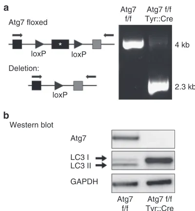
Tyr::Cre-mediated deletion of Atg7 efficiently suppresses autophagy in MCs
To specifically inactivate theAtg7gene in pigment cells, mice carrying a floxed allele of Atg7 (Komatsuet al., 2005) were
mated to the Tyr::Cre mouse line, which triggers recombination in cells of the MC lineage starting from 9.5 days of gestation (Puiget al., 2009). As the Tyr::Cre transgene is located on the X chromosome, hemizygous males and homozygous females were used as sources of MC forin vitro experiments. For in vivo analyses, only male Atg7 f/f (Xtg/Y) mice (here referred to as Atg7 f/f Tyr::Cre) were used in comparison to male Atg7 f/f (Xo/Y) mice (here referred to as Atg7 f/f). Efficient inactivation of theAtg7gene in MC isolated from Atg7 f/f Tyr::Cre mice was confirmed by PCR of genomic DNA (Figure 2a). Western blot analysis confirmed that ATG7 protein was not produced in MC derived from ATG7 f/f
Rapamycin LC3 I LC3 II
GAPDH
hMC mMC
–
– + +
Figure 1. Autophagy is constitutively active in normal human and murine melanocyte (MC).Primary human (hMC) and mouse (mMC) MCs were subjected to rapamycin treatment at 0.5 ugml1for 1 hour. Cell lysates of rapamycin-treated and -untreated cells were subjected to western blot analysis for microtubule-associated protein light chain 3 (LC3) and GAPDH was used as a loading control. Bands corresponding to LC3-I and LC3-II (indicative of active autophagy) are marked by arrows.
Atg7 floxed
Atg7 f/f
4 kb
2.3 kb
loxP loxP
loxP Deletion:
*
Western blot
Atg7
LC3 I LC3 II
GAPDH
Atg7 f/f Tyr::Cre
Atg7 f/f
Atg7 f/f Tyr::Cre
Tyr::Cre animals (Figure 2b, uppermost panel). The conversion of LC3-I to LC3-II was completely blocked in MC of ATG7 f/f Tyr::Cre mice, whereas both LC3-I and LC3-II were readily detected in MC of Atg7 f/f mice (Figure 2b middle panel). Taken together, these data demonstrate that the autophagic machinery was efficiently disrupted in MC of Atg7 f/f Tyr::Cre mice.
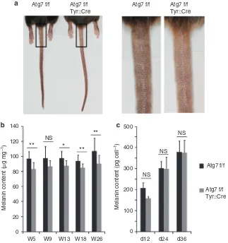
Inactivation of ATG7-dependent autophagy in MC leads to decreased pigmentation of dorsal hair and tail epidermisin situ but does not affect the relative melanin content of cultured MC
Atg7 f/f Tyr::Cre mice were viable and as fertile as Atg7 f/f mice. The pigmentation of tail skin of Atg7 f/f Tyr::Cre mice was consistently lower compared with that of Atg7 f/f mice (Figure 3a) and to a lesser extent at the feet (not shown), whereas, by visual inspection, the coat color of Atg7 f/f Tyr::Cre mice was indistinguishable from that of Atg7 f/f mice. However, photometric quantification of melanin in extracts from shaved hair of the back showed that the melanin content of hair from Atg7 f/f Tyr::Cre mice was about 10–15% lower
compared with that of Atg7 f/f mice (Figure 3b, *Po0.05, **Po0.01). MC numbers per area were consistently but not significantly (P¼0.1) lower in the epidermis of autophagy-deficient mice (Supplementary Figure S1 online).
Ultrastructural investigation revealed that both MC and KC of tail skin contained mature melanosomes, irrespective of the presence of Atg7 in MC, strongly suggesting that autophagy is dispensable for melanosome formation, maturation, and transfer (Supplementary Figure S2 online). Of note, some MCs of mutant but not control mice contained misshapen, swollen, and possibly disintegrating mitochondria.
When isolated and cultured in vitro autophagy-deficient and autophagy-competent MC contained similar amounts of melanin (Figure 3c), and electron microscopy confirmed the presence of mature melanosomes in cells of both genotypes (Supplementary Figure S1 online). However, mutant cells exhibited increased melanosome-associated vacuolation. Tyrosinase and Mitf (microphthalmia-associated transcription factor) mRNAs were expressed at comparable levels in both the absence and presence of autophagy (data not shown).
NS **
** **
*
500
400
300
200
100
0
d12 d24 d36
Atg7 f/f Atg7 f/f
NS
NS
NS
Melanin content (pg cell
–1
)
Melanin content (
µ
g mg
–1
)
Atg7 f/f Tyr::Cre Atg7 f/f
Tyr::Cre
Atg7 f/f Atg7 f/f Tyr::Cre
140
120
100
80
60
40
20
0
W5 W9 W13 W18 W26
Taken together, these results indicate that MCs need Atg7-dependent autophagy to achieve full pigmentation of hair and epidermis but also that melanogenesis can proceed in the absence of an intact autophagy system.
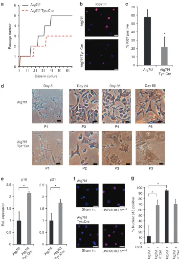
Autophagy deficiency results in decreased proliferation and premature senescence of cultured MC
Next, we characterized the impact of autophagy deficiency on MC in cell cultures. MC derived from Atg7 f/f Tyr::Cre mice showed an altered growth profile as compared with MC from Atg7 f/f mice (Figure 4a). In fact, Atg7-deficient MC stopped proliferation as early as after the third passage at around the fifth week in culture, whereas normal MC continued prolifera-tion and could be maintained up to passage 5. Analysis of Ki-67 expression on day 23 of culture confirmed that the proportion of proliferating cells was strongly decreased in cultures of Atg7 f/f Tyr::Cre MC (Figure 4b and c). In addition, mutant MC started to change their morphology early in the second passage at around week 3 and acquired senescent morphotypes with distended cytoplasms. The premature senescent phenotype became even more prominent at the end of passage 2 and beginning of passage 3 (Figure 4d). By contrast, similar changes in cell morphology were observed in control MC only at around 9 weeks in culture, when the cells had been passaged five times. The changes in the morphology of autophagy-deficient MC were accompanied by significantly higher expression of p16Ink4a (CDKN2A) and p21 (CDKN1A) mRNAs (Figure 4e, Po0.05), and a significantly higher proportion of mutant cells exhibited nuclear p16Ink4a protein as determined by immunofluorescence staining (Figure 4f and g). Nuclear translocation of p16Ink4a increased upon UVB irradiation (20 mJ cm2) (Piepkorn, 2000) in
autophagy-competent cells, whereas it could not be elevated above the high basal level in autophagy-deficient cells (Figure 4f and g). Taken together these data suggest that the absence of autophagy leads to premature senescence of MC.
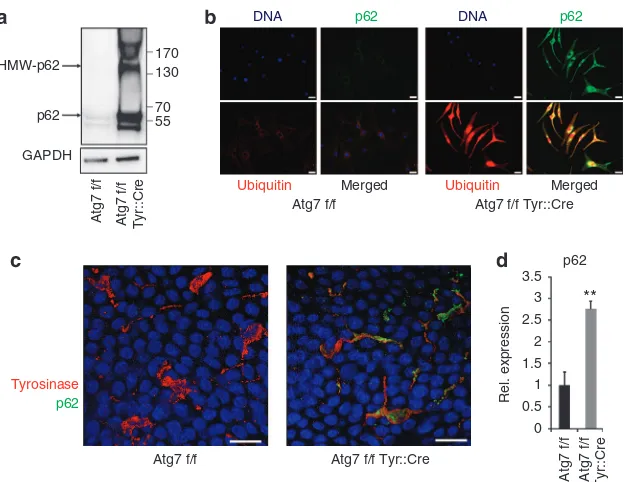
Autophagy deficiency leads to accumulation of high-molecular-weight protein aggregates containing p62/SQSTM1
As autophagy controls cellular processes via the modulation of distinct multifunctional proteins such as the autophagy cargo adapter protein p62/SQSTM1 (Pankiv et al., 2007), we determined the abundance of p62/SQSTM1 and the activity of p62-related processes in normal and Atg7-deficient MC. Immunoblotting showed massive accumulation of free p62/ SQSTM1 and high-molecular-weight p62-positive protein species in lysates of autophagy-deficient MC indicative of p62 oligomerization (Kirkin et al., 2009; Riley et al., 2010) (Figure 5a). Double immune-labeling of cultured cells showed strong increase in both p62/SQSTM1 and ubiquitinylated proteins in mutant MC (Figure 5b). In the cytoplasm, but not the nucleus, these two proteins largely co-localized. Accumu-lation of p62/SQSTM1 also occurredin vivoas demonstrated by double labeling of MC for p62/SQSTM1 and tyrosinase in epidermal sheet preparations of tail skin (Figure 5c). The massive accumulation of p62/SQSTM1 protein was accom-panied by an upregulation of p62/SQSTM1 mRNA in cultured MC (Figure 5d), suggesting that not only a block in the
degradation of the protein but also enhanced transcription contributed to the increase in p62/SQSTM1 protein in autophagy-deficient MC.
Persistently enhanced Nrf2 activity does not prevent increased ROS formation and lipid oxidation in autophagy-deficient MCs
6
4
3
1
0
1 11 21 31
Day 8
P1 P3 P4 P5
P3 P3
100
80
70
60
50
40
30
20
10 *
% Nuclear p16 positive
*
0
UVB – – + +
90 P2
P1
p16
* *
Rel. expression
2.5
1.5
1
0.5
0 2
2.5
1.5
1
0.5
0 2
p21 Atg7f/f
Atg7f/f
Atg7f/f
Atg7f/f
KI67 IF
70
60
50
40
30
20
*
10
0
Atg7f/f
Atg7f/f
% KI67 positive
Atg7f/f Tyr::Cre
Atg7f/f Tyr::Cre
Atg7f/f Tyr::Cre
Atg7f/f Tyr::Cre
Atg7f/f Tyr::Cre
Sham irr.
Sham irr.
UVB20 mJ cm–2
UVB20 mJ cm–2
Atg7f/f Tyr::Cre
Atg7f/f Atg7f/f
Tyr::Cre Atg7f/f Atg7f/f
Tyr::Cre Atg7f/f Atg7f/f Tyr::Cre
Passage number
Day 36 Day 65
Day 24 41 51 61
Days in culture 2
5
Figure 4. Autophagy deficiency leads to decreased proliferation and premature senescence in cultured primary melanocytes (MCs).(a) Proliferation of Atg7 f/f and Atg7 f/f Tyr::Cre cells, passage number plotted against days in culture. (b) Immunofluorescence analysis of Ki-67 expression (red) in the MC from Atg7 f/f and Atg7 f/f Tyr::Cre mice. Bar¼10mm and (c) quantification (n¼5). (d) Representative photos of MC from Atg7 f/f and Atg7 f/f Tyr::Cre mice at the age of day 8, day 24, day 36, and day 65. Phase contrast micrographs, bar¼250mm. (e) Primary autophagy-deficient and -competent MCs were cultured for 22 days and expression of p16Ink4a and p21 was determined using quantitative reverse transcriptase in real time–PCR (n¼3; relative quantification, normalized to expression of beta-2-microglobulin; error bars indicate±SD * indicatesPo0.05 (t-test)). (f,g) Primary autophagy-deficient and -competent MCs were cultured for 22 days, sham irradiated or irradiated with 20 mJ cm2of UVB, and subjected to immunofluorescent staining for p16Ink4a. Bar
1-palmitoyl-2-linoleoyl-sn-glycero-3-phosphocholine (16:0-18:2 PC, m/z 758) were significantly increased relative to their unoxidized precursors in lipid extracts from cultured autophagy-deficient MC, whereas the levels of unoxidized di-palmitoyl-sn-glycero-3-phosphocholine (16:0-16:0 PC m/z 734) was not significantly changed (Figure 6c). These data indicate that deficiency in autophagy, in addition to premature senescence, results in increased oxidative stress in MC, despite the observed increase in Nrf2 activation.
DISCUSSION
Proteins of the autophagic machinery, including beclin 1 (Funderburk et al., 2010), ULK1 (Kalie et al., 2013), and LC3-II have been previously suggested to participate in melanogenesis (Ganesan et al., 2008), and autophagy has been hypothesized to also participate in melanosome formation and maturation (Ho and Ganesan, 2011). Here we have inactivated the central regulator of autophagy, Atg7, specifically in MC and studied pigmentation and MC homeostasis in transgenic mice and MC isolated from these mice. Our findings that mutant mice showed only a slight, although consistent, reduction in hair and skin pigmentation and that mutant MCs are able to produce mature melanosomes suggest that autophagy is neither essential for melanin production by MC nor for the delivery of melanin to hair and epidermal KC. Thus, the results of this study establish that, at least under standard animal care conditions, autophagy is not essential for MC survival and function in vivo.
When analyzing isolated MC in tissue culture we could confirm that the absence of autophagy neither impacted pigment production nor formation and maturation of melano-somes. Murase and coworkers, studying human cellsin vitro, have recently suggested that KCs contribute to skin pigmenta-tion by degrading melanosomes (Muraseet al., 2013). As we have previously reported, mice deficient in KC autophagy did not show differences in skin or hair color (Rossiter et al., 2013), and in preliminary assessment of mice in which Atg7 was deleted in both MC and KC (Atg7 f/f Tyr::Cre, K14::cre) we found no differences with regard to coat and tail skin pigmentation to animals with autophagy-deficient MC only (not shown). Therefore, a mechanism by which autophagy in KC contributes to skin color as suggested by Murase and coworkers (Muraseet al., 2013) is not operantin vivoin mice. By contrast, autophagy-deficient MC isolated from the skin of newborn mice differed considerably from autophagy-competent MC in their proliferative capacity. In fact, Atg7-deficient MC virtually stopped proliferation during the 3rd passage in vitro, acquired a senescent morphology and expressed significantly higher levels of p16ink4 mRNA and proteins compared with control cells. This finding is in line with earlier reports that have shown that autophagy is involved in the regulation of the life span of cells (Vellai, 2009; Lionakiet al., 2013). In cultured fibroblasts, senescence is associated with a decrease in the protein levels of S6K1, 4E-BP1, beclin 1, and Atg7 (Kang et al., 2011). In turn, the impairment of autophagy induces premature senescence, supposedly via mechanisms that involve aberrant oxidation HMW-p62
p62
170 130
70 55
Merged Merged
Atg7 f/f
Atg7 f/f
Atg7 f/f Tyr::Cre
Atg7 f/f Tyr::Cre
p62
**
Rel. expression
3.5
3 2.5 2
1.5 1 0.5 0
Atg7 f/f Atg7 f/f Tyr::Cre Ubiquitin Ubiquitin
DNA p62
p62
DNA p62
GAPDH
Atg7 f/f Atg7 f/f Tyr::Cre
Tyrosinase
Figure 5. Autophagy deficiency leads to accumulation of high-molecular-weight protein aggregates containing p62.(a) Lysates of primary melanocytes (MCs) from Atg7 f/f and Atg7 f/f Tyr::Cre mice were subjected to western blot analysis for p62. (b) Immunofluorescence analysis for p62 (green) and (poly-)ubiquitinated protein (red) in MC isolated from Atg7 f/f and Atg7 f/f Tyr::Cre mice. Bar¼10mm. (c) Epidermal sheet preparations were stained for Tyrosinase (red), p62(green), and nuclei (blue) and analyzed by laser scanning microscopy. Bar¼10mm. (d) Primary deficient and autophagy-competent MCs were cultured for 22 days and subjected to quantitative reverse transcriptase in real time–PCR for p62. Error bars represent mean±SD across
of protein accumulation of misfolded proteins and damaging levels of mitochondrial ROS (Sena and Chandel, 2012). Our finding that autophagy-deficient MC stopped proliferating and acquired a senescent phenotype earlier compared with their autophagy-competent counterparts provides support for a protective role of autophagy for the maintenance of the MC population. However, this in vitrofinding does not translate into a statistically significant difference in the number of epidermal MC between mutant and control animals in vivo.
In addition, the pigmentation maintained in the hair of aging mice showed that autophagy-deficient MCs were able to replenish hair follicles during the hair cycles throughout the life span of the animals. There are several possible explana-tions for the different effects of autophagy suppressionin vitro and in vivo. In culture, the growth medium drives MC to continuous proliferation, whereas in vivo MCs proliferate mainly during the hair cycles (Slominski and Paus, 1993) and after UV exposure(van Schankeet al., 2005). Continuous Hmox1
** NS
Rel. expression
** **
Atg7 f/f
Atg7 f/f
Atg7 f/f Tyr::Cre Atg7 f/f Atg7 f/f Tyr::Cre Atg7 f/f Atg7 f/f Tyr::Cre Atg7 f/f Atg7 f/f Tyr::Cre
Atg7 f/f Tyr::Cre
* *
NS
Normalized rel. intensity
PLPC-OOH/PLPC SLPC-OOH/SLPC DPPC/
PLPC
Nqo1
Gstm1
Hmox1
Gapdh
250
100
50 200
150 2
1.6
1.4
1.2
1
0.8
0.6
0.4
0.2
0 1.8
3.5
2.5
2
1.5
0.5 1
0 3
3.5
2.5
2
1.5
0.5 1
0 3 16
14
12
10
8
6
2
0 4
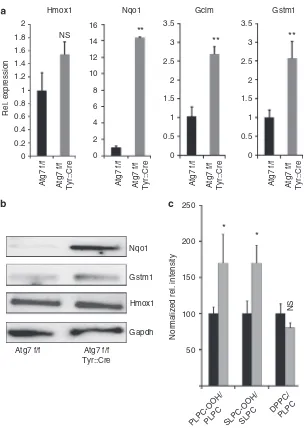
Nqo1 Gclm Gstm1
Figure 6. Autophagy deficiency leads to enhanced expression of nuclear factor erythroid 2–related factor 2 target genes but also to increased phospholipid oxidation.(a) Primary autophagy-deficient and autophagy-competent melanocytes (MCs) were cultured for 22 days and subjected to quantitative reverse transcriptase in real time–PCR for heme oxygenase-1 (Hmox-1), NAD(P)H dehydrogenase, quinone 1 (Nqo-1),g-glutamyl cystine ligase modulatory subunit (Gclm), and glutathione S-transferase Mu 1 (Gstm1). Error bars represent mean±SD (n¼3) and thet-test was performed. **Po0.01; *Po0.05; NS: nonsignificant difference. (b) Lysates of primary MC from Atg7 f/f and Atg7 f/f Tyr::Cre mice were subjected to western blot analysis for NQO-1, GSTM1, HO-1, and GAPDH. (c) Primary autophagy-deficient and autophagy-competent MCs were cultured for 22 days. Relative amounts of
proliferation with increased metabolism in tissue culture might therefore result in an exhaustion of the cellular mechanisms that compensate for the absence of autophagy, whereas such a state may not be reachedin vivoduring the normal life span of mice. Alternatively compensation for the lack of autophagy in MCin vivomight be provided by the surrounding tissue. That such a scenario is conceivable is hinted at by a recent report on a breast cancer tumor model in which serum-starved mesenchymal stem cells activating autophagy supported tumor cell survival by paracrine mechanisms (Sanchezet al., 2011). The recent finding that even aggregated proteins can be transferred to neighboring cells (Guo and Lee, 2014) even opens the possibility that autophagy-deficient MC may dispose of protein aggregates by delivering them to surrounding skin cells. Intriguingly, amyloid-like proteins are present in melanosomes and are consequently delivered to KC (Singh et al., 2010). A potential dependence of autophagy-deficient MC on autophagy in neighboring skin cells will have to be addressed in separate studies.
In addition to the premature cessation of proliferation of autophagy-deficient MC, we have here identified a significant dysregulation of a key stress response system of the cell––i.e., the Nrf2-dependent expression of antioxidant genes. Nrf2 provides adaptive cytoprotection against various ROS by inducing the expression of multiple antioxidant proteins and detoxifying enzymes (Kobayashi and Yamamoto, 2005). However, activation of Nrf2 signaling also requires tight control as evidenced by the observations that sustained high-level activity of Nrf2 in epidermal KC significantly disturbs tissue homeostasis (Schafer et al., 2012). Hyperactivation of Nrf2 in autophagy-deficient liver, brain, and lung tissue (Rileyet al., 2010; Inoueet al., 2011) results in deleterious damage that can be reverted by deleting Nrf2. Recently, a link between Nrf2 and autophagy was uncovered, as the autophagy cargo adapter SQSTM1/p62 is both an activator of Nrf2 activity and Nrf2 target gene (Komatsu et al., 2010). In autophagy-deficient kidney and lung, SQSTM1/p62 accumulation in the cytoplasm competes with Nrf2 for binding to its cytoplasmic anchor KEAP1 and thereby causes enhanced Nrf2 nuclear localization and transcription of its targets, one of them being SQSTM1/p62 itself. Whether this regulatory loop is indeed active in MC requires verification in genetic model systems regulating expression of Nrf2 and p62 together with autophagy; however, it is strongly suggested to be the case by our finding that not only SQSTM1/p62 protein but also mRNA is increased in mutant MC. The Nrf2 pathway contributes to the regulation of the antioxidant response in MC (Marrot et al., 2008), whereas Nrf2 signaling is downregulated after UVB irradiation in MC if not counteracted by alphaMSH(Kokot et al., 2009). Our finding that autophagy-deficient MCs have increased expres-sion levels of several Nrf2 target genes indicates aberrant activation of Nrf2 signaling in these cells. The repertoire of dysregulated Nrf2 target genes seems to be determined by the cell type, as heme oxygenase-1, which is significantly induced in deficient KC, is not overexpressed in autophagy-deficient MC, and this finding merits further investigation. The increased expression of cellular antioxidants does, however,
not result in a reduction in ROS but rather an increase in phospholipid hydroperoxide levels as markers of oxidative stress. As we have observed earlier that also autophagy-deficient KCs display also enhanced lipid oxidation (Zhao et al., 2013), these findings strengthen the concept that functioning macroautophagy may contribute to removal or prevention of excess oxidized lipids. In the case of the MC, it can, however, not be ruled out that the oxidized lipids accumulate as a result of beginning cellular senescence (Girotti and Kriska, 2004).
Our finding that autophagy-deficient MCs undergo prema-ture senescence in vitro resembles the state that has been described for MC from patients with vitiligo, and the role of autophagy in this disease therefore deserves investigation: Not only do cultured MCs from vitiligo patients show signs of sublethal oxidative stress, but they also present a pre-senes-cent phenotype, including increased expression and nuclear staining for p16 (Belleiet al., 2013), both strikingly similar to that which we observed in autophagy-deficient MC. The study by Bellei and another study by the same group (Dell’Anna et al., 2010) further described the detection of oxidized membrane lipids in vitiligo MC. This again is comparable to the significant increase in oxidation products of two major membrane phospholipids, which we detected in autophagy-deficient cells. Finally, Nrf2 signaling is dysregulated in human vitiligo, and, whereas high Nrf2 activation and expression of NAD(P)H dehydrogenase, quinone 1 and g -glutamyl cystine ligase modulatory subunit has been observed in lesional versus non-lesional epidermal samples from patients (Natarajan et al., 2010), a recent report showed that vitiligo MC cannot respond properly to oxidative stress and have an impaired heme oxygenase-1 response to hydrogen peroxide (Jianet al., 2014). Our findings are thus compatible with a model in which autophagy-deficient MC and MC in vitiligo share an ineffective antioxidant defense, cellular redox dysregulation, increased membrane lipid oxidation, and premature senescence. However, as autophagy only margi-nally affects pigmentation and MC numbers in unchallenged mouse skin, we can at the present time neither confirm nor exclude that dysregulated autophagy is relevant in vitiligo.
In conclusion, our study challenges hypotheses that auto-phagy has an essential role in melanogenesis or melanin delivery to KC but uncovers a crucial role of autophagy in the control of MC survival and proliferation. Moreover, the discovery of aberrant Nrf2 signaling in autophagy-deficient MC calls for detailed investigations of the cooperative action of autophagy and Nrf2 in controlling ROS levels in MC biology and human pathologies associated with oxidative damage (Schallreuter, 2007; Swalwellet al., 2012).
MATERIALS AND METHODS Mice and genotyping
and Roder, 2005; Colombo et al., 2007). Breeding schemes are detailed in Supplementary Materials and Methods online. Institutional approval for animal material under decree of the Federal Minstry of Science, Research and Economy (Zl. 1712/115-1997/98-2013).
Human and mouse primary MC culture
Normal human epidermal MCs were obtained from Lonza (Basel, Switzerland) and cultured in MC growth medium (MGM-4, Lonza). Primary mouse MCs were prepared from 1- to 5-day-old pups as described (Bennettet al., 1989) with slight modifications. Briefly, dorsal skin was washed three times with 70% ethanol and sterile phosphate-buffered saline for three times and fat removed. The skin was then cut into 3–4 small pieces and incubated in 2 ml dispase II (Roche, Basel, Switzerland; 2.4 U ml–1) in a 60 mm Petri dish at 371C for 1 hour. The epidermis was separated from the dermis by standard sequential dispase (Roche, 2.4 U ml–1) and trypsin/EDTA (Clonetics, San Diego,
CA, 0.25 mg ml–1) digestion. Medium containing fetal calf serum was
used to stop trypsinization, followed by centrifugation (1500 r.p.m., 2 minutes). The pellet was resuspended in MC growth medium.
Methods to assess proliferation and melanin content are detailed in the Supplementary Materials and Methods section online.
Antibody information, primer sequences and standard procedures for western blotting, quantitative reverse transcriptase in real time– PCR, immunofluorescence microscopy, and ROS assay are detailed in the Supplementary Materials and Methods section online.
UVB irradiation
Irradiation was performed with a Waldmann F15 T8 tube (Waldmann Medizintechnik, Villingen-Schwenningen, Germany). Energy output of the UVB (280–320 nm) source, monitored with a Waldmann UV meter was 1.1 mW cm2at a tube to target distance of 30 cm. MCs
were irradiated with 20 mJ cm2 of UVB. For each experiment,
control MC cultures were treated identically, except for the exposure to UV light.
HPLC-MS/MS quantification of oxidized phospholipids
Analysis of lipids was performed using liquid–liquid extraction procedure followed by quantification using mass spectrometry as described by us recently (Gruber et al., 2012). Cells were washed with phosphate-buffered saline, followed by addition of cold acidified methanol and internal standard (dinonanoyl-phosphatidylcholine, Avanti, Alabaster, AL). Neutral lipids and fatty acids were removed by three extractions with hexane. Analysis of phospholipids was performed using reversed phase chromatography followed by on-line electrospray ionization-MS/MS procedure as described (Gruberet al., 2012) at FTC-Forensic Toxicological Laboratory, Vienna, Austria.
CONFLICT OF INTEREST
The authors state no conflict of interest.
ACKNOWLEDGMENTS
We are grateful to Masaaki Komatsu (Tokyo Metropolitan Institute of Medical Science, Tokyo, Japan) for providing Atg7-floxed mice and to Vincent Hearing (NIH) for the kind gift of aPEP7 antibody that was used in preliminary studies. We thank Minoo Ghannadan and Maria Buchberger for technical advice and helpful discussions. We thank Harald Ho¨ger for the maintenance of the mice and the Core Facility Imaging of the Medical University of Vienna for excellent technical support. This work was supported by research grants from CE.R.I.E.S, Neuilly, France. The financial support of the Federal Ministry of Science,
Research, and Economy (BMWFW) and the National Foundation for Research, Technology, and Development is gratefully acknowledged.
SUPPLEMENTARY MATERIAL
Supplementary material is linked to the online version of the paper at http:// www.nature.com/jid
REFERENCES
Bellei B, Pitisci A, Ottaviani M et al.(2013) Vitiligo: a possible model of degenerative diseases.PLoS One8:e59782
Bennett DC, Cooper PJ, Dexter TJet al.(1989) Cloned mouse melanocyte lines carrying the germline mutations albino and brown: complementation in culture.Development105:379–85
Bonaventure J, Domingues MJ, Larue L (2013) Cellular and molecular mechanisms controlling the migration of melanocytes and melanoma cells.Pigment Cell Melanoma Res26:316–25
Clapcote SJ, Roder JC (2005) Simplex PCR assay for sex determination in mice. Biotechniques38:702, 704, 706
Colombo S, Petit V, Kumasaka Met al.(2007) Flanking genomic region of Tyr::Cre mice, rapid genotyping for homozygous mice.Pigment Cell Res 20:305–6
Dell’Anna ML, Ottaviani M, Bellei Bet al.(2010) Membrane lipid defects are responsible for the generation of reactive oxygen species in peripheral blood mononuclear cells from vitiligo patients. J Cell Physiol 223: 187–93
Delmas V, Martinozzi S, Bourgeois Yet al.(2003) Cre-mediated recombination in the skin melanocyte lineage.Genesis36:73–80
Deretic V (2011) Autophagy in immunity and cell-autonomous defense against intracellular microbes.Immunol Rev240:92–104
Funderburk SF, Wang QJ, Yue Z (2010) The Beclin 1-VPS34 complex–at the crossroads of autophagy and beyond.Trends Cell Biol20:355–62 Ganesan AK, Ho H, Bodemann Bet al.(2008) Genome-wide siRNA-based
functional genomics of pigmentation identifies novel genes and pathways that impact melanogenesis in human cells.PLoS Genet4:e1000298 Girotti AW (1998) Lipid hydroperoxide generation, turnover, and effector
action in biological systems.J Lipid Res39:1529–42
Girotti AW, Kriska T (2004) Role of lipid hydroperoxides in photo-oxidative stress signaling.Antioxid Redox Signal6:301–10
Gruber F, Bicker W, Oskolkova OVet al.(2012) A simplified procedure for semi-targeted lipidomic analysis of oxidized phosphatidylcholines induced by UVA irradiation.J Lipid Res53:1232–42
Guo JL, Lee VM (2014) Cell-to-cell transmission of pathogenic proteins in neurodegenerative diseases.Nat Med20:130–8
Hearing VJ (1993) Unraveling the melanocyte.Am J Hum Genet52:1–7 Hirobe T (1984) Histochemical survey of the distribution of the epidermal
melanoblasts and melanocytes in the mouse during fetal and postnatal periods.Anat Rec208:589–94
Hirobe T (2011) How are proliferation and differentiation of melanocytes regulated?Pigment Cell Melanoma Res24:462–78
Ho H, Ganesan AK (2011) The pleiotropic roles of autophagy regulators in melanogenesis.Pigment Cell Melanoma Res24:595–604
Ichimura Y, Waguri S, Sou YSet al.(2013) Phosphorylation of p62 activates the Keap1-Nrf2 pathway during selective autophagy. Mol Cell 51: 618–31
Inoue D, Kubo H, Taguchi Ket al.(2011) Inducible disruption of autophagy in the lung causes airway hyper-responsiveness. Biochem Biophys Res Commun405:13–8
Jain A, Lamark T, Sjottem E et al.(2010) p62/SQSTM1 is a target gene for transcription factor NRF2 and creates a positive feedback loop by inducing antioxidant response element-driven gene transcription.J Biol Chem285:22576–91
Kalie E, Razi M, Tooze SA (2013) ULK1 regulates melanin levels in MNT-1 cells independently of mTORC1.PLoS One8:e75313
Kang HT, Lee KB, Kim SY et al. (2011) Autophagy impairment induces premature senescence in primary human fibroblasts. PLoS One 6: e23367
Kim ES, Chang H, Choi Het al.(2014) Autophagy induced by resveratrol suppresses alpha-MSH-induced melanogenesis.Exp Dermatol23:204–6 Kirkin V, Lamark T, Sou YSet al.(2009) A role for NBR1 in autophagosomal
degradation of ubiquitinated substrates.Mol Cell33:505–16
Kobayashi M, Yamamoto M (2005) Molecular mechanisms activating the Nrf2-Keap1 pathway of antioxidant gene regulation. Antioxid Redox Signal 7:385–94
Kokot A, Metze D, Mouchet N et al.(2009) Alpha-melanocyte-stimulating hormone counteracts the suppressive effect of UVB on Nrf2 and Nrf-dependent gene expression in human skin.Endocrinology150:3197–206 Komatsu M, Kurokawa H, Waguri S et al.(2010) The selective autophagy substrate p62 activates the stress responsive transcription factor Nrf2 through inactivation of Keap1.Nat Cell Biol12:213–23
Komatsu M, Waguri S, Chiba Tet al.(2006) Loss of autophagy in the central nervous system causes neurodegeneration in mice.Nature441:880–4 Komatsu M, Waguri S, Ueno Tet al.(2005) Impairment of starvation-induced
and constitutive autophagy in Atg7-deficient mice. J Cell Biol 169: 425–34
Lionaki E, Markaki M, Tavernarakis N (2013) Autophagy and ageing: insights from invertebrate model organisms.Ageing Res Rev12:413–28 Luciani F, Champeval D, Herbette Aet al.(2011) Biological and mathematical
modeling of melanocyte development.Development138:3943–54 Marrot L, Jones C, Perez Pet al.(2008) The significance of Nrf2 pathway in
(photo)-oxidative stress response in melanocytes and keratinocytes of the human epidermis.Pigment Cell Melanoma Res21:79–88
Mizushima N (2007) Autophagy: process and function. Genes Dev 21: 2861–73
Mizushima N, Komatsu M (2011) Autophagy: renovation of cells and tissues. Cell147:728–41
Mizushima N, Levine B (2010) Autophagy in mammalian development and differentiation.Nat Cell Biol12:823–30
Murase D, Hachiya A, Takano Ket al.(2013) Autophagy has a significant role in determining skin color by regulating melanosome degradation in keratinocytes.J Invest Dermatol133:2416–24
Natarajan VT, Singh A, Kumar AAet al.(2010) Transcriptional upregulation of Nrf2-dependent phase II detoxification genes in the involved epidermis of vitiligo vulgaris.J Invest Dermatol130:2781–9
Pankiv S, Clausen TH, Lamark Tet al.(2007) p62/SQSTM1 binds directly to Atg8/LC3 to facilitate degradation of ubiquitinated protein aggregates by autophagy.J Biol Chem282:24131–45
Piepkorn M (2000) The expression of p16(INK4a), the product of a tumor suppressor gene for melanoma, is upregulated in human melanocytes by UVB irradiation.J Am Acad Dermatol42:741–5
Puig I, Yajima I, Bonaventure Jet al.(2009) The tyrosinase promoter is active in a subset of vagal neural crest cells during early development in mice. Pigment Cell Melanoma Res22:331–4
Riley BE, Kaiser SE, Shaler TA et al. (2010) Ubiquitin accumulation in autophagy-deficient mice is dependent on the Nrf2-mediated stress response pathway: a potential role for protein aggregation in autophagic substrate selection.J Cell Biol191:537–52
Rossiter H, Konig U, Barresi Cet al.(2013) Epidermal keratinocytes form a functional skin barrier in the absence of Atg7 dependent autophagy. J Dermatol Sci71:67–75
Sanchez CG, Penfornis P, Oskowitz AZet al.(2011) Activation of autophagy in mesenchymal stem cells provides tumor stromal support.Carcinogenesis 32:964–72
Santambrogio L, Cuervo AM (2011) Chasing the elusive mammalian micro-autophagy.Autophagy7:652–4
Schafer M, Farwanah H, Willrodt AHet al.(2012) Nrf2 links epidermal barrier function with antioxidant defense.EMBO Mol Med4:364–79
Schallreuter KU (2007) Advances in melanocyte basic science research. Dermatol Clin25:283–91. vii
Sena LA, Chandel NS (2012) Physiological roles of mitochondrial reactive oxygen species.Mol Cell48:158–67
Singh SK, Kurfurst R, Nizard Cet al.(2010) Melanin transfer in human skin cells is mediated by filopodia–a model for homotypic and heterotypic lysosome-related organelle transfer.FASEB J24:3756–69
Slominski A, Paus R (1993) Melanogenesis is coupled to murine anagen: toward new concepts for the role of melanocytes and the regulation of melanogenesis in hair growth.J Invest Dermatol101:90S–7S
Slominski A, Wortsman J, Plonka PMet al.(2005) Hair follicle pigmentation. J Invest Dermatol124:13–21
Swalwell H, Latimer J, Haywood RMet al. (2012) Investigating the role of melanin in UVA/UVB- and hydrogen peroxide-induced cellular and mitochondrial ROS production and mitochondrial DNA damage in human melanoma cells.Free Radic Biol Med52:626–34
van Schanke A, Jongsma MJ, Bisschop Ret al.(2005) Single UVB overexposure stimulates melanocyte proliferation in murine skin, in contrast to fractionated or UVA-1 exposure.J Invest Dermatol124:241–7
Vellai T (2009) Autophagy genes and ageing.Cell Death Differ16:94–102 Yang Z, Klionsky DJ (2010) Mammalian autophagy: core molecular machinery
and signaling regulation.Curr Opin Cell Biol22:124–31