www.elsevier.com/locate/jinsphys
Enzymatic analysis of uricotelic protein catabolism in the mosquito
Aedes aegypti
Petra von Dungern, Hans Briegel
*Institute of Zoology, University of Zu¨rich, CH-8057 Zu¨rich, Switzerland Received 8 February 2000; accepted 30 May 2000
Abstract
Excess protein ingested by blood meals of mosquitoes is catabolized by a uricotelic pathway. We have established enzyme activity profiles for xanthine dehydrogenase (XDH), the enzyme that catalyzes uric acid synthesis, and related it to intestinal proteolytic activities in femaleAedes aegyptimosquitoes.
During the first day after eclosion the meconium containing urate and urea of larval/pupal origin is discharged, together with XDH activity. Females of constant body size and of defined age were given measured blood meals by enema. XDH activity and uric acid synthesis correlate with the size of the blood meals. Upon completion of protein digestion and catabolism, XDH is excreted in an active form and its activity returns to the residual level. Maximal XDH activity always precedes intestinal proteolytic activities by a few hours. Regulation of XDH activity appears to be purely metabolic, independent of endocrine factors.
Small females fed identical volumes of blood produce fewer eggs than their larger sisters and consequently catabolize a higher proportion of blood protein to uric acid.
Old females are less fecund and show smaller investments of protein into yolk than younger ones. Despite reduced XDH activities, they excrete equal amounts of urate as young females. Obviously in young females XDH activity is in excess of biochemical requirements.2000 Elsevier Science Ltd. All rights reserved.
Keywords: Aedes aegypti; Protein catabolism; Xanthine dehydrogenase; Uricotely; Urate synthesis
1. Introduction
Female mosquitoes feed on blood to meet their protein requirements for reproduction. Haematophagy often has coevolved with a vector potential for many communi-cable diseases. Therefore, the reproductive physiology of
Aedes aegypti, the yellow fever mosquito, has incited enormous scientific interest. When feeding on vertebrate blood, females are able to ingest large amounts of pro-tein within a few minutes. This short act of feeding initiates a series of metabolic processes: the ingested protein is degraded, the nutrients are absorbed by the midgut and transferred to the fatbody for vitellogenesis and typically one batch of eggs is matured. At the same time excess nitrogen is catabolized and excreted.
Mosquito fecundity depends on the amount of blood
* Corresponding author. Tel.:+41-1635-4891; fax:+41-1635-6872. E-mail address:[email protected] (H. Briegel).
0022-1910/00/$ - see front matter2000 Elsevier Science Ltd. All rights reserved. PII: S 0 0 2 2 - 1 9 1 0 ( 0 0 ) 0 0 0 9 5 - 0
ingested as well as on its composition (Briegel, 1985). Under all circumstances mosquitoes ingest more protein with a blood meal than can be used for reproductive pur-poses and the surplus protein is completely catabolized and excreted. Complete nitrogen budgets have been established for Ae. aegypti, accounting for all the nitro-gen ingested with a blood meal. Uric acid predominates, but appreciable amounts of urea and ammonia are also discharged together with free amino acids (Briegel, 1969, 1985, 1986).
uricotely in female Ae. aegypti, aiming at a broader knowledge of excretory physiology in insects (Cochran 1975, 1985).
Detailed analysis of urate synthesis in the haema-tophagous bug Rhodnius prolixusrevealed identical bio-chemical mechanisms to those known from sauropsid vertebrates (Barrett and Friend, 1966); obviously this represents an old evolutionary pathway. The last two synthetic steps involve the oxidation of hypoxanthine to xanthine and of xanthine to uric acid, both catalyzed by xanthine dehydrogenase (XDH) which thereby reduces 2 moles of NAD+to NADH. The enzyme XDH has been isolated and purified from many bird and insect species, and comparisons of the respective enzyme molecules reveal similar properties (Urich, 1990). XDH is a molyb-denum-containing dimeric flavoprotein of around 280 kDa and oxidizes a wide range of purines, pteridines and other heterocyclic compounds. Biochemical and struc-tural properties of this enzyme system have been explored in some detail for Drosophila melanogaster
(Parzen and Fox, 1964; Edwards et al., 1977), Locusta migratoria (Hayden and Duke, 1979a,b), the blowfly
Aldrichina grahami(Higashino et al., 1977; Huyn et al., 1979), and Manduca sexta(Buckner et al., 1993). In all these insects it has been shown that certain amino acids, purines, molybdenum, and particularly protein diets enhance the activity.
In mosquitoes protein catabolism and vitellogenesis are tightly interwoven with the intestinal proteolytic sys-tem because degradation of the ingested blood is the pre-requisite for vitellogenesis. In Ae. aegypti trypsin and aminopeptidase are the two dominant proteases in the digestion of blood (Briegel and Lea, 1975; Graf and Briegel, 1982). Given the intimate relationship between digestion, vitellogenesis, and urate synthesis and excretion, XDH activity is considered a key enzyme sys-tem. An activity profile for XDH will be presented before, during and after blood-induced reproductive cycles. It is also related to trypsin and aminopeptidase activities and to the incorporation of yolk material into maturing oocytes. Furthermore, modulation of uricotely by age, size, and sugar-feeding is assessed to yield a more comprehensive picture of protein catabolism in mosquitoes.
2. Material and methods
2.1. Mosquitoes and morphometry
Aedes aegypti (L.) strain UGAL (Briegel, 1990) was used in all experiments. The larvae were reared on a high protein standard diet at 27±0.5°C (Lea, 1964; Timmer-mann and Briegel, 1993). Adults were maintained at the same temperature and at 85±5% relative humidity under long-day conditions (14L:10D) with permanent access
to 10% sucrose solution. For colony maintenance females were routinely fed on an unanaesthetized, restrained guinea-pig. Mated females were selected according to body size the second day after eclosion. At their third day 3µl of heparinized rat blood were injected by enema according to Briegel and Lea (1975). This was the standard blood meal. Subsequently each female was kept in a test-tube (10×75 mm), allowing for the collec-tion of individual excreta; no water or sugar was sup-plied. For temporal fractionation of faecal material females were regularly transferred into new test-tubes at fixed time intervals. For oviposition, 48 h after a blood meal, females were brought into individual round cages (30×32 mm; 23 ml) and supplied with cellucotton pads soaked with distilled water.
To determine body size experimental mosquitoes were immobilized briefly on ice. The linear measurements were converted to cubic values as an expression of respective body size (Briegel, 1990). In this study the wing length was measured from the axillary incision to the distal end of the wing excluding the fringe, different from the readings of Briegel (1990). Thus, females with wing lengths of 2.7–2.9 mm correspond to 3.2–3.4 mm as determined by Briegel (1990) and represent our stan-dard. In pupae the standard was a cephalothorax of 2.05– 2.15 mm. To record ovarian development the length of the yolk mass was measured in several follicles and expressed in mean µm per follicle (Lea, 1967).
2.2. Biochemical analyses
For enzyme determinations all tissue preparations were performed on ice. Fatbody preparations consisted of abdomens from which the intestine, the gonads, the Malpighian tubules, and the ventral nerve cord were removed and then rinsed in buffer. The samples were collected into cooled centrifuge test-tubes (Eppendorf, 1.5 ml) with a total volume of 500µl ice-cold Tris–HCl buffer (0.05 M, pH 8.0), which was supplemented with 1% bovine serum albumin (Sigma, A 7030). For whole mosquitoes we used a motor-driven hand homogenizer (Pellet Pestle Motor, Contes), whereas midguts, fatbod-ies, and Malpighian tubules were sonicated for 1–2 s (Branson B15; Microtip).
For crude purification the homogenates were incu-bated for 1 h with activated charcoal (Sigma C-5260) at a final concentration of 1% with occasional shaking. The mixture was then centrifuged (14 000g, 5 min), the supernatant was removed and centrifuged for another 5 min. This second supernatant is referred to as crude enzyme solution and was stored at 220°C for up to 2
127981) and 100–250µl crude enzyme solution in a total volume of 3 ml. For the blank the substrate solution was substituted by Tris–HCl buffer (0.05 mM, pH 8.0). Under the above conditions the reaction rates were con-stant for up to 60 min and the enzyme activities were proportional to the amount of crude enzyme solution used.
The assay for xanthine dehydrogenase (E.C. 1.1.1.204) is based on the methods of Glassman (1962), Parzen and Fox (1964) and Hayden and Duke (1979a) and has been further modified in our laboratory. In this assay the oxidation of xanthine to uric acid and the accompanying reduction of NAD+ to NADH were recorded concomitantly in the same sample. The increase in absorption at 294 nm (uric acid) and 340 nm (NADH) was followed alternately every minute for a total of 16 min at 37°C in a spectrophotometer equipped with a thermo-cuvette (Spectronic 1201, Milton Roy).
XDH activity was competitively inhibited by allopuri-nol (Sigma A-8003), a hypoxanthine analogue. Crude enzyme solutions were pre-incubated with allopurinol for 6 min at 37°C at a final concentration of 2.5µM. At this concentration XDH activity was completely inhibited. The inhibition of XDH by allopurinol was verified for every test series.
Enzyme activities are obtained in µmol of urate for-med per minute, based on the molar absorption of uric acid of 1.22×104 cm21 (Anon., 1972). Under all
con-ditions the corresponding increase in NADH, based on the molar absorption for NADH of 6.22×103 cm21 at
340 nm (Sakai et al., 1997), equalled the molar increase in uric acid. Therefore, for every mole of uric acid exactly one mole of NADH was obtained. To distinguish between the xanthine dehydrogenase (EC 1.1.1.204) and xanthine oxidase (EC 1.1.3.22) form of the enzyme, NAD+was omitted from the standard assay mixture. The same activities were observed with or without NAD+.
Usually XDH activity is given in absolute terms, i.e. activity per sample, but for comparisons activities are also expressed as a percentage of the mean maximal activity in the fatbody observed after a standard blood meal. For comparisons between sexes or insects of dif-ferent body sizes, XDH activities were normalized to size. This was achieved by dividing enzyme activities by the cubic value of the wing length.
Specific activity per protein content of the samples was inappropriate because the buffer was routinely sup-plemented with 1% bovine serum albumin. After incu-bation the protein concentrations of buffer and XDH samples were similar: 3.92 mg per homogenate for the fourth instar, 4.27 mg for female pupae, 4.41 mg for the teneral female and 4.14 mg for the same amount of buffer; there were no significant differences between these four samples (P=0.363). Another test with various organs of blood-fed females at different times after enema again showed no correlation (P=0.120).
Midguts were also dissected on ice and collected into micro-centrifuge test-tubes in a total volume of 250 µl Tris–HCl buffer (0.05 M, pH 8.4), sonicated (Branson B15, Microtip) and subsequently stored at 220°C until
analysis. Aliquots corresponding to 0.05–0.5 midguts were used throughout. Trypsin activity was measured at 25°C against TAME (tosyl-arginine methylester; Sigma T-4626) according to Graf and Briegel (1982). Amino-peptidase activity was determined with LPNA
(leucine-p-nitroanilide/HCl; Sigma L-9125) as a substrate follow-ing the procedure of Graf and Briegel (1982). Proteolytic activities are given as U/midgut, based on millimolar extinction coefficients of 0.409 for TAME and 9.62 for LPNA (Ho¨rler, 1995). The activities were also expressed as a percentage of their mean maximal values observed with a standard blood meal.
Protein was measured for individual mosquitoes, body parts or ovaries as total nitrogen. Kjeldahl digests (330°C, 3 h) were processed by Nesslerization (Minari and Zilversmit, 1963; Briegel, 1990; Merck 9028) and the amount of nitrogen calculated based on an ammonium sulfate standard (0.1% in 0.1 N H2SO4). The
protein content of homogenates for XDH determinations was determined by Lowry et al. (1951) with crystalline bovine serum albumin (0.1%; Sigma A-7030) as a stan-dard, dissolved in 0.05 M Tris–HCl (pH 8.0).
Faecal samples were eluted in 1 ml of lithium carbon-ate (1%) and analysed for uric acid (Van Handel, 1975) and haematin (Briegel, 1980a). The haematin values allow one to retrospectively determine the blood con-sumption (Briegel, 1980a). To determine urate in tissues, the method of Van Handel and Klowden (1996) was used and slightly modified by homogenizing whole mos-quitoes or organs in 1 ml of hot distilled water (90°C). After centrifugation (14 000g, 5 min) the pellet was washed once with hot distilled water and the combined supernatants analyzed for uric acid (Van Handel, 1975). The terms uric acid and urate are used indiscrimi-nately throughout this text.
For statistical analyses, regressions and ANOVA we used Statview 4.02. Sample differences were tested for significance by t-test (Sokal and Rohlf, 1981). The fig-ures include the standard error of the means (S.E.M.).
3. Results
3.1. XDH activity in sugar-fed mosquitoes
“standard” body size were used, i.e. females with wing lengths of 2.7–2.9 mm, unless stated otherwise. In females with access to 10% sucrose ad libitum for 72 days, which is far beyond their 50% survival time, XDH activity decreased exponentially in their fatbody:
y=2.42×x20.41 (N=119, r2=0.888). The teneral XDH
activity of 2 µmol/min/female falls to 50% within the first 2 weeks and thereafter declines more slowly to a minimum of 15–20%. In moribund females, i.e. individ-uals that appear motionless but still responsive to physi-cal disturbances, XDH activities were near 0.5
µmol/min, but no activity was recorded shortly after death. Obviously females retain a certain minimal cata-bolic potential in their fatbodies throughout their lives.
The exponential decrease in XDH activity conforms to a gradual loss of body protein. During the first 12 days of imaginal life, whole-body protein is reduced by 22% (=13 µg nitrogen, Table 1); 3 µg of nitrogen are voided in the form of meconial urate and urea (2:1). Sub-sequently catabolism is slower, removing another 15% (=9 µg) until day 84. This reduction is localized in the abdominal fatbody, which loses 68% of its total protein, while the teneral titre of the head–thorax complex is maintained during 84 days (Table 1).
3.2. XDH activity in blood-fed mosquitoes
If undisturbed, a blood-feeding female may ingest her own weight or more within a few minutes. For experi-mental purposes, however, constant blood volumes were desired. We administered enemas of 3µl of rat blood to females of identical body size at 3 days after eclosion. On average such blood meals contained 536±15µg pro-tein or 85.9±2.5 µg of nitrogen (N=26). Besides XDH activity in the fatbody, cumulative faecal uric acid, yolk length and proteolytic activities in the midgut were determined for the same individuals. Females that failed to initiate oogenesis were discarded (less than 5% of the cohorts). Given these standardized conditions, the data from various experiments were combined.
Tissue distribution of XDH activity was monitored for 36 h after a blood meal. XDH activities in whole-body
Table 1
Total nitrogen content of sugar-fed females ofAedes aegyptiat differ-ent times after eclosion, separated into various body segmdiffer-ents. Data are means of 4–16 nitrogen determinations per body segment (µg/segment; percentage in parentheses)
Days Whole body Head–thorax Abdomen Fatbody
0 60 (100) 33 (100) 25 (100) 19 (100) 2 47 (78) 33 (100) 15 (60) 9 (47) 3 49 (81) 35 (106) 17 (68) 10 (53) 12 47 (78) 34 (103) 14 (56) 8 (42) 30 45 (76) 34 (103) 12 (48) 7 (37) 84 38 (63) 32 (97) 8 (32) 6 (32)
homogenates and fatbody homogenates display an ident-ical temporal pattern. The enzyme activity in the fatbody accounted for about 70% of the total activity; another 20% is attributed to the head–thorax complex, while in the Malpighian tubules and in the midgut XDH activity remained below 4%.
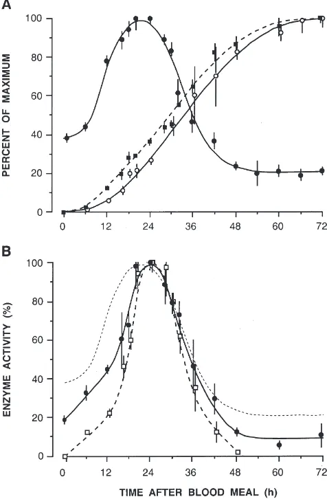
The course of XDH activity throughout a complete reproductive cycle, i.e. 0–72 h until oviposition, is presented in Fig. 1A. Accumulation of yolk in the oocytes and faecal uric acid are included and both reveal remarkable synchrony. Within 18 h the enzyme activity in the fatbody rises from 40% to its maximum of 100% (=4.42±0.01µmol/min), by 48 h it has fallen below pre-“blood meal” levels (to about 20%). Urate synthesis and its excretion increase continuously until 60–72 h, when this process is completed. A total of 18.96±0.95µg
nitro-Fig. 1. (A) XDH activity in the fatbody (I), faecal uric acid (s) and
yolk length (j) during the course of a gonotrophic cycle ofAedes aegypti. (B) Aminopeptidase (I) and trypsin activity (h) in the midgut
of the same females as in A. The dotted line has been traced from the XDH curve in A. All females were of the same size and received 3
gen is eliminated as uric acid, corresponding to 22% of the blood meal protein. Yolk deposition recorded as increase in yolk length per oocyte displays the same rate but precedes urate excretion by 4–6 h. Maximal follicle length of 425 µm (=100%) is reached at 60–72 h.
The relationship between intestinal digestion and pro-tein catabolism is demonstrated in Fig. 1B on a percent-age basis. Profiles for trypsin and aminopeptidase activity are very similar to XDH; proteolytic activities have shorter peaks and appear about 6 h later than the XDH peak. After reaching peak activities, the two pro-teases and XDH decline at the same rate. By 48 h, tryp-sin activity is zero as before the blood meal, while amin-opeptidase and XDH have fallen to their residual levels clearly different from zero but below their initial values. The activity patterns before and after a reproductive cycle, therefore, reveal similarities between aminopepti-dase and XDH.
After oviposition at 72 h, XDH activity returned to pre-“blood meal” levels within 3 days (Fig. 2). Since upon completion of digestion proteases are excreted in their active forms (Briegel, 1975), faecal samples were tested for XDH activity: 25–59% of the maximal XDH activity could be recovered. Residual activity in the female fatbody was 21%. Faecal XDH values varied gre-atly, presumably as a result of the presence of excreted proteases. Proteinase inhibitors were required in the XDH buffer system, followed by immediate analysis; without inhibition faecal XDH was undetectable.
3.3. Effect of blood meal volume
To quantify the effect of increasing protein input, females of identical size received enemas of 1, 3, and 5
µl of blood from the same rat, 3 days after eclosion and sugar-feeding. This corresponds to an input of 28.6,
Fig. 2. Restoration of residual XDH activity (I) after oviposition to
the level found in sugar-fed femaleAedes aegypti(h). Data are percent of maximal activity as in Fig. 1; N=9–57. The arrow denotes ovi-position.
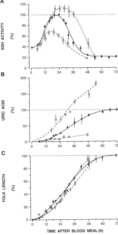
85.8, and 142.7 µg protein nitrogen, approximating the range of blood meals taken in nature. XDH activity and protein intake correlate positively, but are not entirely proportional (Fig. 3A). With large blood meals the dur-ation of peak activity was extended from 6 h to approxi-mately 14 h. Absolute peak activities were 2.94, 4.42, and 4.91µmol/min/female. Concomitant urate excretion increased remarkably (Fig. 3B): faecal urate nitrogen was 2.9 µg, 18.9 µg and 34.5 µg, corresponding to a 1:6:12 proportion in urate synthesis. Protease activities also showed linear correlations with protein intake; they
Fig. 3. XDH activity, uric acid excretion and yolk deposition in femaleAedes aegyptifed increasing volumes of rat blood (1µlh, 3
µlI, 5µls). Data are in percent of the respective maxima as in Fig.
peaked with 0.042, 0.067 and 0.09 U/midgut for trypsin and 0.026, 0.042 and 0.077 U/midgut for aminopeptidase (not shown). Thus, except for XDH maxima, all para-meters tested increase linearly with blood meal size. The rates of yolk deposition are identical, irrespective of the volume ingested (Fig. 3C), while fecundity shows a posi-tive but not entirely proportional correlation with blood volumes.
3.4. Effect of body size
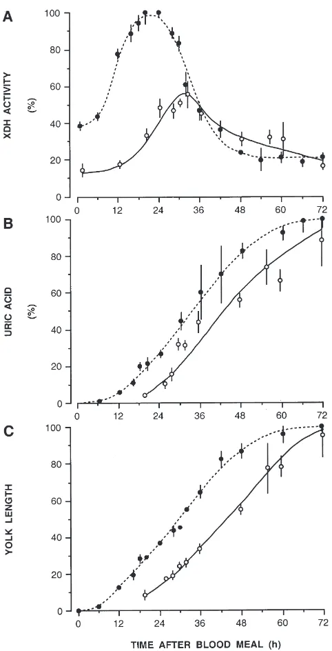
Teneral reserves, blood consumption and fecundity are heavily affected by mosquito body size (Briegel, 1990). Therefore, XDH activity was investigated in females of two size classes: small, with wing lengths of 2.2–2.4 mm, and standard size, with 2.7–2.9 mm wing length. Enemas of 3 µl were given to all of them. It turned out to be the maximal volume small females could withhold. The results are presented in Fig. 4. XDH activity is consistently and substantially lower in small-sized females (Fig. 4A). The activity patterns are similar for small and large females. XDH peaked simultaneously in both groups but in small females maximal activity is maintained for 12 h instead of 6 h. Therefore, lower XDH activity does not prevent increased uricotely. Uric acid discharge by small mosquitoes was 1.5 times higher, while yolk deposition proceeded at the same rate in both groups (Fig. 4B,C). Proteolytic enzymes of the midgut are also reduced in their maxima and delayed for 6–8 h (not shown). This delay might explain the approxi-mately 6 h retardation in vitellogenesis in small females as compared to standard females (Fig. 4C).
3.5. Effect of age
In view of the reduced and retarded fecundity of age-ing females (Briegel, 1983), we investigated the effects of the absolute age of females on their catabolism of protein from identical blood meals. Females aged 30 days were given 3 µl of rat blood by enema and the same parameters were recorded as before.
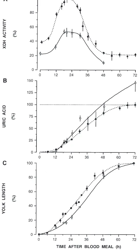
In 30-day-old females, XDH activity has a much lower residual level to start with (about 15% versus 40% in young ones), and maximal activities reach only about 55% of the maxima observed in young ones (Fig. 5A). Furthermore, a clear delay of 12 h in urate and vitellog-enin synthesis is observed in old females (Fig. 5B,C). This is complementary to earlier results on proteolytic processes (Briegel, 1983). It is interesting to note that old mosquitoes, despite their reduced XDH activity, syn-thesize and excrete as much urate within 72 h as their younger sisters. Obviously the lower XDH activity is sufficient for the synthesis of 57 µg of urate, equivalent to 19µg of nitrogen, the same amount as in young ones. The same experiment with 12-day-old females revealed the same activity patterns but delays were
inter-Fig. 4. Effect of body size on XDH activity, uric acid excretion and vitellogenesis inAedes aegyptifed identical volumes of blood. Stan-dard and small body size classes were compared: wing lengths 2.7– 2.9 mm (I) and 2.2–2.4 mm (s). Data are in percent of mean maximal
values as in Fig. 1;N=5–49 for standard andN=5–11 for small females.
mittent to those of 3- and 30-day-old females. Obvi-ously, ageing causes gradual delays of enzyme activities and end-product formation. In an attempt to relate XDH activity to the fatbody protein at different age periods, the peaks of absolute enzyme activity were divided by the content of fatbody nitrogen, taken from Table 1. This gives an estimate of the specific activities for 3-, 12-, and 30-day-old females with values of 0.17, 0.12, and 0.08 µmol/min/µg nitrogen respectively, indicating a gradual decrease of specific activity with progressing age.
mech-Fig. 5. Protein catabolism and vitellogenesis in young (I) and old
(s) females of Aedes aegypti given 3 µl of rat blood. (A) XDH activity; (B) faecal uric acid; (C) yolk length. Data are in percent of the mean maximal values observed in young females as in Fig. 1;N=5– 49 for young andN=5–12 for old females.
anical stress of frequent handling and to the application of the enemas. Their midguts rupture much more fre-quently, and after enema mortalities often exceeded 20%; usually they are below 10%. A “free-feeding” experiment was conducted to determine the physiologi-cal volume of the blood meals taken by older females. Cohorts of 3- and 30-day-old females of equal body size were allowed to feed to repletion on a human forearm, judged by withdrawal of the proboscis. The amount of haematin discharged was measured to compute the vol-ume of blood ingested. The results reveal no significant
differences in haematin defecation: 26.2±1.6µg (N=33) in 3-day-old versus 26.6±2.3 µg (N=19) in 30-day-old females, equal to blood meals of 3.8 µl and 3.9µl per female respectively. Therefore, the standard enemas of 3 µl rat blood were within the physiological range and the gut tissue probably ruptured for reasons other than mechanical stress.
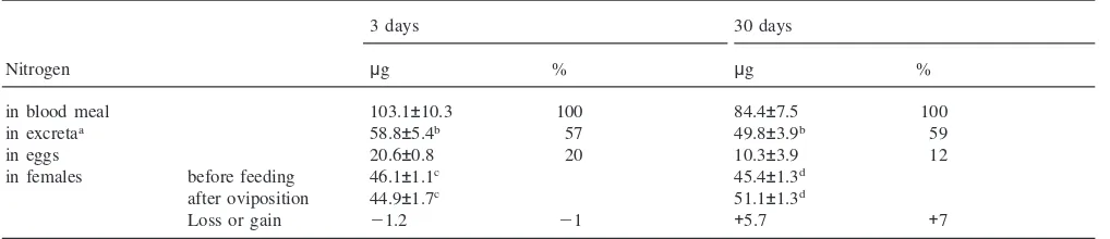
In old females fecundity is reduced by 45% and 39% of the females do not even initiate oogenesis, while in young females only 3% are non-oogenic. With human blood, young females invest 20% of the blood nitrogen into vitellogenesis, the rest is catabolized and excreted (Table 2; Briegel, 1986). In both age groups 57–59% of blood nitrogen are recovered from the faeces (Table 2), and according to Briegel (1986) 22% can be accounted for as evaporative ammonia loss. Despite their smaller investment of only 12% into oogenesis, 30-day-old females excrete similar amounts of nitrogen as their younger sisters and retain a significant amount of 5.7µg or 7% of the blood meal nitrogen, supposedly as extra ovarian protein deposits (Table 2). The tendency to channel some portion of blood nitrogen into maternal reserves is even more evident in old, non-oogenic females which retain 12% of the ingested protein.
3.6. Effect of decapitation
Blood meal utilization for oogenesis is regulated by endocrine factors (Lea, 1975) and trypsin synthesis is also under partial hormonal control (Briegel and Lea, 1979; Graf et al., 1998). We tested such a possibility for XDH activity. Females were decapitated immediately after applying blood enemas of 3 µl. Uricotely was strongly affected. XDH activity showed a similar pattern but its maximal peak was 182% of that of intact controls (range 130–230%), and its period of activity lasted about 30 h longer, thus roughly doubled in both experi-ments.The total urate output was four times higher than in intact controls with the same enemas.
4. Discussion
Table 2
Distribution of blood meal nitrogen between whole body, eggs and excreta ofAedes aegypti. Females were fed on human blood until repletion at day 3 or 30. The blood volume was calculated from the amount of haematin discharged and converted toµg nitrogen. Total nitrogen of females before feeding and after oviposition, of eggs and excreta is presented inµg/mosquito; only oogenic females were considered. (Means±S.E.M.; N=6–20)
3 days 30 days
Nitrogen µg % µg %
in blood meal 103.1±10.3 100 84.4±7.5 100
in excretaa 58.8±5.4b 57 49.8±3.9b 59
in eggs 20.6±0.8 20 10.3±3.9 12
in females before feeding 46.1±1.1c 45.4±1.3d
after oviposition 44.9±1.7c 51.1±1.3d
Loss or gain 21.2 21 +5.7 +7
aA further 22% is excreted as ammonia (Briegel, 1986). b Not significant (P
.0.1)
cNot significant (P .0.5)
d Significant (P=0.0001).
acid. This is accompanied by a gradual loss of up to 68% in fatbody protein during her life-time. This loss of body protein is restricted to the fatbody while the flight muscles in the thorax are conserved, enabling the mos-quito to seek a blood meal. Flight performance, on the other hand, is highest within the first 2 weeks and then falls, caused by a depletion of glycogen reserves (Rowley and Graham, 1968).
The situation is quite different after a blood meal. The massive protein input stimulates a steep increase in catabolism through XDH activity. The bulk of this enzyme activity is confined to the abdominal fatbody. The response of XDH to blood meal protein is dose-dependent, while the total amount of nitrogen excreted relates to the proportion channelled into vitellogenesis. When body size, female age, pre-“blood meal” con-ditions and type of blood donor are constant, the efficiency of blood protein utilization for oogenesis depends on the volume ingested (Briegel 1985, 1990). The rate of yolk deposition into maturing oocytes is not affected (Briegel and Lea, 1975; this report) but its extent is reflected in variable numbers of maturing oocytes. Maximal fecundity in mosquitoes is limited by their finite number of ovarioles (Clements, 1992). A larger blood meal therefore does not necessarily increase fecundity; rather, the larger a blood meal the more nitro-gen needs to be disposed of. For this reason, uric acid excretion shows a linear increase with blood meal size. On the enzymatic level, the rise in XDH activity follows the same kinetics, but the peaks are reached later, as more protein is present. In addition, the peaks do not increase entirely proportional to the amount of nitrogen ingested but their duration is extended. This leads to pro-portional urate excretion. A comparable situation has been found for intestinal trypsin (Briegel and Lea, 1975). Surprisingly, intestinal trypsin and aminopeptidase appear with a delay of 6 h after blood meal when
com-pared to XDH, although with the same rates of secretion. Since the proteases have narrow peaks their decrease from the midgut coincides with the decrease of XDH. All three enzymes are excreted in an active form. After oviposition, i.e. after termination of the reproductive cycle, XDH is restored to the residual level before the blood meal.
and Briegel, 1989). Consequently, amino acids take longer to reach the fatbody and therefore delay vitellog-enesis while excess nitrogen is synchronously fed into the uricotelic pathway.
Vitellogenesis is under rigid endocrine control (Lea, 1975; Hagedorn, 1985; Dhadialla and Raikhel, 1994) and tightly interwoven with the intestinal proteolytic sys-tem and protein catabolism. On the other hand, the initial stimulus for trypsin synthesis is mechanical and/or osmotic stress acting independently of the nervous sys-tem (Graf and Briegel, 1989; Graf et al., 1998), while peak activities are under the control of the neuro-endo-crine system via the ovaries (Briegel and Lea, 1979; Graf et al., 1998). Therefore, decapitation immediately after a blood meal completely blocks vitellogenesis (Gillett 1956, 1957), while trypsin synthesis is reduced only par-tially (Briegel and Lea, 1979). XDH activity was roughly doubled, its expression was extended considerably in time and caused a dramatic increase in urate excretion, removing nitrogen that normally is utilized for vitellog-enesis (Briegel 1980b, 1986). Once again, XDH activity and uric acid excretion reveal a compensatory relation-ship between dietary nitrogen and vitellogenic require-ment. Thus we are clearly dealing here with a quantitat-ive regulation of XDH activity, independent from neuro-endocrine factors that govern proteolytic digestion and vitellogenesis. The final steps in the uricotelic pathway represent a stoichiometric process.
As the three enzymes are excreted in their active forms and are also found in excess of actual require-ments for protein digestion or catabolism, it is likely that excessive enzyme secretions are of advantage for the organism. They enable the female to proceed with her reproductive cycle as quickly and efficiently as possible. The excretion of XDH moreover points to a de novo synthesis, similar to the case with trypsin (Graf and Brie-gel, 1989; Ho¨rler, 1995) and was reported for Droso-phila melanogaster (Edwards et al., 1977).
Mosquitoes grown under suboptimal larval conditions eclose as small imagoes with considerably less protein and lipid reserves (Briegel, 1990; Timmermann and Briegel, 1993). Such females reveal rather complex metabolic interactions. They ingest smaller amounts of blood and mature less eggs because they have fewer ovarioles. They display lower XDH activities, even after large blood meals of 3µl, and yet their urate excretion is increased. Because of reduced fecundity, small females catabolize a larger proportion of the blood protein resulting in a 1.5-fold increase in uric acid synthesis which is not matched by a comparable rise in XDH lev-els. On the contrary, maximal XDH activity is 50% below the value for standard-sized females presumably because of less fatbody tissue. Apparently, however, this is compensated for by doubling the time span of maxi-mal activity. The catabolic system of these smaxi-maller females therefore responds to high protein inputs in two
ways, increasing maximal activities and extending their duration. Despite all these constraints the rate of vitel-logenesis is the same, only the number of maturing oocytes being reduced.
Besides its role in uric acid synthesis, XDH is also a key enzyme in the synthesis of pteridines, a group of pigments found in the organs of many insects (Ziegler, 1961; Kayser, 1985). Being a multifunctional enzyme, the comparison of its distribution in several insect spec-ies has revealed different time- and stage-related patterns as well as different catalytic functions, namely the syn-thesis of uric acid and/or of pteridines. In the last larval instar ofPieris brassicae, XDH activity is found in the fatbody, the digestive tract and in the epidermis cells of the wings, where it is implicated in the ensuing depo-sition of pteridines (Mauchamp and Lafont, 1975). There is a clear functional separation of XDH activity. The fat-body is essentially concerned with uric acid synthesis and storage while pteridine synthesis occurs mainly in the wing discs of the pharate imago (Lafont et al., 1976). In D. melanogaster specific XDH activity in the Mal-pighian tubules equals the level found in fatbody while low activity is reported for the intestine. Diets sup-plemented with amino acid or protein lead to an increase in fatbody activity only (Collins et al., 1970). Extrapol-ation from these data on blood-fed mosquitoes suggests a sole involvement of XDH in urate synthesis because after feeding on blood only the fatbody responds to the high protein diet.
Acknowledgements
These investigations were supported by the Swiss National Science Foundation (to H.B.). We appreciate the rearing of mosquito colonies by I. Flu¨ckiger. We express our gratitude to K. Yoon for results obtained during an earlier part of this project, and to Dr S.E. Tim-mermann for carefully reading this manuscript. Compu-tational help, kindly given by Y. Choffat and Dr R. Stid-will, is greatly appreciated.
References
Anon., 1972. Worthington Enzyme Manual. Freehold, New Jersey, USA.
Barrett, F.M., Friend, W.A., 1966. Studies on the uric acid concen-tration in the haemolymph of fifth-instar larvae of Rhodnius pro-lixus(Sta˚l.) during growth and metamorphosis. J. Insect Physiol. 12, 1–7.
Briegel, H., 1969. Untersuchungen zum Aminosa¨uren- und Pro-teinstoffwechsel wa¨hrend der autogenen und anautogenen Eireifung vonCulex pipiens. J. Insect Physiol. 15, 1137–1166.
Briegel, H., 1975. Excretion of proteolytic enzymes byAedes aegypti after a blood meal. J. Insect Physiol. 21, 1681–1684.
Briegel, H., 1980b. Stickstoffexkretion bei Weibchen von Aedes aegypti (L.) ist nicht endokrin reguliert. Rev. Suisse Zool. 87, 1029–1033.
Briegel, H., 1983. Manipulation of age dependent kinetics on the induction of intestinal trypsin in the mosquito Aedes aegypti (Diptera: Culicidae). Entomol. Generalis 8, 217–223.
Briegel, H., 1985. Mosquito reproduction, incomplete utilization of the blood meal protein for oogenesis. J. Insect Physiol. 31, 15–21. Briegel, H., 1986. Protein catabolism and nitrogen partitioning during
oogenesis in the mosquito Aedes aegypti. J. Insect Physiol. 32, 455–462.
Briegel, H., 1990. Metabolic relationship between female body size, reserves, and fecundity of Aedes aegypti. J. Insect Physiol. 36, 165–172.
Briegel, H., Lea, A.O., 1975. Relationship between protein and proteo-lytic activity in the midgut of mosquitoes. J. Insect Physiol. 21, 1597–1604.
Briegel, H., Lea, A.O., 1979. Influence of the endocrine system on tryptic activity in female Aedes aegypti. J. Insect Physiol. 25, 227–230.
Buckner, J.S., Otto, P.E., Newman, S.M., Graf, G., 1993. Xanthine dehydrogenase in the fat body of Manduca sexta: purification, characterization, subcellular localization and levels during the last larval instar. Insect Biochem. Mol. Biol. 23, 549–559.
Clements, A.N., 1992. Development, Nutrition and Reproduction. The Biology of Mosquitoes, vol. 1. Chapman and Hall, London. Cochran, D.G., 1975. Excretion in insects. In: Candy, D.J., Kilby, B.A.
(Eds.), Insect Biochemistry and Function. Chapman and Hall, Lon-don, pp. 191–215.
Cochran, D.G., 1985. Nitrogenous excretion. In: Kerkut, G.A., Gilbert, L.I. (Eds.), Comprehensive Insect Physiology, Biochemistry and Pharmacology, vol. 4. Pergamon Press, Oxford, pp. 467–506. Collins, J.F., Duke, E.F., Glassman, E., 1970. Nutritional control of
xanthine dehydrogenase. I. The effect in adult Drosophila mel-anogaster of feeding a high protein diet to larvae. Biochim. Biophys. Acta 208, 294–303.
Dhadialla, T.S., Raikhel, A.S., 1994. Endocrinology of mosquito vitel-logenesis. In: Davey, K.G., Peter, R.E., Tobe, S.S. (Eds.), Perspec-tives in Comparative Endocrinology. National Research Council of Canada, Ottawa, pp. 275–281.
Edwards, T.C.R., Candido, E.P.M., Chovnick, A., 1977. Xanthine dehydrogenase fromDrosophila melanogaster. Mol. Gen. Genet. 154, 1–6.
Gillett, J.D., 1956. Initiation and promotion of ovarian development in the mosquitoAedes (Stegomyia) aegypti(Linnaeus). Ann. Trop. Med. Parasitol. 50, 375–380.
Gillett, J.D., 1957. Variation in the time of release of the ovarian devel-opment hormone inAedes aegypti. Nature 180, 656–657. Glassman, E., 1962. Convenient assay of xanthine dehydrogenase in
singleDrosophila melanogaster. Science 137, 990–991.
Graf, R., Briegel, H., 1982. Comparison between aminopeptidase and trypsin activity in blood-fed females ofAedes aegypti. Rev. Suisse Zool. 89, 845–850.
Graf, R., Briegel, H., 1989. The synthetic pathway of trypsin in the mosquitoAedes aegyptiL. (Diptera: Culicidae) and in vitro stimu-lation in isolated midguts. Insect Biochem. 19, 129–137. Graf, R., Raikhel, A.S., Brown, M.R., Lea, A.O., Briegel, H., 1986.
Mosquito trypsin: immunocytochemical localization in the midgut of blood-fedAedes aegyptiL. (Diptera: Culicidae). Cell Tissue Res. 245, 19–27.
Graf, R., Lea, A.O., Briegel, H., 1998. A temporal profile of the endo-crine control of trypsin synthesis in the yellow fever mosquito, Aedes aegypti. J. Insect Physiol. 44, 451–454.
Hagedorn, H.H., 1985. Role of ecdysteroids in reproduction. In:
Ker-kut, G.A., Gilbert, L.I. (Eds.), Comprehensive Insect Physiology, Biochemistry and Pharmacology, vol. 8. Pergamon Press, Oxford, pp. 205–262.
Hayden, Th.J., Duke, E.J., 1979a. Purification and characterization of xanthine dehydrogenase from Locusta migratoria L. Insect Biochem. 9, 583–588.
Hayden, Th.J., Duke, E.J., 1979b. Ontogeny and control of xanthine dehydrogenase inLocusta migratoriaL. Insect Biochem. 9, 589– 594.
Higashino, T., Wadano, A., Miura, K., 1977. Properties and regulation of xanthine dehydrogenase of a blowflyAldrichina grahami. Insect Biochem. 7, 317–322.
Ho¨rler, E., 1995. Proteinases and proteinase inhibitors in mosquitoes. PhD thesis, University of Zu¨rich.
Huyn, Q.K., Wadano, A., Miura, K., 1979. Studies on nitrogen metab-olism in insects: regulation mechanism of xanthin dehydrogenase in the blowfly, Aldrichina grahami (Dipt. Calliphoridae). Insect Biochem. 9, 287–292.
Kayser, H., 1985. Pigments. In: Kerkut, G.A., Gilbert, L.I. (Eds.), Comprehensive Insect Physiology, Biochemistry and Pharma-cology, vol. 10. Pergamon Press, Oxford, pp. 367–415.
Lafont, R., Mauchamp, B., Pennetier, J.-L., Tarroux, P., Blais, C., 1976. Biochemical correlations during metamorphosis in Pieris brassicae. Insect Biochem. 6, 97–103.
Lea, A.O., 1964. Studies on the dietary and endocrine regulation of autogenous reproduction inAedes taeniorhynchus(Wied.). J. Med. Entomol. 1, 40–44.
Lea, A.O., 1967. The medial neurosecretory cells and egg maturation in mosquitoes. J. Insect Physiol. 13, 419–429.
Lea, A.O., 1975. The control of reproduction by a bloodmeal: the mos-quito as a model for vector endocrinology. Acta Trop. 32, 112–115. Lowry, O.H., Rosebrough, N.J., Farr, A.L., Randall, R.J., 1951. Protein measurement with the Folin phenol reagent. J. Biol. Chem. 193, 265–275.
Mauchamp, B., Lafont, R., 1975. Developmental studies inPieris bras-sicae(Lepidoptera). II. A study of nitrogenous excretion during the last larval instar. Comp. Biochem. Physiol. B 51, 445–449. Minari, O., Zilversmit, D.B., 1963. Use of KCN for stabilization of
color in direct Nesslerization of Kjeldahl digest. Anal. Biochem. 6, 320–327.
Parzen, Sh.D., Fox, A.S., 1964. Purification of xanthine dehydrogenase fromDrosophila melanogaster. Biochim. Biophys. Acta 92, 465– 471.
Rowley, W.A., Graham, Ch.L., 1968. The effect of age on the flight performance of femaleAedes aegyptimosquitoes. J. Insect Physiol. 14, 719–728.
Sakai, K., Miyasako, Y., Nagatomo, H., Watanabe, H., Wakayama, M., Moriguchi, M., 1997.l-Ornithine decarboxylase fromHafnia alvei has a novel l-ornithine oxidase activity. J. Biochem. 122,
961–968.
Sokal, R.R., Rohlf, F.J., 1981. Biometry. Freeman, San Francisco. Timmermann, S.E., Briegel, H., 1993. Water depth and larval density
affect development and accumulation of reserves in laboratory populations of mosquitoes. Bull. Soc. Vec. Ecol. 18, 174–187. Urich, K., 1990. Der N-Stoffwechsel der Aminosa¨uren. In:
Vergleich-ende Biochemie der Tiere. G. Fischer Verlag, Stuttgart, pp. 366– 412.
Van Handel, E., 1975. Direct determination of uric acid in fecal material. Biochem. Med. 12, 92–93.
Van Handel, E., Klowden, M.J., 1996. Defecation by the mosquito, Aedes aegypti, is controlled by the terminal abdominal ganglion. J. Insect Physiol. 42, 139–142.