Nuclear Import Analysis of Two Different Fluorescent Marker Proteins
into Hepatocyte Cell Lines (HuH-7 Cell)
Aris Haryanto
1*and Michael Kann
21. Department of Biochemistry, Faculty of Veterinary Medicine, Gadjah Mada University. Jl. Olahraga, Karangmalang, Yogyakarta 55281, Indonesia.
2. Institute of Medical Virology, Justus-Liebig-University, Giessen, Germany. Frankfurterstrasse 107, 35392 Giessen, Germany
Abstracts
The application of fluorescent proteins as expression markers and protein fusion partners has proved immensely valuable for resolving the organization of biological events in living cells. EGFP and DsRed2 are commonly fluorescent marker protein which is used for biotechnology and cell biology research. The present study was designed to identify the expression vector that suitable to ligate with DNA encoding HBV core protein for intracellular localization study in hepatocyte cell, which were expressed as fusion proteins. We also compared and quantified the expressed fluorescent protein which predominantly localized in the cell compartment. The results indicated that DsRed2 shown as less than ideal for intracellular localization study of than EGFP, because of its tetrameric structure of the fluorescent protein and when fused to a protein of interest, the fusion protein often forms aggregates in the living cells.In contrast, EGFP fluorescent protein shown a much higher proportion of cytoplasmic localization, thus being more suitable for analysis of intracellular localization than DsRed2 fluorescent protein.EGFP fluorescent protein is also capable to produce a strong green fluorescence when excited by blue light, without any exogenously added substrate or cofactor, events inside living cell can thus be visualized in a non-invasive way. Based on our present quantitative data and some reasons above shown that EGFP is more suitable than DsRed2 as a fluorescent marker protein for intracellular localization study into HuH-7 cell.
Keywords: EGFP, DsRed2 fluorescent protein , HuH-7 cell, HBV, intracellular localization
Introduction
The enhanced green fluorescent protein (EGFP) is a marker protein that comprised of 238 amino acids with molecular weight 27 kDa. This marker protein was isolated from the jellyfish Aequorea victoria which fluoresces green color when exposed to blue light (Prendergast and Mann, 1978; Tsien, 1998).The EGFP gene is frequently used as a reporter expression (Philip, 2001). In modified forms it has been used to make biosensors, and many animals have been created that express EGFP as a proof of
concept that a gene can be expressed throughout a given organism. The availability of EGFP has thoroughly redefined fluorescent and the way it is used in cell biology and other biological disciplines (Yuste, 2005).
EGFP are usually much less harmful when illuminated in living cells. This has triggered the development of highly automated live cell fluorescence microscopy systems which can be used to observe cells over time expressing one or more proteins tagged with fluorescent proteins. Another powerful using of EGFP is to express the protein in small sets of specific cells. This allows researchers to optically detect specific types of cells in vitro or even in vivo
(Chudakov, 2005). The EGFP gene can be introduced into organisms and maintained in their genome through breeding, or local injection with a viral vector can be used to introduce the gene. Currently, many bacteria, yeast and other fungal cells, plant, fly, and mammalian cells have been created using EGFP as a protein marker.
DsRed is the first coral-derived fluorescent marker protein which to be extensively utilized for biotechnology and cell biology as a spectrally distinct companion or substitute for the green fluorescent protein (GFP) from the Aequorea
jellyfish. It was derived from Discosoma striata (Matz et al., 1999). Wild type DsRed is an obligate tetramer. When fused to a protein of interest, the fusion protein often forms aggregates in living cells, hampering normal localization, trafficking and protein-protein interactions of the protein-protein of interest. Although these features are inconsequential for the use of DsRed as a reporter of gene expression, the usefulness of DsRed as an epitope tag is severely limited. In contrast, to the jellyfish fluorescent proteins, which have been successfully used to tag hundreds of proteins, DsRed conjugates have proven much less successful and are often toxic. The evolution of DsRed from a scientific curiosity to a generally applicable and robust tool has been hampered by several critical problems, including a slow and incomplete maturation and obligate tetramerization (Baird et al., 2000).
The second generation of DsRed is called DsRed2. This fluorescent marker protein contains several mutation at the peptide amino terminus which prevent formation of protein aggreates. The mutation also reduce toxicity effect to the transfected cell. As DsRed, the fluorescent marker protein of DsRed2 still forms a tetramer. Red fluorescence emission from DsRed2 can be observed approximately 6 hours after transfected. It is faster than DsRed which
can be observed after 11 hours posttransfection.
The present study was designed to identify the expression vector that suitable to ligate with DNA encoding HBV core protein for intracellular localization study of HBV core protein into hepatocyte cell, which were expressed as fusion proteins. We also compared and quantified the expressed fluorescent protein which predomonantly localized in the cell compartment.
Materials and Methods
Main materials of this work are plasmid vector encoding red fluorescent marker protein pDsRed2-C1 (Clonetech), plasmid vector encoding green fluorescent marker protein pEGFP-C3 (Clonetech) and hepatocyte cell line HuH-7.
Preparation of pDsRed2-C1 and pEGFP-C3
To clone plasmid vector encoding red fluorescent and green fluorescent marker protein, pDsRed2-C1 and pEGFP-C3 were transformed into competent E. coli XL-1 blue. Into each 1.5 ml E-cup, 1 µl DNA pDsRed2C1 and pEGFP-C3 were added into 25 µl competent E. coli. After gentle mixing, the transformation reactions were cooled down on the ice for 10 min, then the transformation was accomplished by a heat shock at 42°C for 2 min. Afterwards 1 ml LB medium was added and mixed gently. The E. coli were incubated at 37°C for 1 h and platted onto an agar dish containing LB medium with 30 mg/ml Kanamycin. The dish was incubated at 37°C until the liquid has been absorbed. Then the dish was inverted and incubated over night at 37°C.
Maxipreparation DNA plasmid isolation
in 250 ml Kanamycin (30 mg/ml) containing LB medium. The colonies were incubated in shaking incubator at 37°C overnight. The next day, 250 ml bacteria in LB medium was harvested and sedimented. The plasmid DNA was isolated by a Maxipreparation DNA plasmid isolation kit (Qiagen). This plasmid isolation was performed according to the manufacture recommendations.
Preparation of hepatocyte cell line (HuH-7 cell)
Hepatocyte cell lines (HuH-7 cells) were seeded on the culture dish and incubated over night in the incubator CO2 at 37°C. The growing cells were harvested and washed with PBS. The PBS was replaced by 1x trypsin in PBS (Gibco-BRL), which has been preincubated at 37°C. While the dish with the cells was incubated at 37°C for 2-5 min, 0.5 ml 10% FCS containing medium (Gibco-BRL) were added to the each well of 24 well dish containing collagen-treated cover slips (Nunc Inc.). The trypsin in PBS was removed and washed the cells with PBS. The PBS was changed with fresh 10% FCS containing medium. Each cover slip were filled with 2-5 drops cell suspension. Then the cells were allowed to grow over night at 37°C in humidified incubator at 5% CO2.
Transfection into HuH-7 Cell
For transfection, Tfx-20 (Promega) was used as transfection agent, 1 ml DNA (1 mg/ml) of pDsRed2-C1 and pEGFP-C3 were mixed gently with 3 ml Tfx-20, then added into 300 ml FCS-free medium (Gibco-BRL). The mixture was incubated at room temperature for 5-10 min. During the incubation, the medium in the 24 well dish were removed and replaced with FCS free medium. The DNA/Tfx-20 reagent/FCS-free medium mixture were vortexed briefly, then added the mixture to the cells (300 ml per well) and incubated in the incubator at
5% CO2 and 37 °C for 1 h. During the incubation 10% FCS containing medium was warmed at 37°C in the waterbath. After 1 h the FCS free medium was replaced with the 10% FCS containing medium and the dish was returned into the incubator for 24 h.
Indirect immune fluorescence staining
were placed on them. The glass slides were kept over night in the darkness at room temperature. For transformed HuH-7 cells by pDsRed2-C1, indirect immune fluorescence staining were performed using primary antibody mixture containing monoclonal antibody (mAb) 414 anti NPC from mouse (1: 300 dilution) and as secondary antibody used goat Alexa 488-labelled anti mouse antibody (Dianova). Immune fluorescence procedure was performed as described above.
Immune fluorescence confocal laser microscopy
A drop of immersion oil was dripped on each of cover slips in the slide. The slide was put on the microscope. The cells were analyzed using a 63x apochromat objective by a LEICA DM IRBE. For the confocal analysis the cells were scanned by a laser power of 30%, a signal amplification of 85% and a pinhole size of 0.9 with a two-fold magnification. Eight pictures were merged. For the representation of Alexa 488, the FITC filter settings were used, for depicting Texas Red, the TRITC filter setting were used. The z position in all picture was adjusted to the equatorial level of the nuclei. The pictures were arranged using the ADOBE-PHOTOSHOP software program.
Quantification of the Intracellular Localization
Red fluorescent and green fluorescent marker protein which found localized in the compartment of HuH-7 cells, was quantified manually using confocal laser microscope. The Amount of fluorescent proteins that found localized in the cytoplasm, nucleus or both of cell compartment were quantified in the absolute and relative values.
Results and Discussion
Analysis expression of the fluorescent proteins by E. coli transformed with plasmids encoding EGFP and DsRed2 marker protein could clearly analyze in the transfected HuH-7 cells 12 h after transfection for efficient visualization using confocal laser scan microscope. In the cells expressing EGFP, the fluorescent proteins found localized in the cytoplasm, nucleus and both of the cell compartment (Figure 1). In contrast DsRed fluorescences found distributed only in cytoplasm and nucleus (Figure 2). It might indicate that EGFP fluorescent protein which monomer form can passively imported into the nucleus, even very rare and only minority amount. Whereas DsRed2 fluorescent protein can not passive imported into the nucleus because this fluorescent protein is tetramer with molecular weight higher than EGFP fluorescent protein.
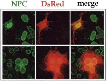
Figure 1. Intracellular Localization of DsRed2 marker protein in HuH-7 cells. DsRed2 (middle column) was found to be localized inside the cytoplasm (upper row) and within the nucleus (lower row) in a similar amount of cells. The nuclear pore complexes are shown in green; the overlays (merge) in the right column.
fluorescence) and EGFP (green fluorescence) by transient expression of the corresponding expression vectors pDsRed2-C1 and pEGFP-C3 in HuH-7 cells (Figure 1 and 2). Both fluorescent marker proteins were expressed under the control of the cytomegalovirus promoter (PCMV). The transfection and cell culture conditions were identical to those used for the expression of the HBV capsid in the experiment before. As in this experiment, the staining of the NPCs was done using the same primary antibody. In pDsRed2 transfection an FITC-labelled secondary antibody was used. For localisation of EGFP the secondary antibody was Texas Red labelled. Both transfections were done in parallel to exclude differences caused by the pre-cultivation of the cells prior to transfect into HuH-7 cells.
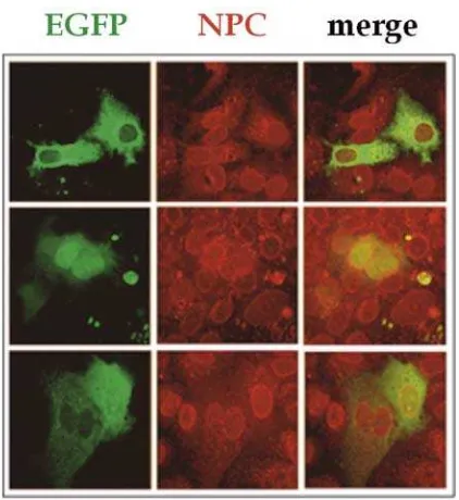
Figure 2. Intracellular Localization of EGFP marker protein in HuH-7 cells. In the majority of cells EGFP (green) localized predominantly in the cytoplasm (upper row). In a minority of cells EGFP was localized nucleus (midlle row) or throughout cytoplasm and nucleus (lower row, right cell). The nuclear pore
complexes are shown in red; the overlays (merge) in the right column.
Absolute and relative numbers of fluorescent marker protein distribution in the cell compartment showing an equal distribution or a predominantly cytoplasmic or nuclear localization of the fluorescent proteins (Tabel 1.). Both transfections of DsRed2 and EGFP fluorescent proteins were performed in parallel work.
Tabel 1. Distribution of DsRed and EGFP in HuH-7 cells after transient expression into HuH-7 cells.
marker cytoplasm nucleus both total
DsRed2 7 10 0 17
41% 59% 0 100%
EGFP 100 39 1 140
71% 28% 1% 100%
partner, and the complex fluorophore maturation kinetics via a green fluorescent intermediate were tardy (half-time; 1 day), incomplete, and spectrally inconvenient for multicolor labeling applications, in which cross talk with GFP-like proteins remained a problem, even after prolonged maturation (Baird et al., 2000; Gross et al., 2000; Mizuno
et al., 2001).
EGFP fluorescent protein shows a much higher proportion of cytoplasmic localization, thus being more suitable for analysis of intracellular localization than DsRed2 fluorescent protein. However, the significant proportion of nuclear EGFP fluorescent protein showed that determination of the ratio of cells showing nuclear and/or cytoplasmic fusion protein is required. EGFP fluorescent protein shown a powerful tool for identifying the intracellular localization of fusion protein and also to monitor gene expression. This marker protein is capable of producing a strong green fluorescence when excited by blue light, without any exogenously added substrate or cofactor, events inside living cell can thus be visualized in a non-invasive way (Chalfie et al., 1994; Mayer et al., 1999; Niedenthal et al., 1996). Based on our present quantitative data and some reasons above shown that EGFP is more suitable than DsRed2 as a fluorescent marker protein for intracellular localization study in the HuH-7 cell.
Acknowledgements
The authors wish to express gratitude to Prof. Dr. Wolfram H. Gerlich, the Head Institute of Medical Virology, Justus-Liebig-University Giessen, Germany for the opportunity to do this work. This research was supported by Sonder Forschung Bereich (SFB) 535, Justus-Liebig-University Giessen, Teilproject B-6 (AG Kann) and Deutsche Akademische Austauschdienst (DAAD) Referat 422.
References
Baird, G. S., Zacharias, D.A., and Tsien, R.Y., 2000. Biochemistry, mutagenesis, and oligomerization of DsRed, a red fluorescent protein from coral. Proc. Natl. Acad. Sci. USA., 97, 11984– 11989.
Chalfie, M., Tu, Y., Euskirchen, G., Ward, W.W., and Prasher, D.C., 1994. Green fluorescent proteoin as marker for gene expression. Science, 263, 802-803. Chudakov, D., Lukyanov, S., and Lukyanov
K., 2005. Fluorescent proteins as a toolkit for in vivo imaging. Trends Biotechnol., 23 (12), 605-13.
Dingwall, C. and Laskey, R., 1992. The nuclear membrane. Science, 258, 942-947.
Gross, L.A., Baird, G.S., Hoffman, R.C., Baldridge, K.K., and Tsien, R.Y., 2000. The structure of the chromophore within DsRed,a red fluorescent protein from coral. Proc. Natl. Acad. Sci. USA., 97, 11990–11995.
Matz, M.V., Fradkov, A.F., Labas, Y.A., Savitsky, A.P., Zaraisky, A.G., Markelov, M.L., and Lukyanov, S.A., 1999. Fluorescent proteins from nonbioluminescent Anthozoa species.
Nat. Biotechnol., 17, 969-973.
Mayer, G., Launhardt, H., and Munder, T., 1999. Application of the green fluorescent protein as a reporter for AceI-based, two hibrid studies. Bio-Techniques, 27, 84-86.
localization in budding yeast. Yeast,
12, 773-786.
Phillips, G., 2001. Green fluorescent protein—a bright idea for the study of bacterial protein localization. FEMS Microbiol. Lett., 204 (1), 9-18.
Prendergast, F. and Mann, K., 1978. Chemical and physical properties of aequorin and the green fluorescent
protein isolated from Aequorea forskålea. Biochemistry, 17 (17), 3448-53.
Tsien, R.Y., 1998. The green fluorescent protein. Annu. Rev. Biochem., 67, 509– 544.