Volume 45 Number 4 December 2012
Research Report
The role of
Actinobacillus actinomycetemcomitans fimbrial
adhesin on MMP-8 activity in aggressive periodontitis
pathogenesis
rini devijanti ridwan Department of Oral Biology
Faculty of Dentistry, Universitas Airlangga Surabaya – Indonesia
abstract
background: Actinobacillus actinomycetemcomitans (A. actinomycetemcomitans) is Gram negative and a major bacterial agent associated with aggressive periodontitis in young adult, this bacteria was an important factor in pathogenesis of aggressive periodontitis. A. actinomycetemcomitans possesses fimbriae with an adhesin protein that was the first bacterial molecules to make physical contact with host. Purpose: The objective of this research was to analyzed the influence of A. actinomycetemcomitans fimbrial adhesin protein induction on MMP-8 activity. Methods: The research was an experimental laboratory study, the step in this study were isolation and identification A. actinomycetemcomitans, characterize A. actinomycetemcomitans adhesin and study the role of A. actinomycetemcomitans adhesin in Wistar rats. results: The result of this research on the role of adhesin in Wistar rats after analysis with Analysis of Variance (ANOVA) showed significant differences in the control group with group induction with A. actinomycetemcomitans, A. actinomycetemcomitans plus adhesin and adhesin. MMP-8 activity increased with induction A. actinomycetemcomitans and 24 kDa A. actinomycetemcomitans adhesin. This fimbrial adhesin protein showed that A. actinomycetemcomitans has the ability to adhesion, colonization and invasion for host in aggressive periodontitis pathogenesis. conclusion: A. actinomycetemcomitans fimbrial adhesin protein induction increasing MMP-8 activity for aggressive periodontitis pathogenesis.
Key words: A. actinomycetemcomitans, adhesin, MMP-8, aggressive periodontitis
abstraK
latar belakang: A. actinomycetemcomitans merupakan salah satu bakteri Gram negatif yang terkait dengan periodontitis agresif yang menyerang penderita usia muda dan merupakan faktor penting dalam patogenesis periodontitis agresif. A. actimycetemcomitans mempunyai fimbriae dengan protein adhesin yang merupakan molekul pertama dari bakteri untuk melakukan kontak fisik dengan host. tujuan: Tujuan penelitian ini adalah menganalisis pengaruh induksi adhesin A. actinomycetemcomitans terhadap aktivitas MMP-8. Metode: Penelitian ini merupakan studi eksperimental laboratori, langkah dari penelitian ini adalah isolasi dan identifikasi A.isolasi dan identifikasi A.identifikasi A. actinomycetemcomitans, isolasi dan karakterisasi adhesin A. actinomycetemcomitans dan uji peran adhesin A. actinomycetemcomitans di tikus Wistar. hasil: Hasil analisis menunjukkan adanya aktivitas MMP-8 yang meningkat bermakna di kelompok kontrol dibandingkan dengan kelompok dengan induksi A. actinomycetemcomitans, A. actinomycetemcomitans + adhesin dan adhesin. Hal ini menunjukkan bahwa A. actinomycetemcomitans berkemampuan melakukan adesi, kolonisasi dan invasi pada host dalam patogenesis periodontitis agresif. Kesimpulan: Induksi protein adhesin A. actinomycetemcomitans dengan berat molekul 24 kDa meningkatkan aktivitas MMP-8 pada patogenesis periodontitis agresif.
Kata kunci: A. actinomycetemcomitans, adhesin, MMP-8, periodontitis agresif
introduction
Aggressive periodontitis is a disease of the tooth supporting tissues characterized by rapid destruction of periodontal ligament and alveolar bone in young patients, usually occurs at the age under 30 years. In the process of aggressive periodontitis tissue attachment loss and gingival recession four times faster than chronic periodontitis.1,2
The national survey results (Riskesdas data 2007) showed that 90% of Indonesia population suffers from periodontal disease are quite aggressive, since 20.3% of East Java residents had problems of the teeth and mouth.3
The pathogenesis of aggressive periodontitis are caused by the interaction between host and bacterial factors. Major bacterial in aggressive periodontitis are dominated by Actinobacillus actinomycetemcomitans and now better known as Aggregatibacter actinomycetemcomitans (A. actinomycetemcomitans). The presence of these bacteria in dental plaque associated with aggressive periodontal tissue damage and aggravated by the presence of genetic and environmental factors.4 Matrix metalloproteinase
(MMP) levels of saliva increased in patient with aggressive periodontitis.5 MMP is an endopeptidase which is an
important mediator of the inflammatory tissue damage that breaks most of the extracellular matrix and basement membrane proteins at physiological pH and temperature, especially as tissue damage in periodontitis. Among MMPs, collagenase (ie: MMP-1, MMP-8, and MMP-13) are the largest matrix metalloproteinases and have the intertitial capacity on collagen damage. MMP-8 is secreted by neutrophils in inactive form and become active when the periodontal tissues inflamed. Activation of MMP-8 can be induced by inflammatory mediators such as IL-1β, TNFα and derivatives microbial proteases, and reactive oxygen species (ROS) produced by neutrophils induced.6
In chronic periodontitis collagenase activity of MMP-8 about 90-95% of the gingival crevicular fluid.2 At the time
of being aggressive periodontitis, the activity of MMP-8 in gingival crevicular fluid (GCF) and significantly increased MMP-8 causes damage to periodontal tissues and alveolar bone. It is not yet known the role of adhesin
A. actinomycetemcomitas in the pathogenesis of aggressive periodontitis, so it is necessary to investigate the role of A. actinomycetemcomitans adhesin against MMP-8 activity.
MaterialsandMethods
The bacterial strain used in this study was A. actinomycetemcomitans clinical isolate. Bacteria were grown in Actinobacillus actinomycetemcomitans growth medium (AAGM) and incubated at 37 ºC anaerobically for 24 hours. Identification of A. actinomycetemcomitans on AAGM plates based on grossmorphology such as adherence to themedium surface, a starlike inner structure and positive catalase.The identification of A. actinomycetemcomitans
confirmed using the Microbact system and PCR. From the
cellular bacteria pellet, genomic DNA was extracted using PCR which was performed by adding1 μl DNA to a reaction mixture (50 μl final volume) containing 20 nmol ofeach primer, 40 nmol of deoxynucleotide triphosphates and 1U of Taq polymerase. Thefollowing cycling conditions were used: denaturation at 94 ºC for 1 minutes, annealing at42 ºC for 2 minutes, and elongation at 72 ºC for 3 minutes. Finally, 10 minutes elongation at 72 ºCfollowed 22 cycles of amplification. The PCR products were purified with the Qiaquick PCR purification kit (Qiagen, Valencia, CA). For clinical evaluation, subgingival plaque samples from patients with periodontitis were obtained by inserting a sterileendodontic paperpoint into the subgingival site for 10 seconds. The paper point was transferred into 200 ml of phosphate buffersaline (PBS) and centrifuged at 15.000 rpm at 4 ºC for 5 minutes. After denaturation at 96 °C for 2 minutes, a total of 25 PCR cycles were performed; each cycleconsisted of 15 seconds of denaturation at 94 °C, 30 seconds of annealing at 54 °C, and 60 seconds of extension at 72 °C. Amplification productswere loaded into 1.8% (wt/vol) agarose gels by electrophoresis, stained with ethidium bromide (0.5 mg/ml), and photographedunder UV light.7
A. actinomycetemcomitans culture in 250 ml AAGM was added with 10 ml of 3% tricholoro acetic acid (TCA) and allowed to stand for 30–60 minutes, then centrifuged at 6,000 rpm at 4 ºC for 15 minutes. Supernatant was discarded and the sediment suspended in 50 ml PBS pH =
7.4 and fimbriae was cut using Omnimixer cutting tools. When cutting, the suspension of bacterial fimbriae was
placed in cooled vessel. Cutting was performed for 1 minute and resting for 30 seconds (for cooling). This process was done for 5 times. Each of these pieces were centrifuged at 12.000 rpm for 15 minutes and the supernatant (pieces
fimbriae) were stored.
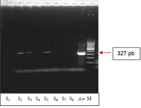
figure 1. PCR profiles of A. actinomycetemcomitans clinical isolates. 12
Lane 1-8 : S1 - 8: Sampel 1 – 8 A.a : A. actinomycetemcomitans M : Marker: Fermentas 100
Lane 1-8 : S1 - 8 : Sampel 1 – 8
S1 S2 S3 S4 S5 S6 S7 S8 A.a M
A 20 mL sample plus 20 μl reducing sample buffer (RSB) included in Eppendorf was heated for 5 minutes in boiling water. Enter the sample on the electrophoresis gel wells running the sample at 120 V for 90 minutes. Lift the gel, perform staining with coomassie brilliant blue R 250 on shaker for 20-30 minutes. After that, gel was transfered into the destaining solution overnight in a shaker until the gel looks clean and then calculated the protein molecular weight in protein bands that appear on the gel. The result of SDS-PAGE in the form of the protein bands was performed hemagglutination test and continued with elution of mice erythrocytes to demonstrate the presence of protein haemagglutinin. Several studies have shown that bacterial adhesin played by hemagglutinin protein.
The hemagglutination test was done to found the hemagglutinin protein on A.actinomycetemcomitans
bacteria with fimbrial adhesin proteins from SDS-PAGE results. A.actinomycetemcomitans fimbrial adhesin protein was reacted with the erythrocytes of mice then seen the hemaglutination titers. At first step, the mice erythrocytes were washed 3 times with PBS pH 7.4 and then made into 0.5% suspension and included 50 mL PBS. Into the first well was added 50 mL protein fimbriae, subsequent serial dilution was made into the next wells, except wells-12 is used as a control (without protein sample). Then into each well was added 50 mL of erythrocyte suspension, shaken in 15 minutes and then left in the room temperature until visible results was obtained. Hemagglutination assay results for the sample is read when the control wells had visible results. Proteins with the highest titer used for subsequent studies. To determine the presence of certain protein in the gel used Western blotting method with antibody anti adhesin.
A. actinomycetemcomitans bacteria was cultured in AAGM medium, at 37 °C for 4-5 days. Liquid culture was centrifuged at 6000 rpm, at 4 °C for 15 min. The precipitate was suspended in PBS containing BSA 1%. Hemaglutin fimbriae protein dose were divided respectively into 0 mg (control), 25 mg, 50 mg, 100 mg, 200 mg and 400 mg. Furthermore, for each dose protein fimbriae added enterocytes suspension of 300 ml and shaken on water bath at 37 °C for 30 minutes. Then the mixture was added to the
bacterial suspension (108/ml) of 300 μl. The mixture was
incubated on the ‘shaking incubator’ for 30 min at 37 °C. Furthermore centrifuged 1500 rpm, at 4 °C for 3 minutes, then washed sediment using PBS twice. The precipitate was taken, and stain with Gram staining. Preparations were observed under a microscope 1000x magnification, and the number of bacteria that attach to the enterocytes were counted. Adhesion index was the average number of bacteria that attach to HeLa cells and was calculated until on 100 HeLa cells for every observation.8
Ten weeks old male Wistar rats with 120–150 grams weight were divided into 4 groups, each group consists of 10 rats. In group one was the control group, induced with 0.9% NaCl, the group 2 induced with adhesin, group 3 induced with adhesin + A. actinomycetemcomitans and group 4 induce with A. actinomycetemcomitans whole cell.9 Before
the treatment, it was examined A. actinomycetemcomitans
in the rat oral cavity. Adhesin was induced in rat by giving 200 mL A. actinomycetemcomitans adhesin with a protein content of 200 ug/ml at 108 A. actinomycetemcomitans
density and given at least 7 days to get real aggressive periodontitis symptoms.10 Induction done on the upper
right of first molar gingival sulcus of Wistar rats.11 Then
examination to determine the severity of periodontal tissue destruction and alveolar bone through MMP-8 activity with Zymogram analysis. ANOVA are used to data analysis for MMP-8 activity.
results
The profile result of A. actinomycetemcomitans
identification in this study using polymerase chain reaction (PCR) (Figure 1). PCR examination for A. actinomycetemcomitans was done after identification tests using microbiology, biochemistry and performed with scanning electron microscopy (SEM). Three from eight samples of A.actinomycetemcomitans DNA from PCR profiles showed positive reaction, i.e, sample number two (S2), three (S3) and five (S5). We used sample number 2 (S2) to isolated and identified as A. actinomycetemcomitans adhesin since this bacteria has rough colony and fimbriae.
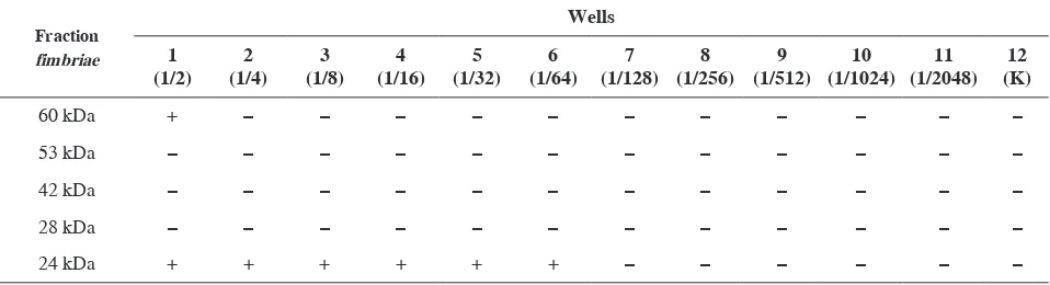
table 1. Haemaglutination test result of fimbriae adhesin protein A. actinomycetemcomitans
In this study, there were five fimbriae protein adhesin, at 60 kDa, 53 kDa, 42 kDa, 28 kDa and 24 kDa. Protein profile on SDS-PAGE of five pieces A. actinomycetemcomitans
fimbriae protein using a stratified omnimixer showed a picture of the most prominent protein bands i.e. the protein with a molecular weight of 60 kDa, 53 kDa, 42 kDa, 28 kDa and 24 kDa. The five proteins that exhibit prominent picture was collected and purification for electroelution then performed to obtain the protein solution.
The protein with highest titer results from electroelusion test used to fimbriae adhesin protein hemagglutination test. Table 1 showed the result of hemagglutination test performed to find proteins hemagglutinin (HA) from A. actinomycetemcomitans fimbriae after bacterial culture was cut by using a modification omnimixer for 5 (five) times. For the hemagglutinin protein of fimbriae fraction preceded by hemagglutination assay using rat erythrocytes,
½ titer obtained from fimbriae protein fraction with 60 kDa molecular weight, 1/128 from the 24 kDa fraction fimbriae protein and negative from 53 kDa, 42 kDa and 28 kDa fractions fimbriae protein.
Further, analysis of the result from haemaglutination test for fimbriae adhesin protein with 60 kDa and 24 kDa done using western blotting test. Western blotting assay was the specific method to determine the presence of certain proteins in the gel by using anti adhesin antibodies that was obtained from 24 kDa fimbriae protein. The 24 kDa fimbriae protein was a specific protein of fimbriae
A. actinomycetemcomitans because on Western blotting assay results obtained fimbriae protein bands at 24 kDa. This band showed fimbriae protein 24 kDa can be detected
its existence because they are specific and have a high sensitivity. At 6.7 kDa protein fimbriae there was no band, it shows that the 6.7 kDa fimbriae protein did not have a strong ability to bind to anti adhesin antibodies because could not be reacted with the substrate. This condition also showed there was no cross-reaction between the protein fimbriae with 24 kDa and 6.7 kDa molecular weight because Western blotting assay use polyclonal antibody from protein fimbriae with 24 kDa and 6.7 kDa molecular weight. Result of adhesion test in HeLa cell culture in some dose i.e 400 μg/ml, 200 μg/ml, 100 μg/ml, 50 μg/ml and 0 μg/ml showed the decrease of A. actinomycetemcomitans
amount that was attached to the surface of HeLa cells and obtained results were a significant reduction of the A. actinomycetemcomitans amount on HeLa cells with the increasing dose of fimbrial adhesin protein.
The activity of MMP-8 in Zymogram was aimed to identify MMP-8 through the degradation of the substrate and by molecular weight. The MMP-8 activity was performed by measuring the density of bands on SDS-PAGE by Silver staining. The higher band density in Zymogram showed the higher of MMP-8 activity. The result of Zymogram assay with Silver staining to measure MMP-8 activity is showed in Figure 2.
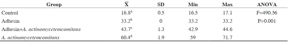
It was shown that mean of MMP-8 activity in group with A.actinomycetemcomitans induction was higher (60.4) than the group with adhesin + A. actinomycetemcomitans
induction (43.7), adhesin induction (33.2) and control group (16.8) (Table 2).
One way ANOVA test results showed the value of the activities of MMP-8 in the control group and the group with adhesin, adhesin + A. actinomycetemcomitans and
A. actinomycetemcomitans induction were significantly different, p=0.001 (p<0.005) and then analyzed by Tukey HSD test. Analysis of Tukey HSD found a significant differences from the activity of MMP-8 in the control group and the group treated with induction adhesin, adhesin + A. actinomycetemcomitans and A. actinomycetemcomitans
and between the treatment groups.
discussion
Five fimbriae adhesin protein have been identified from clinical isolates of A. actinomycetemcomitans by molecularby molecular mass at SDS-PAGE. The fimbrial adhesin were 60 kDa, 53 kDa, 42 kDa, 28 kDa and 24 kDa, After haemaglutination
table 2. The mean and standart deviations number of MMP-8 activity
Group X sd Min Max anoVa
Control 16.8a 0.5 16.5 17.1 F=490.56
Adhesin 33.2b 0 33.2 33.2 P=0.001
Adhesin+A. actinomycetemcomitans 43.7c 1.3 42.9 44.6
A. actinomycetemcomitans 60.4d 1.9 59 71.7
Note: There different superscript indicates that there are significantly differences between groups (p <0.05) figure 2. MMP-8 activity of the group induction with
A.actinomycetemcomitans showed the highest activity
M Adhesin+A.a C A.a Adhesin
200 kDa
140 kDa
100 kDa
70 kDa 50 kDa
test we identified two fimbrial adhesin with positive test, that were 60 kDa and 24 kDa and the titre were ½ and 1/128. In Western blotting assay result obtained fimbrial adhesin band at 24 kDa molecular weight, this suggests that fimbrial adhesin protein could be detected its existence because it is specific and have a high sensitivity.
Adhesion index was calculated by counting the number of A. actinomycetemcomitans that was attached to the surface of HeLa cells. The results of the adhesion index showed significant reduction of the number of A. actinomycetemcomitans in HeLa cells with the increasing dose of fimbrial adhesin protein. The more protein that causes the point saturated on HeLa cell receptors will caused decrease ability A. actinomycetemcomitans to attach in Hela cell. This condition could be said that the fimbrial adhesin protein of A.actinomycetemcomitans
could prevent the attachment of A. actinomycetemcomitans
on HeLa cells with methode to binding the 24 kDa A. actinomycetemcomitans fimbrial adhesin protein to HeLa cell receptors. The result of haemaglutination, westernblotting and adhesion assay indicating that fimbrial adhesin protein at 24 kDa molecular weight detected as a fimbrial adhesin protein for A. actinomycetemcomitans
clinical isolate.
MMP-8 activity was processed with Zymogram to identify MMP-8 through the degradation of the substrate and by molecular weight. MMP-8 activities performed by measuring the density of bands on SDS-PAGE by Silver staining, band density in Zymogram results indicate a bond between the enzyme and the substrate to form a bond of enzyme-substrate complex (ES complex). At ES complex bonding, substrate bound to the active side region depicting the activity of enzymes that examined 8. MMP-8 activity can be measured when it is done by looking further reaction products produced or residual undigested substrate. The higher density obtained band showed that the activity of MMP-8 were higher. The results showed there was an MMP-8 activity increase in Zymogram was significantly differed compared A. actinomycetemcomitans,
A. actinomycetemcomitans + adhesin, adhesin induction and control groups. This suggests that the colonization and invasion of A. actinomycetemcomitans role in the stimulation of proinflammatory cytokine IL-8 is secreted by monocytes, keratinocytes, endothelial cells and fibroblasts, this spending will stimulate MMP-8 by neutrophils. MMP-8 as a collagenase-2 which is a potential and important role in the degradation of connective tissue in the area of inflammation. MMP-8 is secreted in the form of not glycosylated one with 55 kDa molecular weight or glycosylated secreted by 75 kDa molecular weight and after activation will decrease with 10–20 kDa of the molecular weight.12,13 MMP-8 is secreted in a latent form in the 75–80
kDa and 55 kDa molecular weight, become an active form in the 65 kDa and 45 kDa molecular weight.14
MMP-8 is released from neutrophils in a latent, inactive proform and becomes activated during periodontal inflammation by independent and/or combined actions of
host-derived inflammatory mediators, such as TNFα and IL-1β, and microbial-derived proteases and reactive oxygen species (ROS) produced by triggered neutrophils. The molecular mass of MMP-8 varies in different publications between 50 and 85 kDa, and forms as small as 20 kDa have been reported reflecting different degrees of MMP-8 glycosylation and/or whether the enzyme is in latent or activated/truncated form. Naturally activated MMP-8 obtained from peripheral neutrophils can be detected by immunoblotting at 65–70 kDa, and the MMP-8 in gingival crevicular fluid migrates primarily as a 60 kDa form with smaller amounts of 78 kDa species, corresponding to active and latent forms of the enzyme, respectively. MMP-8 is also the major collagenase present in inflamed human gingival tissue. Extracts of periodontitis patients untreated gingival tissue in contrast to healthy subjects gingiva contain pathologically elevated levels of MMP-8 in a catalytically active form. MMP-8 is also the major MMP present in human mature dental plaque.15
MMP-8 is released in a latent form in periodontal inflammation as a result of stimulation by host derived inflammatory mediators such as IL-1β, TNF-α, various periopathogenic bacteria and their virulence factors. The molecular weight of MMP-8 differs a lot according to cell source varying from 85 kDa(sometimes even >100 kDa), to smaller than 20 kDa sizes. The proform of PMN typed MMP-8 can be detected in 75-80 kDa and converted to 65 kDa active form, whereas non-PMN type MMP-8 is detected in 55 kDa and 45 kDa for latent and active forms, respectively. Activation can be proteolytic (e.g. by MMP-3) or non-proteolytic (initial activation by oxygen radicals).16
In conclusion, 24 kDa fimbrial adhesin protein A. actinomycetemcomitans has a role in the increased of MMP-8 activity in aggressive periodontitis pathogenesis.
acKnoWledGMent
This research is supported by Universitas Airlangga (DIPA Universitas Airlangga, Penelitian Unggulan Perguruan Tinggi Tahun Anggaran 2012).
references
1. Velden V, Abbas F, Armand S, Loos BG, Timmerman MF, Weijden V. Java project on periodontal diseases. The natural development of periodontitis: risk factor, risk predictors and risk determinants. J Clin Periodontol 2006; 33(8): 540–9.
2. Newman MG, Takei N, Klokkevold P, Carranza F. Carranza’sCarranza’s clinical periodontology. 10th ed. Philadelphia, New �ork, London:Philadelphia, New �ork, London:
WB Saunders Co; 2006. p. 168–81, 409–14, 675–88.
3. Laporan Riset Kesehatan Dasar Propinsi Jawa Timur. Jakarta: Badan Penelitian dan Pengembangan Kesehatan Departemen Kesehatan Republik Indonesia; 2007. p. 143.
5. Levine L, Baev V, Lev R, Stabholz A, Ashkenazi M. AggressiveAggressive periodontitis among young Israeli army personennel. J Periodontol 2006; 77(8): 1392–6.
6. Sorsa T, Tjdcrhanc L, Salo T. Matrix metalloproteinases (MMPs) in oral apoptosis in cancer. Am J Pathology 2004; 153(4): 1041–8. 7. Suzuki N, Nakano �, �oshida �, Ikeda D, Koga T. Identification
of Actinobacillus actinomycetemcomitans serotypes by multiplex PCR. J Clin Microbiol 2001; 39(5): 2002–5.
8. Santoso S. Protein adhesin Salmonella Typhii sebagai faktor virulensi berpotensi imunogenik pada produksi s-IgA protektif. Dissertation. Surabaya: Program Pascasarjana Universitas Airlangga; 2002. p. 85–107.
9. Schreiner H, Markowitz K, Miryalkar M, Moore D, Diehl S, Fine DH. Aggregatibacter actinomycetemcomitans induced bone loss and antibody response in three rat strain. J Periodontol 2011; 82(1): 142–50.
10. Zhou Q, Desta T, Fenton M, Graves DT, Amar S. LPS cytokinesLPS cytokines profiling of macrophage exposed to Porphyromonas gingivalis, its lipopolysachcharide, or its Fim A protein. Infect Immun 2005; 73(2):2005; 73(2):73(2): 935–43.
11. Dumistrescu AL. Histological comparison of periodontal inflammatory changes in two models of experimental periodontitis the rat: a pilot study. TMJ 2006; 56(2): 211–7.
12. Moilanen M, Pirila E, Grenman R, Sorsa T, Salo T. Expression and regulation of collagenase-2 (MMP-8) in head and neck squamous cell carcinomas. J Pathol 2002; 197(1): 72–81.
13. Moilanen M, Sorsa T, Stenman M, Nyberg P, Lindy O, Vesterinen J, Paju A, Konttinen �T, Stenman UH, Sato T. Tumor-associated trypsinogen-2 (trypsinogen-2) activates procollagenases (MMP-1, -8, -13) and stromelysin -1 (MMP-3) and degrades type I collagen. Biochemistry 2003; 42(18): 5414–20.
14. Korpi J. Collagenase-2 (matriks metalloproteinase-8) in tongue squamous cell carcinoma, bone osteosarcoma, and wound repair. Dissertation. Acta University, 2010. p. 31–41.
15. Mantyla P. The scientific basis and development of a matrix metalloproteinase (MMP) -8 specific chair-side test for monitoring of periodontal health and disease from gingival crevicular fluid.
Dissertation. University of Helsinki, 2006. p. 25–33.
16. Tanzer AB. Characterization of cytokines, matrix metalloproteinases