Screening of
Lactobacillus Strains Against Salmonella
Both Isolated from
Malaysian Free-Range Chicken Intestine for Use as Probiotic
(Penyaringan Strain Lactobacillus Melawan Salmonella yang Kedua-duanya Dipencilkan daripada Usus Ayam Kampung Malaysia untuk Kegunaan Sebagai Probiotik)
ANDRI HUTARI, WALEED SHAKER JASEEM, AIDIL ABDUL HAMID & WAN MOHTAR WAN YUSOFF*
ABSTRACT
A total of eight strains of Lactobacillus and two strains of Salmonella were isolated from free-range Malaysian chickens intestine. Evaluation based on in vitro studies included aggregation, co-aggregation, growth with bile salts, tolerance to acidic pH, and inhibitory activity were carried out. The isolated Lactobacillus were Lactobacillus fermentum IA, Lactobacillus fermentum IB, Lactobacillus fermentum IC, Lactobacillus fermentum ID, Lactobacillus salivarius subsp. salicinus IE, Lactobacillus salivarius subsp. salicinus IF, Lactobacillus salivarius subsp. salivarius IG, and Lactobacillus spp. IH. The corresponding isolated Salmonella were Salmonella spp. 3B21 and Salmonella spp. 1A12. The ability of aggregation and also tolerance to pH 2.5 are found in Lactobacillus fermentum ID, Lactobacillus salivarius subsp. salicinus IF, Lactobacillus salivarius subsp. salivarius IG, and Lactobacillus spp. IH. The isolate most resistance to 1% bile salts is Lactobacillus fermentum ID but observed to be weak in inhibitory activity against Salmonella spp. The best co-aggregation and strongest inhibitory activity against Salmonella spp. was observed in Lactobacillus salivarius subsp. salivarius IG. Despite being not so resistant in the presence of bile salts 0.5 and 1% (w/v), the lag time in the presence of bile salts 0.3% (w/v) of Lactobacillus salivarius subsp. salivarius IG and also for Lactobacillus spp. IH are the shortest. Based on good aggregation properties, the best co-aggregation, tolerance to acidic pH 2.5 and bile salts 0.3% (w/v) and strongest inhibitory activity against Salmonella spp., Lactobacillus salivarius subsp. salivarius IG comes out as the best candidate as probiotic for chicken.
Keywords:Inhibitory activity; Lactobacillus; Malaysian free-range chicken; Salmonella
ABSTRAK
Sebanyak lapan strain Lactobacillus dan dua strain Salmonella dipencilkan daripada usus ayam kampung Malaysia. Penilaian berdasarkan kajian in vitro seperti ujian agregasi, koagregasi, kerintangan terhadap garam hempedu, kerintangan terhadap pH asid, dan ujian aktiviti perencatan telah dilakukan. Pencilan Lactobacillus tersebut ialah Lactobacillus fermentum IA, Lactobacillus fermentum IB, Lactobacillus fermentum IC, Lactobacillus fermentum ID, Lactobacillus salivarius subsp. salicinus IE, Lactobacillus salivarius subsp. salicinus IF, Lactobacillus salivarius subsp. salivarius IG, dan Lactobacillus spp. IH. Sedangkan pencilan salmonella yang didapatkan ialah Salmonella spp. 3B21 and Salmonella spp. 1A12. Kemampuan agregasi dan juga ketahanan terhadap pH 2.5 dijumpai pada Lactobacillus fermentum ID, Lactobacillus salivarius subsp. salicinus IF, Lactobacillus salivarius subsp. salivarius IG, dan Lactobacillus spp. IH. Pencilan yang paling tahan terhadap garam hempedu 1% ialah Lactobacillus fermentum ID, tetapi Lactobacillus tersebut menunjukkan aktiviti perencatan yang lemah terhadap Salmonella spp. Koagregasi terbaik dan aktiviti perencatan yang paling kuat terhadap Salmonella spp. dijumpai pada Lactobacillus salivarius subsp. salivarius IG. Meskipun tidak begitu tahan di dalam kehadiran garam hempedu 0.5 dan 1% (w/v), masa lag Lactobacillus salivarius subsp. salivarius IG dan juga Lactobacillus spp. IH di dalam kehadiran garam hempedu 0.3% (w/v) adalah yang paling singkat. Berdasarkan ciri-ciri agregasi yang baik, koagregasi yang terbaik, kerintangan terhadap pH 2.5 dan garam hempedu 0.3% (w/v), serta aktiviti perencatan yang paling kuat terhadap Salmonella spp., Lactobacillus salivarius subsp. salivarius IG keluar sebagai calon terbaik probiotik ayam.
Kata kunci:Aktiviti perencatan; ayam kampung Malaysia; Lactobacillus; Salmonella
INTRODUCTION
The reason for the isolation and identification of Lactobacillus strains from free-range Malaysian chicken were to screen for potential probiotic strains that have the
speciic association with Salmonella species prevalent
means. For the past four decades, antibiotics have been supplemented to animal and poultry feed to improve growth performance and eficiency and protect animals from adverse effects of pathogenic and non-pathogenic enteric microorganisms (Ferket et al. 2002). There are also reports that antibiotics could increase the colonization of the chicken gut by salmonellae, creating a potential public health problem (Fuller 1999). The feeding of antibiotics also resulted in the retention of antibiotics in animal tissue, imbalances in normal intestinal lora, reduced beneicial intestinal microbial populations, and the generation of antibiotic-resistant bacteria (Reid & Friendship 2002; Schneeman 2002). To overcome these problems, efforts have been directed towards the development and use of probiotics in food animals (Reid & Friendship 2002).
A probiotic is a “live microbial feed supplement which beneicially affects the host animal by improving its intestinal microbial balance” (Fuller 1989). Probiotics reacts to intestinal pathogens by the production of antibacterial compounds, including lactic and acetic acid antibiotic-like substances, competition for nutrients, and adhesion sites, increased and decreased enzyme activity, increased antibody levels and increased macrophage activity (Hose and Sozzi 1991). Probiotic supplementation of the intestinal
microlora in poultry, especially with Lactobacillus species,
showed beneicial effects on resistance to infectious agents
such as Escherichia coli (Jin et al. 1996), Salmonella sp. (Pascual et al. 1999), Campylobacter sp. (Stern et al. 2001) & Eimeria acervulina (Dalloul et al. 2005).
Lan et al. 2003 have demonstrated that there is a difference in probiotic characteristics of Lactobacillus strains within the same species but from different origin. As such we believe that probiotic strains for chicken should originated from chicken in the same environment.
Screening and application of probiotic Lactobacillus isolated from chicken have been widely studied, but the study using free-range Malaysian chicken has never been fully investigated. This paper reports the potential of a local Lactobacillus isolate as probiotic for chicken based on aggregation, co-aggregation, bile resistance, and tolerance to acidic pH were tested. Inhibitory activity of Lactobacillus strains against two strains of Salmonella spp. based on an agar spot test, well diffusion assay, and blank disc method were also investigated.
MATERIALS AND METHODS
ISOLATION AND IDENTIFICATION OF LACToBACILLuS The entire intestine of a Malaysian chicken was removed from the body cavity, aseptically minced, diluted 1/10 (peptone 0.1% w/v, NaCl 0.85% w/v) and homogenized using a blender for 2 min. Serial 10-fold dilution from the homogenate were made and plated on deMan, Rogosa and Sharpe (MRS) agar (Merck). The incubation was carried out anaerobically for 2 d at 37°C. After incubation, bacterial isolates were randomly sampled and subcultured
on MRS broth (Merck) at 37°C in anaerobic conditions. Bacterial isolates that were gram-positive and catalase-negative rods were selected for further identiication by the API 50CHL kit system (BioMerieux, France). All
isolates were identiied by using the API WEB software
version 5.0 from BioMerieux and Bergey’s Manual of Determinative Bacteriology (Buchanan & Gibson 1974) for comparison of assimilation and/or fermentation
pattern. All identiied strains were kept at -70°C in MRS
broth with glycerol (30% v/v). Lactobacilli were activated and grown in a MRS medium.
ISOLATION AND IDENTIFICATION OF SALMonELLA
The homogenate were incubated for 24 h at 37°C. One portion of the homogenate was diluted 1/10 into 10 mL of tetrathionate broth and incubated at 37°C for 24 h, and a second portion was diluted 1/10 into 10 mL Rappaport-Vassiliadis broth and incubated at 37°C. After 24 h, 10 μL of each culture were streaked onto one plate each of brillian green agar (Oxoid) and xylose lysine desoxycholate agar (Oxoid). Plates were incubated for 18 to 24 h at 37°C before assessment for the presence of characteristic presumptive Salmonella colonies. At least one presumptive Salmonella colony was chosen from every plate containing presumptives. These colonies were grown overnight on brain heart infusion (BHI) agar (Oxoid) at 37°C prior to conirmation. Isolates were screened using the urease and oxidase test, followed by API 20E analysis (BioMerieux,
France) for all urease- and oxidase-negative isolates (Plummer et al. 1995). All identiied strains were kept at -70°C in BHI broth with glycerol (30% v/v). Subculture
in BHI medium (24 h, 37°C ) were made before use in the
experiment.
AGGREGATION TEST
Aggregation test was performed as described by Jankovic et al. (2003). The aggregation phenotype was scored positive if the overnight cultures were clear with cells clumped at the bottom of the tube (Figure 2). The strains were considered nonaggregating if the overnight cultures were turbid.
CO-AGGREGATION TEST
The co-aggregation test was performed as described by Handley et al. (1987). Suspensions of Lactobacillus spp. and Salmonella spp. were adjusted to an optical density (OD) of 0.5 ± 0.02 measured at 600 nm. A suspension (0.5
mL) of the Salmonella, and a similar suspension (0.5 mL) of the test Lactobacillus sp., were mixed in a glass test tube, mixed thoroughly using a vortex. Control tubes contained 1.0 mL of a suspension of each bacterial species. The OD
(A+B)/2 - C
––––––––––– × 100. (A+B)/2
where A and B represent the OD (600 nm) in control tubes of containing only Lactobacillus spp. or Salmonella spp., respectively, after 4 h incubation; C represents the OD
(600 nm) of the mixed culture after the same period of incubation. The experiment was repeated three times with duplicates each time.
TOLERANCE TO LOW PH
Tolerance of the isolates to low pH was tested as follows: overnight cultures of the isolated strains were centrifuged at 4000 rpm for 15 min at 4°C. After resuspending the pellet in the same buffer of saline solution, it was diluted 1/10 in sterile phosphate-buffered saline (PBS) at pH2.0,
2.5, 3.0 and 3.5. After 3 h at 37°C, the appropriate dilutions were plated on MRS agar and incubated at 37°C for 48 h. This method was a modiication based on Gusils et al. (2002) and Ehrmann et al. (2002). All tests were carried out in triplicate.
BILE SALTS RESISTANCE
The Lactobacillus strains were grown overnight in MRS
broth and 10 mL of the culture suspensions adjusted the optical density to 0.5 ± 0.02 at 600 nm were inoculated
into 250 mL Erlenmeyer lasks containing 100 mL of MRS
broth with 0.1, 0.3, 0.5 and 1% (w/v) bile salts (Oxoid) or without bile salts (which acted as controls). The absorbance at 600 nm was adjusted to 0.5 ± 0.02 in order to standardise the number of bacteria (107-108CFU mL-1). The cultures
were incubated in shaker incubator (Infors HT, Multitron) at 37°C. Absorbance was measured at 600 nm at 2, 4, 6, 8, 10, 12, 18 and 24 h against the corresponding non inoculated blanks. The experiment was repeated twice and each reading represents the mean of three observations.
INHIBITORY ACTIVITY
For detection of inhibitory activity, the agar spot test, the well diffusion assay, and the paper blank disc method were used. These methods were modiication based on those described by Schillinger & Lucke (1989), Jin et al. (1996), and Nowroozi et al. (2004). For the agar spot test, overnight culture of Lactobacillus spp. were spotted (3 mm) onto the surface of MRS agar (Merck) plates and
incubated anaerobically in anaerobic jar for 24 h at 37°C to allow colonies to develop. Approximately 5 × 10⁹CFUs
of Salmonella spp. in 15 mL of BHI agar (Merck) were
poured on the plate in which Lactobacillus was grown. After incubation for 24 h at 37oC, the radius of the clear
inhibition zone around Lactobacillus was recorded. The test for each Lactobacillus strain against Salmonella spp. was carried out three times with duplicates each time.
For the well diffusion assay, plates containing solidiied Nutrient Agar (20 mL) overlaid with 12 mL of
soft Nutrient agar (1% agar in Nutrient Broth, Merck) were
inoculated with 75 μL of an overnight culture of Salmonella
spp. Five wells, four at periphery and one at the centre, each 5 mm in diameter, were made in the agar using a cork
borer and 100 μL of cell-free culture supernatant (CFCS) of
a Lactobacillus strain were transferred into each periphery
well. At the centre, 100 μL MRS broth was transferred
into the well as a control. The plates were incubated aerobically for 24 h at 37°C, and then observed for clear inhibition zones around the well. The test for each cell-free culture supernatant (CFCS) of Lactobacillus strain against Salmonella spp. was carried out three times with duplicates each time.
For the blank disc method, ive sterile paper blank discs, four at periphery and one at the centre, were placed on the Nutrient agar (Merck) in which 50 μL of an overnight culture of Salmonella spp. was inoculated. Fifty microlitres of cell-free culture supernatant (CFCS) of a Lactobacillus strain were dropped into each periphery paper blank disc.
At the centre, 50 μL MRS broth was transferred into the
paper blank disc as a control. The plates were incubated aerobically for 24 h at 37°C, and then observed for clear inhibition zones around the paper blank discs. The test for each cell-free culture supernatant (CFCS) of Lactobacillus strain against Salmonella spp. was carried out three times with duplicates each time.
PREPARATION OF CELL-FREE CULTURE SUPERNATANT (CFCS)
The Lactobacillus strains were grown anaerobically in
MRS broth (Merck) for 24 h at 37°C. Bacterial cells were
removed by centrifuging the culture at 4000 rpm for 15 min at 4°C. The supernatant was sterilized by iltration (0.22 μm-pore size, cellulose acetate ilter, Millipore) and used immediately or stored at – 70°C until use within 24 h.
RESULTS AND DISCUSSION
LACToBACILLuS AND SALMonELLA ISOLATION
Twenty-eight isolates of Lactobacillus were isolated on
MRS medium from chicken intestine. Eight of them were
selected for further assays because of their ability to inhibit indicator strains (Salmonella spp.). They were four strains of Lactobacillus fermentum, two of Lactobacillus salivarius subsp. salicinus, one of Lactobacillus salivarius subsp. salivarius, and one of Lactobacillus spp. (Table 1). For isolation of Salmonella from the same source, a total two Salmonella spp. isolates (Table 1) were isolated.
Therefore, our indings are in agreement with the above references.
AGGREGATION TEST
Of the 8 lactobacilli isolated from gastrointestinal (GI) tracts of free-range Malaysian chicken, aggregation activity was found in 6 isolates (Table 2). The results suggest that these Lactobacillus strains have the ability to aggregate in the GI tract. Huis in’t Veld et al. (1994) suggested that one of the proposed mechanisms that could increase the potential of bacteria to survive and persist in the GI tract is their ability to aggregate. Therefore, the six isolates have potential to survive. These capabilities may be due to a secreted protein of 32 kDa as reported by Reniero et al. (1992). This protein is found in supernatant and acts as aggregation-promoting factor (APF).
CO- AGGREGATION TEST
Co-aggregation between Lactobacillus and Salmonella shows low percentage (Table 2). The co-aggregation percentage of Lactobacillus salivarius subsp. salivarius
IG with the two Salmonella strains showed the highest percentage (12.4 and 13.8%).
Co-aggregation tests represent simple and reliable methods applicable to a large number of the test strains for screening lactobacillus as reported by Gusils et al. (1999). These properties are thought to be linked to the ability to interact closely with undesirable bacteria. Because of co-aggregation percentage is high, Lactobacillus salivarius subsp. salivarius IG is thought to be very potential as probiotic for chicken comparing to the others. Co-aggregation data may explain that Lactobacillus salivarius subsp. salivarius IG have the ability to trap Salmonella in the GI tract. fermentum ID, Lactobacillus salivarius subsp. salicinus IF, Lactobacillus salivarius subsp. salivarius IG, and
Lactobacillus spp. IH. Jacobsen et al. (1999) suggested
that the pH of 2.5 seemed to be damaging to the bacteria. Thus, the four strains exhibited good pH tolerance for probiotic use.
BILE SALTS RESISTANCE
The results show that bile salts exerted a slight inhibitory effect on the growth of the 8 Lactobacillus strains. Lactobacillus fermentum ID is the isolate most tolerant to bile salts 0.5 and 1% because its lag time is the shortest, whilst the lag time for Lactobacillus salivarius subsp. salicinus IE is the longest. In the presence of 0.3% bile salts, the growth of Lactobacillus salivarus subsp. salivarius IG
and Lactobacillus spp. IH showed shorter lag time than the others (Figure 1-3). This result further support our previous data, thus Lactobacillus salivarius subsp. salivarius IG has good probiotic characteristic. This is because resistance TABLE 1.Isolated Lactobacillus and Salmonella strains
Code Biochemical identiication
IA Lactobacillus fermentum
IB Lactobacillus fermentum
IC Lactobacillus fermentum
ID Lactobacillus fermentum
IE Lactobacillus salivarius subsp. salicinus
IF Lactobacillus salivarius subsp. salicinus
IG Lactobacillus salivarius subsp. salivarius
IH Lactobacillus spp.
3B21 Salmonella spp.
1A12 Salmonella spp.
TABLE 2.Aggregation, co-aggregation and tolerance to low pH of Lactobacillus strains isolated from free-range Malaysian chicken intestine
Lactobacillus strains
Incubation time (h)
Incubation time (h) Incubation time (h)
A
bs
orba
nc
e a
t 600 nm
A
bs
orba
nc
e a
t 600 nm
A
bs
orba
nc
e a
t 600 nm
(c)
(a) (b)
–– control –– 0.10% –– 0.30% –o– 0.50% –– 1%
FIGURE 1. The growth proiles of Lactobacillus strains (a-c) on MRS broth as measured by the absorbance at 600 nm in the presence of 0.1-1.0% (w/v) bile salts. Control cultures without bile salts.
(a) Lactobacillus fermentum IA, (b) Lactobacillus fermentum IB and (c) Lactobacillus fermentum IC
to pH and bile salts is of great importance in survival and growth of probiotic in the intestinal tract (Havenaar et al. 1992). As it is related to speciic enzyme activity-bile salt hydrolase (BSH) which helps hydrolyze conjugated bile, thus reducing its toxic effect (Du Toit et al. 1998).
INHIBITORY ACTIVITY
The inhibitory activity of Lactobacillus isolates against Salmonella spp. is presented in Table 3 and Figure 4. A total of eight Lactobacillus strains isolated were found to produce inhibition zones against the two strains of Salmonella spp. Based on an agar spot test, the radii of their inhibition zones ranged from 4.8 to 12.4 mm. Lactobacillus salivarius subsp. salivarius IG was found to be the most effective in inhibiting the growth of Salmonella spp. with 10.6 and 12.4 mm of clear inhibition zone against the two Salmonella strains, whereas Lactobacillus fermentum IC
was the least effective.
The growth of the two Salmonella strains was also inhibited by the cell-free culture supernatant (CFCS) of the eight Lactobacillus strains. Based on well diffusion assay and blank disc method data in Table 3, the result also showed that the CFCS of Lactobacillus salivarius subsp.
salivarius IG exhibited the highest inhibition to growth of the two Salmonella strains based on the size of inhibition zones. This indicates that inhibitory factor(s) secreted into environment.
Our results are in agreement with the report of Gariga et al. (1998) and Walter (2005) that Lactobacillus salivarius as the predominant species among gastrointestinal microbiota of young chickens. Pascual et al. (1999) too reported good potential of Lactobacillus salivarius to reduce Salmonella enteritidis colonization in vivo. Together with its ability to colonize the gastrointestinal tract of chicken after a single inclusion in the feed mixture, highlights it as a suitable strain for widespread use in the avian industry in order to minimize Salmonella colonization.
Incubation time (h)
A
bs
orba
nc
e a
t 600 nm
(a)
Incubation time (h)
A
bs
orba
nc
e a
t 600 nm
(b)
Incubation time (h)
A
bs
orba
nc
e a
t 600 nm
(c)
–– control –– 0.10% –– 0.30% –o– 0.50% –– 1%
FIGURE 2. The growth proiles of Lactobacillus strains (a-c) on MRS broth as measured by the absorbance at 600 nm in the presence of 0.1-1.0% (w/v) bile salts. Control cultures without bile salts. (a) Lactobacillus fermentum ID;
(b) Lactobacillus salivarius subsp. salicinus IE and (c) Lactobacillus salivarius subsp. salicinusIF
A
bs
orba
nc
e a
t 600 nm
Incubation time (h)
A
bs
orba
nc
e a
t 600 nm
(a)
Incubation time (h) (b)
–– control –– 0.10% –– 0.30% –o– 0.50% –– 1%
FIGURE 3. The growth proiles of Lactobacillus strains (a-b) on MRS broth as measured by the absorbance at 600 nm in the presence of 0.1-1.0% (w/v) bile salts. Control cultures without bile salts.
However, characterization of the inhibitory substance(s) was not carried out in this study. Makras et al. (2006) reported that the antibacterial activity of L. acidophilus
IBB 801, L. amylovorus DCE 471, L. casei Shirota and L. rhamnosus GG was solely due to the production of lactic acid. The same result has been reported by Jin et al. (1996), that inhibitory activities of Lactobacillus culture supernatant against pathogen bacteria were not due to the production of hydrogen peroxide or bacteriocins, but probably due to the production of organic acids. Makras et al. (2006) suggested that lactic acid produced was responsible for signiicant inhibitory activity upon invasion of Salmonella into Caco-2/TC7 cells.
CONCLUSION
Eight of twenty eight isolates of Lactobacillus isolated from free-range Malaysian chickens intestine were identiied and
evaluated based on in vitro studies that include aggregation, co-aggregation, growth with bile salts, tolerance to acidic pH, and inhibitory activity. Lactobacillus salivarius subsp. salivarius IG were found as the best candidate as probiotic for chicken based on good aggregation properties, strongest co-aggregation, tolerance to acidic pH (2.5) and bile salts (0.3%) and strongest inhibitory activity against Salmonella spp.
REFERENCES
Buchanan, R.E. & Gibson, N.E. 1974. Bergey’s Manual of Determinative Bacteriology. 8th ed. Baltimore: William &
Wilkins Company. p. 1246.
Dalloul, R.A., Lillehoj, H.S., Tamim, N.M., Shellem, T.A. & Doerr, J.A. 2005. Induction of local protective immunity
to Eimeria acervulina by a Lactobacillus-based probiotic.
Comparative Immunology, Microbiology & Infectious
Diseases 28: 351-361
TABLE 3.Inhibitory activity of intestinal Lactobacillus spp. against Salmonella spp. of Malaysian free range chicken intestine
Lactobacillus isolates
Radius of clear inhibition zones (mm) of Salmonella spp.
Agar spot test Well diffusion assay Blank disc method
Salmonella
3B21
Salmonella
1A12
Salmonella
3B21
Salmonella
1A12
Salmonella
3B21
Salmonella
1A12
Lact. fermentumIA 5.2 6.3 5.7 5.3 5.3 5.7
Lact. fermentumIB 5.3 6.1 6.0 6.4 5.0 5.6
Lact. fermentumIC 4.8 5.5 4.9 5.8 4.2 5.1
Lact. fermentumID 5.7 5.6 5.5 6.0 5.2 5.9
Lact. salivarius ss. salicinus IE 10.3 10.9 7.8 8.3 6.5 7.0
Lact. salivarius ss. salicinusIF 9.9 10.1 8.0 8.8 6.3 6.5
Lact. salivarius ss. salivariusIG 10.6 12.4 8.4 8.9 7.3 8.7
Lact. spp. IH 10.0 11.5 6.9 7.8 6.2 7.2
Each value was the mean of three repeat experiments with a duplicate each
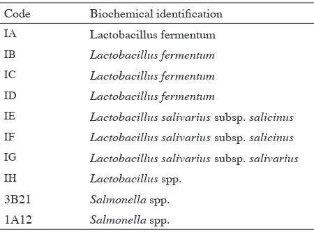
(a) (b) (c)
FIGURE 4. Inhibition zone of Salmonella spp. 3B21 against Lactobacillus strains by agar spot test (a) well diffusion assay, (b) blank disc method (c). a. Lactobacillus salivarius subsp. salicinus IF; b. Lactobacillus spp. IH; c. Lactobacillus fermentumID;
Du Toit, M., Franz, C., Schillinger, U., Warles, B. & Holzappfel, A. 1998. Characterization and selection of probiotic lactobacilli for a preliminary minipig-feeding trail and their effect on serum cholesterol level, faeces pH and faeces moiture contents. Int. Food Microbiol. 40: 93-104.
Ehrmann, M.A., Kurzak, P., Bauer, J. & Vogel, R.F. 2002. Characterization of lactobacilli towards their use as probiotic adjuncts in poultry. Journal of Applied Microbiology 92: 966-975.
Ferket, P.R., Parks, C.W. & Grimes, J.L. 2002. Beneits of dietary
antibiotic and mannanoligosaccharide supplementation for poultry. In Multi-State Poultry Meeting, May 14-16, Atlanta, GA, USA.
Fuller, R. 1989. Probiotics in man and animals. J. Appl. Bacteriol.
66: 365-378.
Fuller, R. 1999. Probiotics for farm animals. In: Tannock, G.W. (ed.). Probiotics: A Critical Review. Wymondham, England:
Horizon Scientiic Press, pp. 15-22.
Gariga, M., Pascual, M., Monfort, J.M. & Hugas, M. 1998. Selection of lactobacilli for chicken probiotic adjuncts.
Journal of Applied Microbiology 84:125-132.
Gusils, C., Chaia, A.P., Gonzales, S. & Oliver, G. 1999. Lactobacilli isolated from chicken intestines: Potential use as probiotics. J. Food.Protect. 2(3): 252-256.
Hammes, W.P. & Hertel, C. 2006. The Genera Lactobacillus and
Carnobacterium. Prokaryotes 4: 320-403
Handley, P.S., Harty, D.W.S., Wyatt, J.E., Brown, C.R., Doran, J.P. & Gibbs, A.C.C. 1987. A comparison of the adhesion, coaggregation and cell-surface hydrophobicity properties
of ibrillar and imbriate strains of Streptococcus salivarius.
Journal of General Microbiology 133: 3207-3217.
Havenaar, R., Ten Brink, B. & In‘T veld, J.H.J.H. 1992. Selection of strains for probiotic use. In: Fuller, R. (ed.). Probiotics: A
ScientiicBasis. London, Chapman & Hall, pp. 209-221. Hose, H. & T. Sozzi. 1991. Biotechnology group meeting:
probiotics – fact or iction? J. Chem. Technol. Biotechnol.
36: 379-383.
Huis in’t Veld, J.H.J., Havenaar, R. & Marteau, P. 1994.
Establishing a scientiic basis for probiotic R&D. Trends
Biotechnol. 12:6-8.
Jacobsen, C.N., Nielsen, V.R., Hayford, A.E., Moller, P.L., Michaelsen, K.F., Paerregaard, A., Standstrom, B., Tvede, M. & Jacobsen, M. 1999. Screening of probiotic activities of forty-seven strains of Lactobacillus spp. by in vitro techniques
and evaluation of the colonization ability of ive selected
strains in human. Appl. Environ. Microbiol. 65: 4949-4956. Jankovic, I., Ventura, M., Meylan, V., Rouvet, M., Elli, M. &
Zink, R. 2003. Contribition of aggregation-promoting factor to maintenance of cell shape in Lactobacillus gasseri 4B2.
J.Bacteriol. 185(11): 3288-3296.
Jin, L.Z., Ho,Y.W., Abdullah, N., Ali, M.A. & Jalaludin, S. 1996. Antagonistic effect of intestinal Lactobacillus isolates on pathogens of chicken. Letters in Applied Microbiology 23: 67-71.
Juven, B.J., Schved, F. & Lindner, P. 1992. Antagonistic compounds produced by a chicken intestinal strain of
Lactobacillus acidophilus. J. Food Protect 55: 157-161.
Lan, P.T.N., Binh, L.T. & Benno, Y. 2003. Impact of two probiotic
lactobacillus strains feeding on fecal lactobacilli and weight
gains in chicken. J. Gen. Appl. Microbiol. 49: 29-36. Langhendries, J.P., Detry, J., Van Hees, J., Lamboray, J.M.,
Darimont, J., Mozin, J., Screatin, M.C. & Sentere, J. 1995. Effect of a fermented infant formular containing viable
biidobacteria on the faecal lora composition and pH of
healthy full-term infants. J. Pediatric Gastroenterol. nutr.
21: 177-181.
Makras, L., Triantafyllou, V., Fayol-Messaoudi, D., Adriany, T., Zoumpopoulou, G., Tsakalidou, E., Servin, A. & De Vuyst, L. 2006. Kinetic analysis of the antibacterial activity of probiotic lactobacilli towards Salmonella enterica serovar Typhimurium reveals a role for lactic acid and other inhibitory compounds. Research in Microbiology 157: 241-247. Nowroozi, J., Mirzaii, M., Norouzi, M. 2004. Study of
Lactobacillus as Probiotic Bacteria. Iranian J. Publ. Health
33(2): 1-7.
Pascual, M., Hugas, M., Badiola, J.I., Monfort, J.M. & Garriga, M. 1999. Lactobacillus salvarius CTC2197 prevents
Salmonella enteriditis colonization in chicken. Applied and
Environmental Microbiology 65: 4981-4986.
Plummer, R.A.S., Blissett, S.J. & Dood, C.E.R. 1995. Salmonella
contamination of retail chickens products sold in the UK.
Journal of Food Protection 58(8): 843-846.
Reid, G. & Friendship, R. 2002. Alternatives to antibiotic use: Probiotics for the gut. Anim. Biotechnol 13: 92-97. Reniero, R., Cocconcelli, P., Bottazzi, V. & Morelli, L. 1992.
High frequency of conjugation in Lactobacillus mediated by an aggregation-promoting factor. J. Gen. Microbiol. 138: 763-768.
Schneeman, B.O. 2002. Gastrointestinal physiology and functions. Br. J. nutr. 88(Suppl.2): S159-163.
Schillinger, U. & Lucke, F. 1989. Antibacterial activity of
Lactobacillus sake isolated from meat. Applied and
Environmental Microbiology 55(8): 1901-1906.
Stern N.J., Cox, N.A., Bailey, J.S., Berrang, M.E. & Musgrove, M.T. 2001. Comparison of mucosal competitive exclusion and competitive exclusion treatment to reduce Salmonella
and Campylobacter spp. colonization in broiler chickens.
Poult. Sci. 80: 156-60.
Walter, J. 2005. The microecology of Lactobacilli in the gastrointestinal tract. InProbiotics & prebiotics: Scientiic
aspects, edited by Tannock, G.W. Wymondham, England,