Don Johnson, MD
Carleton University and University of Ottawa, Ottawa, Ontario, Canada
ACL Made Simple
With 163 Illustrations
1 3
Don Johnson, MD
Carleton University and University of Ottawa Ottawa, Ontario, K1S 5B6
Canada
Library of Congress Cataloging-in-Publication Data Johnson, Don
ACL made simple / Don Johnson. p. cm
Includes bibliographical references and index. ISBN 0-387-40146-6 (alk. paper)
1. Anterior cruciate ligament. 2. Anterior cruciate ligament–Wounds and
injuries–Prevention. 3. Ligaments–Wounds and injuries. 4. Sports injuries. I. Title. RD561.J627 2003
617.1¢027–dc21 2003050496 ISBN 0-387-40146-6 Printed on acid-free paper.
© 2004 Springer-Verlag New York, Inc.
All rights reserved. This work may not be translated or copied in whole or in part without the written permission of the publisher (Springer-Verlag New York, Inc., 175 Fifth Avenue, New York, NY 10010, USA), except for brief excerpts in connection with reviews or scholarly analysis. Use in connection with any form of information storage and retrieval, electronic adaptation, computer software, or by similar or dissimilar methodology now known or hereafter developed is forbidden.
The use in this publication of trade names, trademarks, service marks, and similar terms, even if they are not identified as such, is not to be taken as an expression of opinion as to whether or not they are subject to proprietary rights.
While the advice and information in this book are believed to be true and accurate at the date of going to press, neither the authors nor the editors nor the publisher can accept any legal responsibility for any errors or omissions that may be made. The publisher makes no warranty, express or implied, with respect to the material contained herein.
Printed in the United States of America 9 8 7 6 5 4 3 2 1 SPIN 10931349 www.springer-ny.com
Springer-Verlag New York Berlin Heidelberg
A member of BertelsmannSpringer Science+Business Media GmbH
Disclaimer:
Contents
Chapter
1
Introduction . . . .
1
Chapter
2
Diagnosis of the ACL Injury . . . .
12
Chapter
3
Partial Tears of the ACL . . . .
26
Chapter
4
Treatment Options for ACL Injuries . . . .
31
Chapter
5
Graft Selection . . . .
45
Chapter
6
Hamstring Graft
Reconstruction Techniques . . . .
65
Chapter
7
Patellar Tendon Graft Technique . . . .
121
Chapter
8
Rehabilitation . . . .
139
Chapter
9
Complications . . . .
154
Chapter
10
Results . . . .
181
Chapter
11
Readings . . . .
193
Index . . . .
199
1
Introduction
1 During the past decade, the anterior cruciate ligament (ACL) has become a familiar term. Most athletes have heard about it or know someone who has had an injury to the ACL (Fig. 1.1). This book provides comprehensive information about the ACL that will help the caregiver make an informed decision on the best management of any injury.
The ACL is the main stabilizer of the knee for athletic pivotal activ-ities. The first repair of the cruciate ligament was attributed to Hay Groves in 1917. Then, in the 1930s, Ivor Palmer wrote one of the first definitive monographs on the subject, in which he advocated early sur-gical repair by suturing the ligament. Although primary suture repair was eventually found to have a high failure rate in athletes, Palmer had set the stage for the aggressive surgical approach of Swedish surgeons. The modern phase of treatment began when Jones, Erickson, and Macintosh all advocated reconstruction, rather than repair, of the ACL with the patellar tendon. In the 1980s, the extra-articular reconstruc-tions, as pioneered by Macintosh, were replaced with the intra-articular reconstructions as popularized by Erickson. The patellar tendon graft was the gold standard in the 1980s, but during the 1990s, as a result of improved graft preparation and fixation, the semitendi-nosus became more popular.
In the 1970s, the torn ACL was considered the beginning of a pro-gressive deterioration of the knee that often ended an athlete’s career. Now many athletes routinely return to play as soon as three to four months after an injury and certainly by the next season. The medical profession has gained considerable experience in the surgical treatment of ACL injuries, but has made little impact in the prevention of the injury, especially among downhill skiers.
2 1. Introduction
has lessened the morbidity of the operation and extended the operative option to both the recreational and older athlete.
With the increase in sports participation by the baby boomer gener-ation, injury to the ACL has become one of the most common athletic injuries.
Basic Science
Anatomy
The ACL is composed of two separate bundles, the anteromedial and the posterolateral. The intra-articular length of the ligament is between 28 and 31 mm. The attachment sites on the tibia and femur have a fairly small isometric center (Fig. 1.2).
The ACL and posterior cruciate ligament (PCL) are closely inter-twined and are called the “central pivot.” An injury to either ligament disrupts the function of the joint and may lead to late degenerative arthritis. The relationship between the ACL and the PCL is shown on the video on the enclosed CD.
Basic Science 3
Figure1.2. The ACL is the main restraint to anterior motion of the tibia on
Biomechanics
Tensile Strength
Noyes has reported the ultimate failure load to be 1750 N. He also noted that the young cadaver specimens were stronger than the older ones.
Viscoelasticity
The speed of the force applied to the ligament affects the type of tear. The slow, twisting type of low-velocity injury experienced by skiers may elongate the ligament it ruptures up to 30%. There may be partial rupture of the anteromedial bundle, leaving the posterolateral bundle intact or vice versa.
If the patient is involved in low-demand activities, this partial remnant may be adequate for stability. We followed 20 recreational athletes diagnosed as having a partial tear. After two years, only one required a reconstruction of the ACL. This one may have been a partial tear or partial healing of a complete tear, but the result is no demonstrable laxity. In high-demand athletes, the bundle may be insufficient to control the pivotal stress and may have to be reconstructed. The conventional wisdom states that if more than 50% of the ligament is still intact, no reconstruction is necessary.
Contrast that type of injury to that of the football or basketball player who suddenly brakes and changes direction. This high-velocity force will usually produce a midsubstance tear, which has little potential for healing.
The Isometric Points
The surgeon must know the isometric points of insertion of the tibia and femur to drill the proper tunnels for the reconstruction of the ACL. Larson and Siddles have computer mapped these points.
The middle of the femoral tunnel is 7 mm in from the drop-off at the 11 or 1 o’clock position. If a 10-mm tunnel is drilled at this point, then a 1- to 2-mm posterior wall will be left. The tibial site is 7 mm from the leading edge of the PCL at 70° of knee flexion. The ligament of Humphrey and fat on the PCL must be taken into account in this measurement.
Intra-Articular Length
articular length, and the femoral tunnel must be compared to the graft length to avoid mismatch in the endoscopic reconstruction. This calcu-lation avoids protrusion of the graft on the tibial side. The overall length of the graft is 9 to 10 cm.
Natural History of ACL Injuries
In his textbook,Knee Ligaments, Structure, Function, Injury, and Repair, Dale Daniel describes the fate of the unoperated knee ligament injury. The data come from 500 patients who came to the Kaiser emergency room in San Diego with knee injuries.
Many authors have stated that the ACL injury is career ending for the athlete and that the knee will embark on a course of progressive degeneration. Noyes, however, reported in 1983 on a group of sympto-matic patients that he placed on a conservative program of exercise, bracing, and activity modification. From this study came the “rule of thirds.” One-third of the patients were able to compensate and return to light sports without symptoms, one-third had to significantly reduce their activities, and one-third required reconstruction. Only 10% of the patients returned to sports without problems.
In Daniel’s San Diego study of 279 patients with an isolated ACL injury, 20% underwent acute reconstruction, 18% underwent subse-quent reconstruction because of chronic symptoms, and the remainder were treated nonoperatively. This group was followed for at least 5 years. Daniel drew the following conclusions from this study:
• The acute ACL tear is associated with a meniscal tear in 50% of the cases (lateral tears are more frequent than medial tears).
• In the chronic cases, the incidence of tears is 80%. The medial tear is more common in the chronic situation.
• 40% of the tears are repairable.
• Chondral injuries are twice as common (40%) in the chronic cases as compared to the acute cases.
• The patients have minimal pain and no giving way with normal activ-ities of daily living.
The study further concluded that the outcome of the ACL injury is related to the following risk factors:
• The age of the athlete.The younger patient did not fare as well as the older patient.
ticipate after an ACL injury. This was also related to the level of com-petition and the number of hours of sports participation.
• Degree of anteroposterior (a-p) laxity. Daniel also determined that with more a-p laxity, the functional level of the athlete decreased. He advocated an objective measurement of the laxity with the KT-1000 arthrometer. He determined that more than 3 mm of side-to-side dif-ference was diagnostic of an ACL injury in 98% of the cases. With more than 7 mm of difference and a gross pivot shift examination, he suggested surgical reconstruction.
Mechanisms of Injury
Noncontact Pivot, Internal Rotation/External Rotation
The most common injury mechanism involves no contact with others. The athlete is simply running and abruptly changes direction. The ACL is stressed by the rotation of the tibia, resulting in a tear of the ACL, as illustrated in the video on the CD. The athlete lands in the flexed posi-tion, the quadriceps contract, and the tibia is subluxed anteriorly. Then with further flexion, the tibia reduces with a snap. This is the same mech-anism that the pivot-shift test mimics. Ireland has reported that the noncontact mechanism is responsible for 80% of ACL tears.
The Quadriceps Active Mechanism
The quadriceps contraction may be of importance in injuring the ACL. Barrett and coworkers have calculated that an eccentric quadriceps con-traction can generate up to 6000 N. This far exceeds the strength of the ACL at 1700 N. This force can be observed in basketball in the jump-stop landing. Ireland believes that there is a “position of no return” for ACL injury. The body position is body forward-flexed, hip-abducted, knee that is internally/externally rotated with valgus; the foot is pronated. This position can be reviewed in the video of the badminton player.
However, some additional unrecognized factor must be involved in this mechanism. Many athletes have done that particular move a thousand times without injury. Then, one particular time, the ACL tears. Ireland calls this a “heart attack of the knee.” There must be late acti-vation of the hamstrings, because contraction of the hamstrings would normally protect the tibia from subluxing forward.
and the force of pulling on the quadriceps subluxes the tibia forward and ruptures the ACL. The next sequence shows the ruptured ACL. The last sequence demonstrates a positive Lachman test on the specimen.
In summary, the body and knee must be in the correct position, the quadriceps must contract strongly enough to sublux the tibia, and the hamstrings fail to protect the anterior subluxation.
Can this be prevented? Perhaps by neuromuscular, proprioceptive training, some injuries may be prevented.
Contact Mechanism
A common mechanism in football or hockey is the blow to the outside of the knee when the knee is flexed and rotated. This initially injures the medial collateral ligament (MCL), and then, with further valgus, the ACL is torn.
Another variation of the contact injury is the internal rotation skiing injury described by Bob Johnson, a doctor from the University of Vermont. In this mechanism, the skier sits back and the ski carves to the inside, producing an internal rotation stress on the knee and a tear of the ACL.
The elements of a potential ACL tear by the contact mechanism are:
• Uphill arm back.
• Skier off balance to the rear. • Hips below the knees. • Uphill ski unweighted.
• Weight on the inside edge of downhill ski. • Upper body generally facing downhill ski.
Johnson has also described another mechanism that occurs when the tail of the ski hits a bump on the snow and the high ski boot levers the tibial forward, thereby producing an anterior drawer force and tearing the ACL.
Hyperflexion or Hyperextension Mechanisms
These less-common mechanisms of injury are often associated with other injuries to ligaments, such as the posterior cruciate ligament.
Gender Issues
(NCAA) are 2.4 times greater in soccer and 4.1 times greater in bas-ketball for female athletes. The reason is still speculative, but several theories are under investigation. Arendt’s statistics show that the non-contact injury mechanism was the main cause of the ACL tear.
In an article by Traina and Bromberg, the authors listed the follow-ing as possible causative factors:
Extrinsic
• Muscular strength. • Body movement. • Shoe surface interface. • Level of skill.
Intrinsic • Joint laxity. • Limb alignment.
• Notch width and ligament size.
Extrinsic Conditioning
Many authors believe that the novice female athlete is introduced to activities that are beyond her physical conditioning. Tim Hewett has demonstrated that unconditioned females land from a jump with the knee more extended, and, because of the wide pelvis, in a valgus posi-tion. This extended valgus position puts them at risk for an ACL injury. If slight external rotation is added on landing, then they are in a posi-tion of no return (as described by Ireland). Hewett has advocated not only conditioning programs, but also instruction on proper landing posi-tion (i.e., slightly flexed with knees straight ahead). This is one positive step that can be instituted to reduce the incidence of ACL injuries in females.
Muscular Strength
Woitys (in Griffin et al.) has shown that gender differences exist in muscle strength, muscle recruitment order, and hamstring peak torque times. The implication is that women should emphasize hamstring strengthening to protect the ACL.
Body Movement
• Planting and cutting: 29%. • Straight knee landing: 28%.
• Landing with knee hyperextended: 26%.
Hewett has shown that training the female athlete to modify the landing stance to a flexed neutral knee position has reduced the inci-dence of ACL injuries.
Intrinsic Joint Laxity
There are contradictory studies on the role of ligamentous laxities. Daniel’s study with the KT-1000 arthrometer showed no gender differ-ences in the measurable laxity of the ACL. It has been documented that exercise produces laxity of the ACL, but there are no significant differ-ences in gender.
Yu et al. have shown that the ACL has both estrogen and proges-terone receptors. The cyclic variation of estrogen may affect the liga-ment metabolism and make females more prone to injury during the estrogen phase of their cycle. Karangeanes and Vangelos studied the incidence of ACL injury during the cycle of increased estrogen and found no significant difference.
Limb Alignment
Ireland has emphasized limb alignment (the wider pelvis, increased femoral anteversion, and the genu valgum) with decreased muscular support, specifically the hamstrings, as possible causes for the increased ACL injury rates in women
Notch Width
Shelbourne and Klootwyk have documented that women have a smaller notch than men. It has also been reported that athletes who sustain ACL injuries have a narrow notch (Fig. 1.3). It may well be that the narrow notch is only one indication of a small incompetent ligament that is easily torn. Evidence for this is seen after a large notchplasty in which the notch will fill in around the new graft.
Conclusion
At the present time, the best advice to give the female athlete is to be well conditioned and land with a flexed knee.
10 1. Introduction
Figure1.3. The anatomic variation of wide pelvis, valgus knees and reduced
Prevention
Johnson believes that if you are aware of the common mechanism that produces an ACL injury, you can help skiers prevent the injury. He has reviewed thousands of hours of on-hill ski injury video and identified a common mechanism that involves sitting back on the skis and trying to recover as one ski carves inward. The Vermont group has produced a videotape on this mechanism of injury and its prevention. His advice is, do not sit back and then try to recover. Rather, fall to the uphill side. A skier aware of this mechanism may be able to prevent an ACL injury. Johnson has taught the ski patrollers in the area about the mechanism; injury rate has been reduced by 62%.
The phantom foot mechanism and the possible preventive measures have been outlined in a videotape available from Dr. Robert Johnson, University of Vermont, Stafford Hall, Room 426A, Burlington, VT 05405-0084; voice (802) 65-2250; fax (802) 656-4247.
2
Diagnosis of the ACL Injury
12
History
The athlete describes a twisting injury to his knee, associated with a “popping” sensation in the knee. This is followed by immediate pain and swelling of the knee. He may indicate the feeling of the knee coming apart with the “2-fist sign.” Figure 2.1 shows the athlete indicating the “2-fist sign.”
The severity of the symptoms vary a great deal, depending on the degree of meniscal, chondral, and capsular injury. The athlete may come in walking, with minimal swelling, or on crutches, unable to bear weight. It depends on the associated injuries.
In rare situations, the injury that tears the anterior cruciate ligament (ACL) may be so trivial that the athlete returns to the game. But the next time he pivots on his knee, much more damage, such as a tear of the meniscus, is the result.
Physical Examination
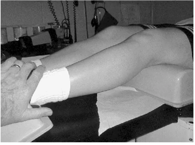
Lachman Test
the lower leg should be lowered over the edge of the table to help relax the hamstrings. This position also works well for examiners with small hands or when examining a very large leg. This is called the drop leg test. The ideal knee flexion angle is 30°.
The Lachman test is a subtle test that requires experience to admin-ister confidently. The knee is flexed to 30°, the femur is stabilized, and the tibia is pulled forward. The test is positive when the endpoint is soft. The main feel is the lack of the endpoint to the anterior translation of the tibia. The comparison to the opposite side is important.
The grading should be, negative, 1+with endpoint, or positive with no endpoint (Table 2.1). It is difficult to differentiate between 2+and 3+or to compare between examiners, so these grades have little meaning.
Physical Examination 13
Pivot-Shift Test
This test is more difficult to perform, but is more consistent in repro-ducing the athlete’s symptoms. Holding the heel in one hand and apply-ing a valgus stress in the other hand, the knee is slowly flexed. The tibia, when in internal rotation, slides anterior when the valgus stress is applied. The tibia, as well as the valgus, subluxes easily if anterior force is applied. After the anterior subluxation of the tibia is noticed, the knee is slowly flexed, and the tibia will reduce with a snap at about 20° to 30° of flexion. This reduction can be augmented with an external rotation of the tibia, as noted in Figure 2.3. This is the “pivot shift.” It is the same mechanism that the athlete experiences when his knee “comes apart” with pivoting. The patient will usually indicate that is the sensation experienced when the knee gave out. The pivot-shift test is graded from 0 to 3 (Table 2.2). The video on the CD demonstrates the pivot-shift test.
14 2. Diagnosis of the ACL Injury
Figure2.2. The Lachman test for ACL laxity.
Table2.1. The Lachman test is graded from 0–3.
Value Interpretation 0 Negative
1+ 0–5 mm of anterior displacement, sometimes with an end point 2+ 5–10 mm of anterior displacement, with no end point
Range of Motion
The physician should always examine the knee for loss of extension by holding both heels clear of the table and comparing the extension of the injured knee against the uninjured knee (Fig. 2.4).
The loss of extension is often the result of the ends of the torn liga-ment impinging anteriorly in the notch. The other common cause of lack of extension is a displaced bucket-handle tear of the meniscus. This may also alert you to a hyperextension and external rotation that indicates an associated posterolateral injury.
Effusion
The tear of the ACL usually produces a hemarthrosis that will appear immediately after the injury. In acute knee injuries, the torn ACL is the Physical Examination 15
Figure2.3. The pivot-shift test.
Table2.2. The grading of the pivot-shift test.
Value Interpretation 0 Negative shift
1+ A glide 2+ A pivot shift
cause of a bloody effusion in 75% of the cases. This right knee above demonstrates the effusion (Fig. 2.5), noted by the lack of the contour of the patella, which is seen the day after injury.
The acute knee should be aspirated of blood to make the patient 16 2. Diagnosis of the ACL Injury
Figure2.4. Both legs are lifted by the heels to examine knee extension.
Physical Examination 17
Figure2.6. Aspiration of the hemarthrosis that results from an ACL tear.
more comfortable (Fig. 2.6). If there are visible fat globules on the surface of the blood, this should make you think of an intra-articular fracture. Appropriate imaging studies should be done to detect a tibial plateau fracture.
Joint-Line Tenderness
Both the medial and lateral joint lines should be palpated for tender-ness (Fig. 2.7). The meniscal injury usually has joint-line tendertender-ness. In 50% of acute cases, an associated meniscal injury is present. The lateral meniscal tear is more common in the acute situation. In chronic cases, the incidence rises to 80% and is more common on the medial side. In acute cases, it is difficult to do a McMurray test described next because of limited flexion.
McMurray Test
meniscus. The McMurray test is performed by fully flexing the knee and rotating the tibia as the knee is slowly extended (Fig. 2.8). A positive test is painful with full flexion and rotation; a clunk or snap is heard or felt when the knee is extended. The medial tear is elicited initially with the internal rotation followed by the external rotation during extension. The lateral tear is done in the reverse fashion. This rotation of the tibial plateau will catch the posterior horn of the meniscus between the tibia and femoral condyle, producing a clunk and causing pain. The meniscus tugging on the pain-sensitive synovium at its peripheral attachments produces the pain. The test is notoriously inaccurate, and in most situ-ations the pain with full flexion and rotation is sufficient to confirm an injury to the meniscus.
The mechanism of the popping with the McMurray test is demon-strated in the video on the CD. It shows the tibial plateau subluxing forward and trapping the posterior horn of the meniscus between the femur and the tibia. This is associated with a clunk. It also illustrates why the unstable knee has a high incidence of meniscal tears.
Collateral Ligament Assessment
The collateral ligaments are assessed by varus and valgus stress testing at 0° and 30° (Fig. 2.9). The grading is “stable” or “no motion.” Grade 18 2. Diagnosis of the ACL Injury
Figure2.7. Palpation of the medial joint line for tenderness compatible with a
Figure2.8. The McMurray test.
Figure 2.9. Valgus stress is applied to the knee to test the medial collateral
20 2. Diagnosis of the ACL Injury
Figure2.10. In the anterior drawer test, the ACL is stressed by pulling the tibia
anteriorly at 90° of flexion.
1 has no motion, but is painful on stress. Grade 2 has laxity with an end-point, and grade 3 is gross laxity at both 0° and 30°. The site of tender-ness on the ligament can determine the site of injury (i.e., on the femur or tibia). The examination of the collaterals is important to determine whether the ACL injury is isolated.
Anterior Drawer Test
Quadriceps Active Test
Figure 2.11 shows the left tibia subluxed forward with a quadriceps con-traction. This is indicative of an ACL-deficient knee. When the quadri-ceps is contracted against resistance with the knee flexed at 30° and without weight bearing, there is an anterior displacement of the tibia (this is an open kinetic chain exercise). Figure 2.11 shows the tibia sub-luxed anteriorly. This nonweight-bearing exercise is called open kinetic chain exercise.
Open kinetic chain exercise is also seen with the patient on the quadriceps machine in a fitness room. The athlete sits on the leg exten-sion machine and extends the knee. The quadriceps pulls the tibia forward if there is no ACL or causes significant strain on the ACL graft. In the early rehabilitation phase, this exercise must be avoided to prevent strain on the recently implanted graft.
Associated Ligament Injuries
It is always important to perform a posterior drawer test (Fig. 2.12). If this is done routinely, you will not miss a posterior cruciate ligament Physical Examination 21
injury. The video on the CD demonstrates the posterior drawer test. The external rotation of the tibia must be measured at both 90° and 30° to rule out associated injury to the posterolateral corner.
Imaging
Plain Radiographs
The screening examination should be a simple anteroposterior and lateral radiograph of the knee. This will reveal open growth plates, ACL bony avulsions, significant osteochondral fractures, tibial plateau frac-tures, or epiphyseal fractures.
Tomograms
If the radiograph is negative, but considerable bony tenderness exists, then tomograms should be done to rule out plateau fractures.
Computed Tomography Scan
The 3-D scan can help plan treatment for associated tibial plateau fractures.
22 2. Diagnosis of the ACL Injury
Bone Scan
If the pain persists, this scan may confirm occult bony injury.
Magnetic Resonance Imaging
In a few situations, magnetic resonance imaging (MRI) will change your management of an injury. The diagnosis of the ACL tear should be made clinically. If the loss of extension persists, the MRI can be performed to determine whether this is a bucket-handle tear or an impingement of the ACL bundle, a cyclops lesion. The meniscus tear should be repaired early and, in some situations, the ACL reconstruction should be delayed until a good range of motion has been achieved after the meniscus repair. In the cyclops lesion, both the debridement of the ligament ends and the ACL reconstruction can be done simultaneously as described by Pinczewski. Remember that a good physical examination by an expe-rienced physician is more reliable than an MRI.
Examination Under Anesthesia and Arthroscopy
The arthroscope has been the key to unlocking the diagnosis of knee pathology (Fig. 2.13). The arthroscope has improved the diagnosis of knee injuries, but the scope examination is only one aspect of the puzzle. One of the mistakes residents make is to go ahead with the arthroscopy before performing a clinical examination of the knee. The examination under anesthesia (EUA) is a valuable adjunct to the diagnostic work-up. At this time, the grading of the laxity may be documented. It is often difficult to examine the very large knee of a football player with multi-ple ligament injuries in the training room. The EUA may be the only means of making the diagnosis.
Arthroscopy of the acute knee presents no more technical prob-lems than with the elective case. The hemarthrosis must first be flushed out. The synovium and ligamentum mucosum around the ACL must frequently be removed to fully assess the degree of liga-ment injury. The hook is used to probe the two bundles, and to assess tension.
The video on the CD shows how the diagnostic arthroscopy must be performed in a similar fashion each time, so that the knee will be completely examined and no region forgotten. This must be done before any surgical procedures are started. The video shows the inside view of the “W” arthroscopy. The “W” procedure enables the physician to view the patellofemoral joint, the medial gutter, the medial compart-ment with the medial meniscus, and then to go over the top of the
ligamentum mucosum to view the ACL and finally the lateral compart-ment with the lateral meniscus.
The video shows the arthroscopic view of the torn ACL. The menis-cus and articular surface should be completely examined. The capsular injury may be seen by inspecting the gutters, and examining over and under the meniscus. If there is significant capsular tearing, then gravity pressure only, rather than a pump, should be used. The ACL tear has produced a stump at the front of the knee that prevents full extension. This mimics a locked knee.
24 2. Diagnosis of the ACL Injury
This ACL tear is only partial or interstitial. The fat pad in front of the ligament has to be removed to visualize the ligament, and the ligament must be probed to assess its status. This ligament tear may have been produced by the narrow stenotic notch.
The diagnostic examination of the knee must be complete to detect any meniscal injuries. In the acute knee, the incidence of meniscal tear is approximately 50%. In the chronic ACL-deficient knee, the incidence of meniscal tears may be as high as 75%.
The video demonstrates the chronic ACL tear. The residual ligament is probed with a hook, and it can be appreciated that it is not attached to the femoral condyle.
3
Partial Tears of the ACL
26
One of the dilemmas facing the sports physician is treatment of the partial tear of the anterior cruciate ligament. The definition of a partial tear is a history of injury to the anterior cruciate ligament, a positive Lachman test with a firm end point, a negative pivot-shift test, KT-1000 side-to-side difference of <5 mm, and arthroscopic evidence of injury to the anterior cruciate ligament.
The natural history of the partial tear is controversial. Reports suggest that both conservative and operative treatment offer good results. Noyes and his colleagues had a 50% incidence of instability in high-demand sports participation athletes who had an anterior cruciate ligament tear of more than 50%. They also had a 75% incidence of reinjury. This suggests that patients in high-demand sports require reconstruction. Freunsgaard and Johnannsen had good results with con-servative treatment in patients who avoided high-demand athletics, and Buckley and colleagues reported that the degree of anterior cruciate tear did not correlate with outcome. Only half of their patients were able to resume their previous level of sports activity.
Physical Examination
Lachman Test
The Lachman test is positive, but there is a firm end point (See Fig. 2.2). This anterior excursion is greater than the opposite side, but less than 5 mm of the side-to-side difference measured on the KT-1000 arthrometer.
Pivot-Shift Test
defi-cient. The pivot shift is the most important assessment of the partial tear.
The KT-1000 Arthrometer
The KT-1000 arthrometer will normally show a side-to-side difference of less than 5 mm (Fig. 3.1). The slope of the curves that are pulled with the KT-2000 demonstrate the difference. Force of 15, 20, and 30 pounds is applied to the vertical axis of the knee; the horizontal axis shows millimeters of displacement. The curve on the left shows the normal anterior cruciate ligament. The middle curve shows that there is initially more displacement, but then a firm restraint to anterior translation. This corresponds to the firm end point to the Lachman test. The third curve on the right is the anterior cruciate deficient knee with complete rupture. The stronger the pull, the more anterior displacement.
Physical Examination 27
Magnetic Resonance Imaging
It is difficult to estimate the degree of ACL injury with the MRI, as the laxity of the ligament cannot be accurately assessed. Therefore, it is not a useful tool for diagnosing partial tears of the anterior cruciate ment. Figure 3.2 shows a small band where the anterior cruciate liga-ment should be. It is difficult to estimate how much of the ligaliga-ment is still present.
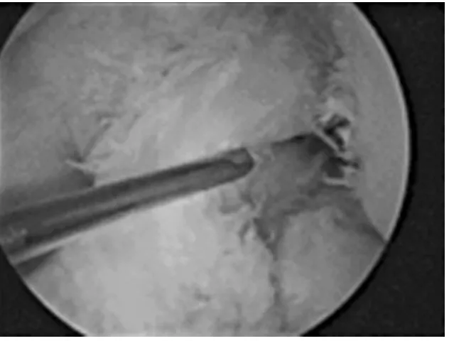
Arthroscopic Assessment
Arthroscopic assessment of the anterior cruciate ligament tear is diffi-cult for two reasons. First, it is hard to see the ligament without remov-ing the synovium and fat pad. Second, it is only an estimate of the degree of tearing of the ligament. It seems to be best to try to estimate 28 3. Partial Tears of the ACL
whether the tear is less than or greater than 50%. A hook probe must be used to examine the ligament proximally to see where the ligament is attached—to the side wall, the roof, or the posterior cruciate ligament. The best position is the side wall at the normal site of the anterior cruciate ligament. The most common situation is to see the ligament attached to the posterior cruciate ligament.
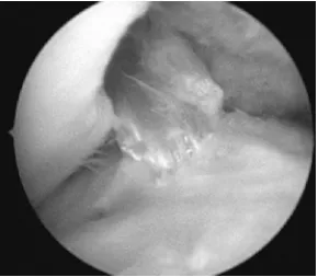
Figure 3.3 shows the appearance of the ligament proximally. It has attenuated to a small band attached to the side wall. This may give a 1+
Lachman test and a negative pivot-shift test, but would not stand up to vigorous pivoting activities. Figure 3.3 also shows the use of the probe to examine the ligament proximally. This example is lax, but is less than 50% tear. This amount of ligament laxity should allow a return to sports without a reconstruction.
Treatment Options
Partial Tears
The treatment options for a patient with partial ACL tear are to give up or modify his or her sports activities. The patient who can modify his Treatment Options 29
sports activities and avoid pivotal sports will do well with a partial ante-rior cruciate ligament injury. This is the only parameter that the indi-vidual has control over, and that point should be emphasized when counseling athletes.
Brace and Arthroscopy
The use of a brace combined with modification of activity can be suc-cessful. Sometimes a meniscal injury will still cause a giving way sensa-tion. The best long-term outcome for the young patient is to have a meniscal repair. The dilemma is whether to reconstruct the ACL. The results of a meniscal repair are much better when the knee has been reconstructed and is stable.
ACL Reconstruction
If there is a positive pivot-shift test or a small bundle attached to the femur, and the athlete wants to be active in pivoting sports, anterior cru-ciate ligament reconstruction should be considered.
Indications for ACL Reconstruction
The patient who is a candidate for reconstruction of the ACL is the com-petitive, pivoting athlete who is involved in sports such as soccer, rugby, and basketball. In addition, the patient should have clinical symptoms of instability, with a history of giving way, a positive Lachman, and pivot-shift test with more than 5 mm side-to-side difference on the KT-1000 arthrometer.
4
Treatment Options for ACL Injuries
31 The treatment of the anterior cruciate injury must be individualized to the patient. Not all tears of the ACL need operative repair. The treat-ment options for the elite athlete, who needs reconstruction, as well as the inactive patient, who needs no reconstruction, are fairly limited. It is the recreationally active individual whose ACL injury requires counseling for the best treatment plan. There are a number of factors to consider in this decision, including, as Shelbourne has emphasized, age, chronicity, activity level, and associated injury to the meniscus and articular surface.
Patient Factors
The treatment of the ACL injury should be determined by the follow-ing factors.
Age of the Patient
The older patient may be more likely to modify his lifestyle and accept a conservative treatment program, while the younger patient, who is involved in competitive sports, wants to return as quickly as possible to high-level sports without the use of a brace.
Activity Level and Intensity
Degree of Instability
In the Kaiser study, the outcome was related to degree of instability. If the KT-1000 arthrometer side-to-side difference was greater than 7 mm, the chance of a better outcome was with surgical reconstruction.
Size of Athlete
The forces that a 300 lb lineman exerts on his knee with pivoting are much more that the 150 lb tennis player. In the case of the former, surgical reconstruction should be considered.
Treatment Choices
There are 3 treatment choices for the ACL tear.
Give Up or Modify Sport Activities
It is important to emphasize that the ACL is asymptomatic with most activities of daily living. If the patient is not involved in sports then he will usually have no giving way episodes, and no surgical treatment is necessary. Giving way in the sedentary patient is more likely the result of meniscal pathology. The meniscus may be treated by arthroscopy, and the patient can continue with the nonoperative treatment program. The patient should be counseled to switch into knee friendly sports, such as cycling and swimming.
Brace and Arthroscopic Meniscectomy
If the patient is recreationally active, a functional brace will often be sufficient to stabilize the knee for low-demand sports, such as doubles tennis. However if he has giving way in the brace, a meniscal tear may be present. Approximately 50% of ACL tears have an associated menis-cal tear. The younger athlete should have a menismenis-cal repair and recon-struction for the ACL. The long-term results of meniscal repair are better with a stable knee, and the meniscal repair without reconstruc-tion is not an opreconstruc-tion. The older patient should have a meniscectomy and use a brace for sports. If the patient still gives way in the brace, then consideration should be given to a reconstruction.
Anterior Cruciate Ligament Reconstruction
ticipate in sports and, hopefully, prevent late degenerative changes. Shelbourne has recently reported that if the meniscus and articular cartilage is normal at the index operation, the X-ray evaluation will be normal at 10 years in 97% of the patients. This means that the athlete who has an early ACL reconstruction will be able to continue to be active without the risk of degenerative osteoarthritic changes in his knee. The patient who continues in sports with recurrent giving way, as a result of ACL laxity, will have a degenerative knee in 10 to 12 years.
Summary
The most important outcome factors are the patient’s age, the activity level, and the degree of instability. The activity level is the only one of these factors that the patient can control. Thus for a nonoperative approach to be successful, the patient’s activity level must be modified. The other treatment options, such as brace and meniscal repair, will only be successful if activity is diminished. Ninety percent of the patients who undergo ACL reconstruction will be able to return to full athletic participation.
Plea for Conservative Treatment
Conventional wisdom states that the ACL does not heal. However, in some instances, especially with downhill skiing injuries, it can. There is little argument that the young competitive, pivoting soccer player with a positive pivot shift and a 7-mm side-to-side difference on the KT arthrometer needs a reconstruction, but consider another example.
Case 1
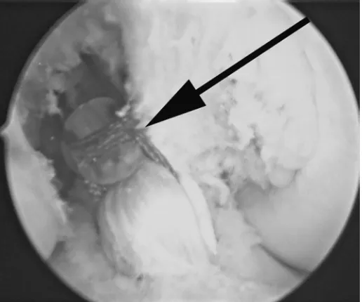
KB, who is a 31-year-old interior designer and an advanced recreational skier, injured her knee downhill skiing. She had an external rotation, valgus injury with an audible pop in her knee. The bindings did not release. She was assessed at the ski hill and diagnosed with an ACL tear. Two weeks later, an examination at the clinic revealed an effusion, joint-line tenderness, positive Lachman, and a positive pivot-shift test. She was advised to have a reconstruction and started therapy to improve the range of motion and reduce the effusion. At six weeks after injury, she had a positive Lachman (no end point), positive pivot shift, and a KT manual maximum side-to-side difference of 6 mm. She was advised to proceed with a reconstruction.
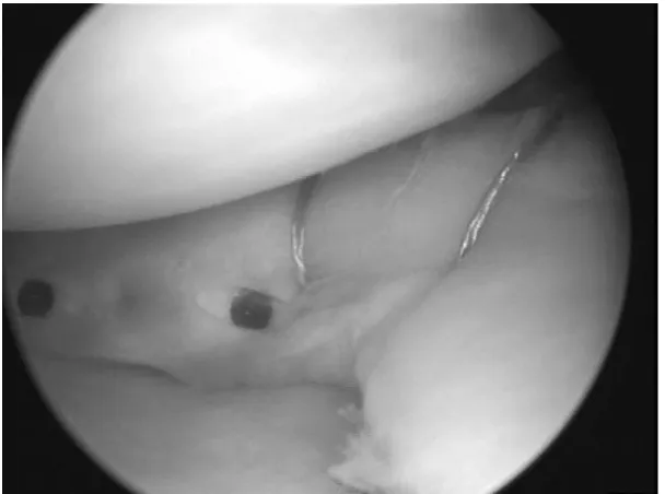
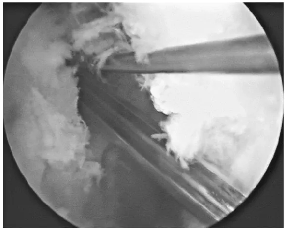
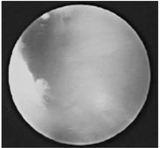
ference of 2 mm. The arthroscopic examination demonstrated normal menisci, normal articular cartilage, and an ACL healed to the femoral condyle. The appearance of the well-healed ACL is shown in Figure 4.1. We have usually divided the degree of injury to the ACL as more than or less than 50%. In reality, this means a normal appearing bulk of ligament present as opposed to this thin strand of ligament. Figure 4.2 shows the arthroscopic appearance of a small incompetent band of ACL left after partial healing of the ACL tear. No physician would have a problem proceeding with a reconstruction in this situation.
Conclusion
On the basis of the clinical examination alone, a physician could prob-ably recommend conservative treatment of this injury. However, what the physician cannot assess is how much of the ligament is present. If the patient described above had only the small strand shown in Figure 4.2, would it be wise to allow her to go back to aggressive skiing, to pos-sibly reinjure her knee and tear her meniscus?
Yet, a number of skiing patients who have torn the ACL have been documented with clinical examinations and with KT arthrometer read-ings that eventually heal and do not require surgery. In the past, surgery 34 4. Treatment Options for ACL Injuries
would have taken place within the first few weeks of the injury, and there would have been no opportunity to see this natural evolution of the healing process.
Timing of Surgical Intervention
The timing of surgical treatment is controversial. Shelbourne has shown that reconstruction done acutely results in more stiffness and greater loss of range of motion. To avoid this, reconstruction should be delayed until a full range of motion is achieved. In the Sports Medicine Clinic, after the diagnosis is made, most patients go to physiotherapy to regain range of motion and to reduce the swelling. No one is reconstructed without full knee extension. If full extension is not gained in physio-therapy, then the torn ACL bundle or a bucket-handle tear of the menis-cus must be treated first. Arthroscopy should be performed to repair the meniscus or excise the cyclops lesion of the ACL before the recon-struction. After the meniscal tear is repaired or excised, physiotherapy is resumed to regain knee extension before the reconstruction. Pinczweski has reported that the cyclops lesion of the ACL may be Timing of Surgical Intervention 35
removed and the ACL reconstruction done at the same time without risk of limited motion postoperatively, but Shelbourne recommends that repair of the bucket-handle meniscus tear and the ACL reconstruction should be staged.
There are no hard and fast rules, such as wait three weeks before operating. Some patients will have good range of motion and no swelling in one week, and they need only to work on the bike preoper-atively. Other patients will take six or eight weeks to be ready for surgery. The physician should read the tissues. This means to look at the effusion, range of motion, and the induration of the capsule. The time to operate is when the tissue is soft and compliant, and the range of motion is good. The treatment options are outlined to the patient, who receives an educational information sheet on the options. If he is unde-cided, then a trial of brace management is suggested. The brace may also be used to try to get the patient through the current season of sport or semester of school. He may be able to participate at a reduced level while waiting to have the reconstruction. Shelton has reported his experience with high school athletes who tear their ACLs early in the season. Thirty of 43 patients returned to play in 6 weeks with a brace, but only 12 had no giving way episodes. Twenty-nine of these patients eventually underwent ACL reconstruction. The downside of this expe-rience is that some of these patients were unable to undergo meniscal repair because of further injury of the meniscus. It is also important to reexamine and remeasure the KT-1000, as some of these patients will partially heal to a 1+Lachman. This partial healing may be adequate stability for the recreational athlete.
Controversial Treatment Decisions
Other factors that influence the decision to treat are associated lesions, such as chondral fractures, meniscal tears, and other ligament tears, but the real controversies center on the age of the patient, the associated injury to the medial collateral ligament, and the patient with medial compartment osteoarthritis.
Older Athlete
tion for surgery. Patient selection may be expanded according to activ-ity level. The younger and more pivotal athlete, who wants to return to sport sooner may be a candidate for the patellar tendon graft. Shel-bourne has reported on return to sports at four months with a contra-lateral patellar tendon graft harvest.
Older, more recreational athletes usually have a semitendinosus auto-graft auto-graft or an alloauto-graft patellar tendon. There have been several authors, including Brandsson, who have reported positive results of ACL reconstruction in patients more than 40 years of age. Remember that the patellar tendon graft is for the surgeon, and the semitendinosus graft is for the patient.
Immature Athlete
The two options to consider with the nine-year-old patient who tears his ACL is restriction of activity and the use of a brace until skeletal maturity. Then consider an intra-articular reconstruction versus an early reconstruction using the semitendinosus graft and button fixation.
ACL/MCL Injuries
The management of the combined ACL/MCL injury is controversial. This is a common injury seen among skiers who catch an inside edge and externally rotate the knee. Shelbourne has advocated initial con-servative treatment of the MCL, followed by ACL reconstruction as indicated. Our current protocol at the Sports Medicine Clinic is to treat the MCL with an extension splint, or brace, until it is stable. Then the patient works to regain range of motion and strength, after which recon-struction of the ACL, if necessary, can be performed. After the medial collateral ligament heals, the degree of partial healing of the ACL is usually sufficiently stable for recreational activities. This patient will often not require surgical reconstruction.
The dilemma occurs when there is residual laxity of both the MCL and the ACL. In this situation, the patient will have significant symp-toms with pivotal activity. The treatment is a custom-made functional brace with double upright support. If there are still instability symptoms, reconstruction of the ACL must be performed. The MCL may be treated in a variety of ways. The course of the ligament may be picked with an awl to produce bleeding and microfracture of the ligament attachment. This produces scarring and shortening of the MCL. This is an option for a mild degree of laxity. The next level of treatment is to plicate the ligament with sutures. The attachment site of the MCL on the femur may be removed with an osteotomy and countersunk into the femur about 1 cm to shorten the ligament. The bone plug is held with a staple. The posterior capsule is plicated to this post of retensioned liga-ment. In severe cases of laxity, the ligament is shortened and reinforced with an autograft or allograft of semitendinosus. A brace must be used in the postoperative protocol to protect this MCL reconstruction for a prolonged period.
Osteoarthritis and the ACL Deficient Knee
laxity. The symptoms are pain and giving way associated with a varus knee and medial compartment narrowing on the standing X-rays. This patient should be managed with a combined ACL reconstruction and tibial osteotomy done at the same sitting. It is acceptable to stage the osteotomy as the initial procedure, followed by the ligament recon-struction six months later. The third scenario is the patient with advanced medial compartment osteoarthritis and residual ACL laxity. The injury usually is long standing; the knee is in varus, but lacks exten-sion. The patient at this point has pain, but not giving way. The best treatment is a tibial osteotomy. The closing wedge osteotomy of Coven-try has been the standard, but the opening wedge osteotomy is becom-ing popular.
Nonoperative Management Protocol
The nonoperative treatment of the acute injury consists of the following:
Extension splint and crutches. The length of time on crutches will depend on the degree of associated meniscal capsular injury.
Cryotherapy with the Cryo-Cuff helps to reduce the swelling and pain. Physiotherapy to regain range of motion and strength.
Nautilus or gym program to strengthen the muscles with machines and to improve the cardiovascular fitness with steppers and bikes.
Functional brace to stabilize the knee in pivotal motions. Note that Martinek has shown that knee bracing is not required after ACL reconstruction.
Counseling concerning knee friendly sports and activities.
Gradual return to sports as the range of motion and strength improves. Follow-up evaluation to assess the success of the conservative program.
The nonoperative program for the chronic ACL deficient knee consists of the following:
The use of a functional custom fitted brace, such as the DonJoy Defiance brace.
A progressive strengthening exercise program for the hamstrings and quadriceps conducted in a gym. Cardiovascular condition-ing should also be done with bicyclcondition-ing, stair climbcondition-ing, and similar activities.
Counseling for activity modification to reduce pivoting sports. Knee friendly sports such as biking and cross country skiing should be encouraged, rather than basketball and soccer.
Surgical Indications
The indications for surgical treatment of the ACL tear are the following:
A young competitive pivotal athlete who wants to return to sports. The failure of a nonoperative program, with persistent pain, swelling,
and giving way.
A desire to increase the level of athletic activity without the use of a brace.
A repairable meniscal tear in a young athlete. The meniscus repair has a high failure rate unless the knee is stabilized with an ACL reconstruction.
Frequently Asked Questions About the ACL
Patients will ask many questions about the surgical procedure. The most frequently asked questions, with appropriate responses, are given below.
What Is the ACL?
The ACL is the main crossed ligament in the middle of the knee that connects the femur (thigh bone) with the tibia (shin bone). It controls the rotation of knee and prevents giving out of the knee with pivotal motions of the leg.
Why Should You Have Surgery to Repair the ACL?
You only need to have an ACL reconstruction if you are physically active in pivotal sports such as basketball, volleyball, or soccer. Only approximately 10% of patients who have injured their ACL can return to these sports without an ACL reconstruction. Some patients can use a brace, modify their activities, and resume sports without surgery. The best option for the young, pivotal athlete is to have a reconstruction to prevent episodes of giving way because of ACL laxity. With each rein-jury, there is risk of further damage to the meniscus and articular carti-lage. The ACL can be reconstructed with fairly predictable results, but the long-term outcome depends on the damage to the meniscus and articular surface. The goal of the ACL reconstruction is to provide a stable knee and prevent further damage to the meniscus and articular cartilage.
Do I Need the Surgery If I Am Not Involved in Pivoting Sports?
The answer is no. The ACL is used only during pivoting motions. Some-times the giving way sensation may be the result of a torn meniscus that may be repaired with a minor operation. An older, recreational athlete may function fine with activity modification and the use of a brace. Every surgical procedure has a risk benefit, and ACL reconstruction is no exception. If the patient can modify activities to avoid pivotal motions, the knee may function well without surgery.
What If I Have an ACL Tear and I Continue Pivoting Sports?
The patient pursuing this approach will probably suffer giving way episodes, accompanied by pain and swelling. In the long term, this will cause wearing of the inside of the knee (osteoarthritis). The patient who wants to carry on with vigorous pivoting sports should have an opera-tion to reconstruct the knee.
How Does the Physician Know That the ACL Is Completely Torn?
It does not matter whether the ligament is partially or completely torn. If the knee is lax, as can be measured by clinical examination or with the KT-1000 arthrometer, the ACL is not functioning to protect the knee against pivotal motions. The MRI can determine if the ligament is com-pletely torn, but cannot differentiate the degree of laxity.
Is It Possible That More Than the ACL Is Injured?
After the initial injury, there is a 50% chance of damage to the menis-cus. In the acute situation, the meniscus tear may be repaired. In the chronic situation, the incidence of meniscal tear is 75%, and the torn portion of the meniscus usually has to be removed.
What Happens to the Knee Joint When the Meniscus Is Removed?
In the long term, the removal of all, or part of the meniscus, is associ-ated with an increased incidence of osteoarthritis.
What Is the Average Time Needed Before the Patient Can Return to Sports After the Surgery?
The answer is four to six months, but sometimes, it may take as long as one year to fully return to a pivotal sport.
How Long Will the Patient Be Out of Work?
It depends entirely on the type of work. If the work involves physical activity, it will take three to four months or until your legs are strong enough. If the work is sedentary, it will probably take two to three weeks.
When Can the Patient Start Driving After Surgery?
Driving can be resumed when weight bearing is comfortable. This usually is sooner when the left knee is involved.
Is Physical Therapy Necessary? How Hard Is It?
Physical therapy is necessary for approximately one to six weeks postoperatively. The therapy goal is to reduce the pain and swelling, regain range of motion, and increase the strength of the muscles. Therapy may have to be modified based on the individual’s progress through the weeks of rehabilitation. To view the rehabilitation program, see Chapter 8.
Which Graft Is Better: the Semitendinosus or the Patellar Tendon?
The choice of a graft is almost immaterial. The outcome of the ACL reconstruction depends not so much on the type of graft, but on the technique of placing the graft in the correct position, the fixation of the graft, and the postoperative rehabilitation. Because of the minimum harvest site morbidity, the most common graft used in our sports clinic is the hamstring graft. The patellar tendon graft is used for the athlete who wants to return to sports quickly, for example, at three months. The earlier return to activities is based on the faster healing of the bone-to-bone healing of the patellar tendon graft when compared to the tendon-to-bone healing with the hamstring graft. The latter may take as long as three months to heal. In a recent metaanalysis of the literature com-paring the hamstring and patellar tendon grafts, no significant difference in outcome was found. However, the patellar tendon grafts were a little more stable, and the patient was able to return to the same level of sports 18% more often than those who received the hamstring graft.
What About Synthetic Grafts?
Synthetic materials are not routinely used to substitute for the ACL because of the higher incidence of failure. These materials are indicated in special situations, such as multiple ligament injuries or some reoperations.
What About the Allograft?
The allograft is obtained from a cadaver, so a minimal risk of disease transmission exists. In addition, the graft takes longer to incorporate and often has tunnel enlargement as a result. Long-term results have shown more failures with the use of the allograft than with other options.
How Long Does the Patient Have to Use the Special Knee Brace?
After the surgery, the patient will have to use a Zimmer extension splint, or a functional brace for four to six weeks to protect the graft until it heals to the bone. The patient can return to sports four to six months after surgery, but with the brace on. The brace can be discarded a year after the procedure.
How Are the Screws Removed?
Surgery is not required to remove the screws. Because the screws now used are made of a special sugar-type compound, they will dissolve within a couple of years after the surgery.
Is the Surgery a Day Care Procedure?
The answer is yes. The patient will spend just a few hours in the hospi-tal day care recovery room after the surgery.
Does the Giving Way Cause Pain after the Acute Episode?
The answer is yes. It also can cause more damage to the articular surface and the meniscus, thereby leading to later osteoarthritis.
Does the Harvest of the Graft Cause a Problem with the Knee Later On?
The answer is yes. There is some weakness of the hamstrings after removal of the semitendinosus and the gracilis tendons. There is usually no weakness after patellar tendon harvest, but pain around the kneecap is common postoperatively.
Will the Knee Wear Out If the ACL Is Not Fixed?
a reconstruction and continue to be active can have a normal knee after 10 years.
What Are the Potential Complications of ACL Surgery?
The complications that may occur after ACL reconstruction are those that are related to any surgical procedure such as infection and deep venous phlebitis (i.e., blood clot in the calf). The complications specifi-cally related to the operation are loss of range of motion, anterior knee pain, persistent pain and swelling, and residual ligament laxity because of graft failure. An injury to the nerves or blood vessels after this type of surgery is extremely uncommon.
5
Graft Selection
45
History
The type of graft that the surgeon chooses for ACL reconstruction has evolved over the past few decades. In the 1970s, Erickson popularized the patellar tendon graft autograft that Jones had originally described in 1960. This became the most popular graft choice for the next three decades. In fact, in a survey of American Academy of Orthopaedic Surgeon members done in 2000, 80% still favored the use of the patel-lar tendon graft.
In the light of harvest site morbidity and postoperative stiffness asso-ciated with the patellar tendon graft, many surgeons began to look at other choices, such as semitendinosus grafts, allografts, and synthetic grafts. Fowler and then Rosenberg popularized the use of the semi-tendinosus. However, even Fowler was not convinced of the strength of the graft. Then, Kennedy and Fowler developed the ligament augmen-tation device (LAD) to supplement the semitendinosus graft. Gore-Tex (Flagstaff,AZ), Leeds-Keio, and Dacron (Stryker, Kalamazoo, MI) were choices for an alternative synthetic graft to try to avoid the morbidity of the patellar tendon graft. The initial experience was usually satisfac-tory, but the results gradually deteriorated with longer follow-up.
Allograft was another choice that avoided the problem of harvest site morbidity. The initial allograft that was sterilized with ethylene oxide had very poor results. Today the freeze-dried, fresh-frozen, and cryo-preserved are the most popular methods of preservation of allografts. The allograft has become a popular alternative to the autograft because it reduces the harvest site morbidity and operative time. However, Noyes has reported a 33% failure with the use of allografts for revision ACL reconstruction.
aggressive program emphasized no immobilization, early weight bearing, and extension exercises.
There was renewed interest in the semitendinosus during the mid-1990s. Biomechanical testing on the multiple-bundle semitendinosus and gracilis grafts demonstrated them to be stronger and stiffer than other options. This knowledge combined with improved fixation devices such as the Endo-button gave surgeons more confidence with no-bone, soft tissue grafts. The Endo-button made the procedure endoscopic, thereby eliminating the need for the second incision.
Fulkerson, Staubli, and others popularized the use of the quadriceps tendon graft. This again reduced the harvest morbidity, especially when only the tendon portion was harvested.
Shelbourne has described the use of the patellar tendon autograft from the opposite knee. He claims that this divides the rehabilitation between two knees and reduces the recovery time. With the contralat-eral harvest technique, the average return to sports for his patients was four months.
With both the patellar tendon and the semitendinosus added to the list of graft choices, the need for the use of an allograft is minimized.
The latest evolution is to use an interference fit screw to fixate the graft at the tunnel entrance. This produces a graft construct that is strong, short, and stiff. It means that the surgeon now has to learn just one technique for drilling the tunnels and can chose whatever graft he or she wishes: hamstring, patellar tendon, quadriceps tendon, or allograft.
Successful ACL reconstruction depends on a number of factors, including patient selection, surgical technique, postoperative rehabilita-tion, and associated secondary restraint ligamentous instability. Errors in graft selection, tunnel placement, tensioning, or fixation methods may also lead to graft failure. Comparative studies in the literature show that the outcome is almost the same regardless of the graft choice. The only significant fact from the metaanalysis, as confirmed by Yunes, is that the patellar tendon group had an 18% higher rate of return to sports at the same level. The most important aspect of the operation is to place the tunnels in the correct position. The choice of graft is really inciden-tal. Studies by Aligetti, Marder, and O’Neill show that the only signifi-cant differences among the grafts is that the patellar tendon graft has more postoperative kneeling pain.
Evolution in Graft Choice at Carleton Sports
Medicine Clinic
tion in the mid-1990s, the hamstring graft became more popular. The swing to hamstring grafts then became largely patient driven. When the patients went to therapy after the initial ACL injury, they saw how easy the rehabilitation was for the hamstring tendon and opted for that graft. The main choices of graft for ACL reconstruction are the patellar tendon autograft, the semitendinosus autograft, and the central quadri-ceps tendon, allograft of patellar tendon, Achilles tendon, or tibialis anterior tendon, and the synthetic graft.
Patellar Tendon Graft
The patellar tendon graft was originally described as the gold-standard graft. It is still the most widely used ACL replacement graft (i.e., it is used in approximately 80% of cases), but it is not without problems.
Shelbourne has pushed the envelope further with the patellar tendon graft. He has recently reported on the harvest of the patellar tendon graft from the opposite knee, with an average return to play of four months postoperative.
The advantages of the patellar tendon graft are early bone-to-bone healing at six weeks, consistent size and shape of the graft, and ease of Patellar Tendon Graft 47
Figure5.1. The evolution of the graft choice. The white bar is the hamstring
harvest. The disadvantages are the harvest site morbidity of patellar tendonitis, anterior knee pain, patellofemoral joint tightness with late chondromalacia, late patella fracture, late patellar tendon rupture, loss of range of motion, and injury to the infrapatellar branch of the saphe-nous nerve. Most of the complications are the result of the harvest of the patellar tendon. This is still the main drawback to the use of the graft.
Patellar Tendon Graft Indications
The ideal patient for an ACL reconstruction is the young, elite, com-petitive, pivotal athlete. This is the young athlete who wants to return to sports quickly and is going to be more aggressive in contact sports for a longer period of time. There is no upper age limit for patellar tendon reconstruction, but the younger athlete has more time to commit to knee rehabilitation. If the patellar tendon is the gold standard of grafts, then this is the graft of choice for the professional, or elite, athlete. Finally, the competitive athlete understands the value of the rehabilita-tion program and will not hesitate to spend three hours a day in the gym. The author’s assessment is that 50% of the success is the opera-tion, and 50% is the rehabilitation program.
Pivoting Activities
The ACL is only required for pivotal athletics. Most nonpivotal athletes can usually cope without an ACL. Cyclists, runners, swimmers, canoeists, and kayakers, for example, can function well in their chosen sport without an intact ACL.
Athletic Lifestyle
This operation should be reserved for the athletic individual. In most activities of daily living the ACL is not essential. If the nonathlete has giving way symptoms, it is probably the result of a torn meniscus and not a torn ACL. The meniscal pathology can be treated arthroscopically, and the patient can continue with the use of a brace as necessary.
Patellar Autograft Disadvantages
Harvest Site Morbidity
ment, and arthrofibrosis. The common long-term problem is kneeling pain.
Kneeling Pain
The most common complaint after patellar tendon harvest is kneeling pain. This can be reduced by harvesting through two transverse inci-sions. This reduces the injury to the infrapatellar branch of the saphe-nous nerve.
Patellar Tendonitis
Pain at the harvest site will interfere with the rehabilitation program. The strength program may have to be delayed until this settles. The problem is usually resolved in the first year, but it can prevent some high performance athletes from resuming their sport in that first year.
Quadriceps Weakness
The quads weakness may be the result of pain and the inability to par-ticipate in a strength program. If significant patellofemoral symptoms develop, the athlete may be unable to exercise the quads.
Persistent Tendon Defect
If the defect is not closed, there may be a persistent defect in the patel-lar tendon. This results in a weaker tendon.
Patella Entrapment
If the defect is closed too tight, the patella may be entrapped, and patel-lar infera may result. This will certainly result in patellofemoral pain, because of an increase in patellofemoral joint compression.
Patella Fracture
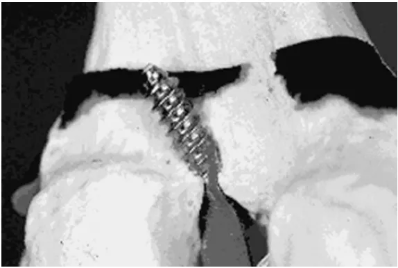
The fracture may occur during the operation or in the early postopera-tive period. Intraoperapostopera-tive patella fracture may be the result of the use of osteotomes. If the saw cuts are only 8-mm deep and 25-mm long, and the base is flat to avoid the deep V cut, an intraoperative fracture is rare. The late fractures are produced by the overruns of the saw cuts. The overruns may be prevented by cutting the proximal end in a boat shape. The left X-ray (Fig. 5.2) shows a displaced transverse patellar frac-ture, at three months postoperative. The right X-ray (Fig. 5.3) shows the postoperative internal fixation with cannulated AO screws and figure-of-eight wire.
Figure5.2. X-ray of displaced transverse patellar
fracture at three months postoperative.
Figure5.3. X-ray of postoperative internal fixation
The proximal transverse saw cut is critical (Fig. 5.4). The stress risers that go beyond the edge of the bone block should be avoided. An overrun of 2 mm may cause a late transverse patellar fracture. If there are overruns, the burr may be used at the corner to round these cuts. The fracture is usually sustained by muscular contraction. Change to making the proximal cuts boat shaped to prevent the stress risers (Fig. 5.5). The graft is usually cut to this shape to pass into the joint; now it is just cut in that shape before removing it.
Tendon Rupture
This may occur if a very large graft is taken from a small tendon. The standard is a 10-mm graft, measured with a double-bladed knife. The bone blocks are trimmed to 9 mm to make the graft passage easier.
Patellar Tendon Graft 51
Figure 5.4. The method of cutting the patellar bone plug to avoid a late