www.elsevier.com / locate / bres
Research report
Time-course and dose–response study on the effects of chronic
L
-DOPA administration on striatal dopamine levels and dopamine
transporter following MPTP toxicity
a,b ,
*
b a aFrancesco Fornai
, Giuseppe Battaglia , Marco Gesi , Filippo S. Giorgi ,
b,c b b,c
Francesco Orzi
, Ferdinando Nicoletti , Stefano Ruggieri
a
Department of Human Morphology and Applied Biology, University of Pisa, Via Roma 55, I-56100 Pisa, Italy b
I.R.C.C.S. I.N.M. Neuromed, Pozzilli (IS), Italy c
Department of Neurological Sciences, University ‘La Sapienza’, Roma, Italy Accepted 19 September 2000
Abstract
Despite a long-lasting therapeutic use of L-DOPA in Parkinson’s disease, doubts still remain concerning the possibility that chronic L-DOPA might accelerate the progression of this movement disorder. To address this point, in the present study we examined the effects
of chronicL-DOPA administration either in intact or MPTP-treated parkinsonian mice. We produced an intermediate striatal dopamine loss
by administering a low dose of MPTP (30 mg / kg); then, we treated mice chronically, for different time intervals, with a daily dose of
L-DOPA (50 mg / kg). In particular, to study the time-course of the effects ofL-DOPA on the recovery of nigrostriatal dopamine axons,
mice were sacrificed at 5, 30, 60, and 90 days after a dailyL-DOPA administration. To evaluate presynaptic integrity of the nigrostriatal
pathway we measured dopamine, metabolite levels, and dopamine uptake sites. In the same animals, we measured striatal serotonin levels and we analysed monoamine content in the olfactory bulb. Administration of MPTP produced a neurotoxic effect, which fully recovered in 2–3 months. Daily L-DOPA administration did not modify this recovery process. Additionally, there was no significant effect of L-DOPA in intact mice, despite a slight decrease in striatal dopamine levels at 5 and 30 days. However, this effect was neither worsened
nor reproduced by administering higher doses ofL-DOPA (up to 400 mg / kg) for the same amount of time. These data rule out neurotoxic
effects induced by prolonged L-DOPA administration, both in intact and MPTP-treated mice. Moreover, administration ofL-DOPA does
not change the recovery process which takes place after a nigrostriatal lesion. 2000 Elsevier Science B.V. All rights reserved.
Theme: Disorders of the nervous system
Topic: Degenerative disease: Parkinson’s
Keywords: L-DOPA; MPTP; Recovery; Experimental parkinsonism; DAT
1. Introduction administration, a number of neurological complications (i.e., dyskinesia), tightly related to the ongoing therapy Since its introduction in the 1960s (see Ref. [18]), the occur [40,4]. It has been postulated that even the natural neurotransmitter precursorL-DOPA, via its decarboxylation course of the disease might be accelerated by chronic
to dopamine (DA), represents the most effective therapy in L-DOPA administration [15]. Thus, an open question still
Parkinson’s disease (PD) [19]. There is concern, however, remains concerning L-DOPA-related symptoms: do they
that the symptomatic relief induced byL-DOPA convoys a represent a functional and potentially reversible behavioral
toxic effect by accelerating the neuronal loss which disorder, or do they reflect an actual progression of the normally takes place during the disease process [19]. disease, substantiated by accelerated neuron and / or axon Indeed, there is no doubt that, after prolonged L-DOPA terminal loss promoted byL-DOPA?
This latter hypothesis was suggested since the late 1970s [15], when it was demonstrated that L-DOPA produces *Corresponding author. Tel.:139-50-835-942; fax:139-50-552-102.
E-mail address: [email protected] (F. Fornai). oxidative compounds like semiquinone and O-quinone
derivatives which might be toxic for DA neurons. In line nigrostriatal DA terminals. In mice, in 2–3 months after with this, a number of in vitro and microinfusion studies the onset of a moderate striatal DA loss, a spontaneous carried out during the last decade, provided evidence for recovery of nigrostriatal DA axons takes place [12]. To
L-DOPA- and / or DA-induced neurotoxicity in non-cat- document the presynaptic integrity of the DA nigrostriatal
echolamine [36,7,27] and catecholamine cell lines pathway we measured both DA, metabolite levels, and DA [22,29,39], in primary mesencephalic cell cultures uptake sites both in intact and MPTP-lesioned striatum, [23,24,30], and via direct microinfusion of DA within the injected chronically either with saline or L-DOPA. To
striatum [10]. evaluate the site-specificity of the effects induced by
Despite these results, obtained with in vitro (and mi- MPTP and / or L-DOPA we also analysed monoamine
croinfusion) studies, in vivo experiments consistently levels in the olfactory bulb. failed to demonstrate neurotoxic effects induced by L
-DOPA on intact nigrostriatal DA system [17,31].
These results are in line with neuropathological studies 2. Materials and methods
carried out in humans with a misdiagnosis of PD who,
despite receiving a high daily dose ofL-DOPA for several 2.1. Animals
years, showed a normal mesencephalic histopathology
[32,33]. Two hundred and sixty C57 Black male mice (Charles
However, it should be considered that during PD, River Calco, CO, Italy), 9 weeks old, were used for the exogenous L-DOPA is handled differently by the few study. Since previous experiments demonstrated that
surviving DA neurones. For instance, during the pro- MPTP toxicity critically depends on age [3,8], all mice gression of the disease only scant striatal DA axons are used in the present work were 9 weeks old at the time of taking up the same amount of L-DOPA, which therefore, MPTP administration. Mice were housed with free access
reaches a higher concentration in each nigrostriatal termi- to food and water and kept under environmentally con-nal. Moreover, in PD surviving DA axons undergo an trolled conditions (12-h light / dark cycle with light on increased neurotransmitter metabolism [1,21] which leads between 07:00 and 19:00). In the present study, we to augmented rate of L-DOPA utilisation and, thereby, to evaluated the potential toxicity ofL-DOPA: it is established
heightened DA oxidation. These effects might converge in that DA neurotoxins (i.e., amphetamines) vary their effica-increasing oxidative stress within surviving DA terminals cy depending on housing conditions (for a review, see Ref. accelerating the progression of PD [38]. [35]); therefore, we did not vary the number of animals per Indeed, a few in vivo experiments carried out in cage (n510) and the size of the cages (38322 cm wide parkinsonian animals provided evidence for a neurotoxic and 15 cm high).
effect of L-DOPA [5,28]; however, in other studies this Experiments were approved by Local Ethical Committee
toxic effect was not observed [9,25]. In particular, in a and animals were treated in accordance with the Guide-recent elegant report, Murer et al. [25] demonstrated the lines for Animal Care and Use of the National Institutes of absence of L-DOPA toxicity, suggesting a trophic role of Health.
L-DOPA administered for 6 months to rats carrying a
partial striatal DA loss induced by 6-hydroxydopamine 2.2. Experimental design (6-OHDA).
In the present study, we examined the effects of chronic 2.2.1. Time-course study
L-DOPA administration in parkinsonian mice by using a In the time-course study, 160 C57 Black mice were
different experimental approach. We administeredL-DOPA divided into four groups each composed of 40 animals
chronically to mice carrying a partial lesion of the nigros- receiving various treatments: Group A received an in-triatal DA pathway induced by the neurotoxin MPTP. The jection of saline and then again, it was administered daily experiment was performed in mice since, among rodents, with saline. Group B was administered saline once and this animal species is more sensitive to experimental than, chronically a daily dose of L-DOPA methyl ester
Parkinsonism induced by DA neurotoxins [14]. In order to hydrochloride (Research Biochemicals, RBI, Natick, MA; evaluate either potential deleterious, or protective effects 50 mg / kg, free base). Group C was treated with a single induced by chronicL-DOPA, we produced an intermediate dose of MPTP hydrochloride (RBI; 36 mg / kg,
corre-striatal DA loss by administering a low dose (30 mg / kg) sponding to 30 mg / kg of MPTP), then it was administered of MPTP. Then, we challenged these animals with a daily with a daily dosage of saline. Group D was treated with dose ofL-DOPA (50 mg / kg). Mice were sacrificed at four MPTP and then received a daily chronic administration of
different time intervals (5, 30, 60, and 90 days) after L-DOPA. In all groups, repeated administration ofL-DOPA
MPTP administration. Apart from disclosing a potential or saline started 12 h following the single MPTP or saline effect depending on the time of exposure toL-DOPA, this injection. This time interval was chosen based on previous
experimental protocol was designed to evaluate the time- studies to start chronic L-DOPA administration when
1
cleared from the striatum [11]. The time-course of the 1.5 ml of ice-cold buffer (50 mM Tris, 5 mM EDTA, 320 recovery process of striatal DA innervation following an mM sucrose; pH 7.5) and centrifuged at 10003g for 10 intermediate dose of MPTP was evaluated sacrificing mice min at 48C. The pellet was discarded and the supernatant from each group at four distinct time intervals. In par- centrifuged at maximum speed in a microfuge for 30 min ticular, the original four groups of mice were further at 48C. The pellet was re-suspended in 1.2 ml of binding divided into four subgroups each composed of 10 animals buffer (50 mM Tris, 300 mM NaCl, 5 mM, 0.1% ascorbic
3 which were sacrificed at 5 days, and 1, 2, and 3 months, acid; pH 7.9) by sonication. After addition of [ H]GBR
respectively, after MPTP administration. 12935 (DuPont NEN; final concentration 3 nM, 40–60
During chronic L-DOPA administration mice were Ci / mmol), membrane preparations were incubated for 45
weighted once a week in order to maintain a constant min at 258C. Non-specific binding was determined in the amount of L-DOPA / body weight. Injections were carried presence of DA 200 mM. Incubation was stopped by
out intraperitoneally (i.p.), using a constant volume (10 centrifugation for 30 min. The pellet was washed twice ml / kg). Mice were sacrificed at 40 h after the last L- with 1 ml of binding buffer and radioactivity was
mea-DOPA / saline injection and their brains were immediately sured by scintillation counting. removed.
The left striatum and the olfactory bulb were dissected 2.5. Data analysis and processed for measuring monoamine levels, whereas
the right striatum from the same animals was dissected and For monoamine assay, a standard curve was prepared processed for measuring presynaptic striatal DA uptake using known amounts of DA, NE, 5-HT and metabolites
sites. (Sigma), dissolved in 0.1 M perchloric acid containing a
constant amount (10 ng / ml) of the internal standard
2.2.2. Dose–response study (DBA), as used for tissue samples. The standard curve for
Since in the time-course study (see Section 3) we each compound (DA, NE, 5-HT or metabolites) was observed a slight decrease of striatal DA levels at early calculated using regression analysis of the peak areas for time intervals (5 days and 1 month) after L-DOPA (50 known concentrations of each compound. For binding
mg / kg) administration to intact mice, we explored this studies data are expressed as percentage of specific bind-effect in detail by increasing the dosage ofL-DOPA. In this ing; the latter was calculated from the ratio between the
additional experimental step, 100 C57 Black mice were cpm / mg protein of specific binding, and the cpm / mg divided into five groups each composed of 20 mice. Group protein of total binding.
A received saline. Group B was administered withL-DOPA For NE, DA, 3,4-dihydroxyphenylacetic acid (DOPAC),
at 50 mg / kg. Group C received L-DOPA at 100 mg / kg. homovanillic acid (HVA), 5-HT and
5-hydroxy-in-Group D was administered with L-DOPA at 200 mg / kg. doleacetic acid (5-HIAA) levels, results are expressed as
Group E was injected withL-DOPA at 400 mg / kg. From the mean6S.E.M. of eight to 10 animals per group. For
each group, 10 animals were sacrificed at 5 days after a binding studies, results are expressed as the mean6S.E.M. daily saline /L-DOPA administration, whereas 10 mice of eight to 10 animals per group. Effects of MPTP and / or
were sacrificed after 1 month of daily treatment. In these L-DOPA on monoamine levels in the striatum and olfactory
mice we measured striatal monoamine levels. bulb as well as on striatal GBR 12935 binding sites, were evaluated using analysis of variance with Sheffe’s post-hoc
2.3. Assay of monoamines analysis.
The striatum and the olfactory bulb were sonicated in
0.6 ml of ice-cold 0.1 M perchloric acid containing 10 3. Results
ng / ml of 3,4-dihydroxybenzylamine (DBA, Sigma, San
Louis, MO) as the internal standard. An aliquot of the 3.1. Effects of MPTP administration on the time-course homogenate (50 ml) was assayed for proteins [20]. After of striatal DA and the DA transporter
centrifugation at 80003g for 10 min, 20 ml of the clear
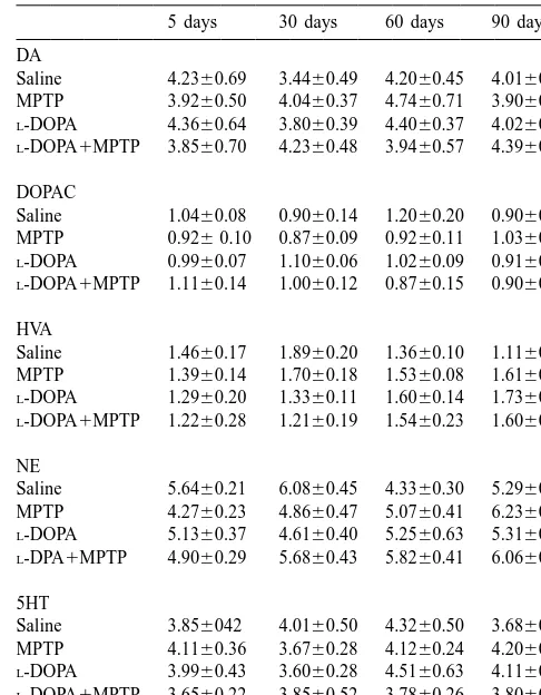
Table 1
Monoamine and metabolite levels (values are given in ng / mg protein) in a
Male C57 Black mice have been sacrificed at different time intervals after a single MPTP (30 mg / kg) administration following a chronic daily dosage of eitherL-DOPA (50 mg / kg) or an equivalent volume of saline.
The olfactory bulb have been processed and assayed for monoamine Fig. 1. Effects of dailyL-DOPA administration at different time intervals
leves. Data are given as the mean6S.E.M of eight to 10 animals per after MPTP injection. Male C57 Black mice were sacrificed at different
group. No differences in monoamine and metabolite levels have been time intervals (5, 30, 60, 90 days) after an i.p. injection of MPTP (30
`
measured by using the ANOVA with Sheffe’s post-hoc analysis. mg / kg) and a daily administration ofL-DOPA (50 mg / kg i.p.). Mice
were sacrificed, the left striata were dissected and processed for HPLC analysis. The levels of dopamine (DA) (A), dihydroxyphenylacetic acid (DOPAC) (B), and homovanillic acid (HVA) (C) were evaluated. For
there was an intermediate decrease in DA binding sites
each time interval results were obtained from eight to 10 animals per
group and are given as the mean6S.E.M. At each time interval compared with controls (saline, 18.3962.59%; MPTP,
differences between groups were evaluated using ANOVA with Sheffe’s 8.2562.46%), whereas at 2 and 3 months striatal DAT post-hoc analysis. *P,0.05 compared with controls.
levels in MPTP-treated mice were close to controls.
although the depleting effect of MPTP for DA metabolites 3.2. Effects of L-DOPA per se
was less pronounced. Levels of 5-HT and its metabolite
5-HIAA within the striatum were not affected by MPTP As shown in Fig. 1A, chronic administration ofL-DOPA
administration at any time interval (data not shown). No did not modify striatal DA levels. At 5 days and 1 month significant difference was measured in saline-injected after daily L-DOPA administration there was a slight
controls during the 3 months observation time (Fig. 1). As decrease in striatal DA levels; however, in the following shown in Table 1, no significant effect was produced in the time intervals, this effect was no longer present (Fig. 1A). olfactory bulb by MPTP administration at any time interval Similarly, striatal DA uptake sites were not affected by compared with controls. Similarly, in the olfactory bulb chronic L-DOPA administration (Fig. 2). Neither striatal
MPTP administration did not modify NE levels (Table 1). 5-HT levels (data not shown) nor levels of monoamines in As shown in Fig. 2, measurement of striatal plasma the olfactory bulb (Table 1) were modified during the membrane DAT matched the data obtained by assaying time-course of L-DOPA administration compared with
3.3. Effects of higher doses of L-DOPA
Since at 5 days and 1 month there was a slight decrease in DA levels of L-DOPA-treated mice, we increased the
daily dose ofL-DOPA up to 8-fold. As shown in Fig. 3, no
effects were observed either at 5 days (Fig. 3A) and 1 month (Fig. 3B) after a daily administration ofL-DOPA up
to 400 mg / kg.
3.4. Effects of a daily administration of L-DOPA on the
recovery following MPTP-induced neurotoxicity
Animals injected with MPTP and then receiving a daily
Fig. 2. Effects of daily L-DOPA on striatal dopamine uptake sites at
L-DOPA administration had the same striatal DA levels as different time intervals after MPTP administration. Male C57 Black mice
MPTP-treated animals receiving a chronic administration
were sacrificed at three different time intervals (5, 60, 90 days) after an
i.p. injection of MPTP (30 mg / kg) and a daily administration ofL-DOPA of saline (Fig. 1). This was confirmed by assaying DAT
(50 mg / kg i.p.). Animals from each group were sacrificed, their right binding sites (Fig. 2). Therefore, L-DOPA administration striata were dissected and processed for the measurement of specific
did not modify the spontaneous recovery process occurring
binding of the dopamine transporter (DAT). For each time interval results
in partially lesioned nigrostriatal DA terminals (Figs. 1 and
were obtained from eight to 10 animals per group and are expressed as
2). In this case, we did not observe the slight decrease in
the mean6S.E.M. At each time interval the differences between groups
were evaluated by using ANOVA with Sheffe’s post-hoc analysis. *P, DA levels which was detected at early time intervals in
0.05 compared with controls. intact mice injected with L-DOPA. Striatal HT and
5-HIAA levels were not affected at any time interval. Similarly, no effect was detected on monoamine levels in the olfactory bulb (Table 1).
4. Discussion
In the present study we confirmed previous data [11] showing that i.p. administration of the DA neurotoxin MPTP at 30 mg / kg to C57 Black mice produces an intermediate degree of toxicity to the nigrostriatal DA pathway (measured by the amount of striatal DA loss and by the decrease of striatal DA uptake sites). Following this kind of lesion, a full recovery process takes place in 2–3 months after MPTP administration.
Results of this study show that a single i.p. daily administration of L-DOPA (50 mg / kg), does not affect
striatal DA levels when administered to intact mice for different time intervals (5, 30, 60, or 90 days). We observed only a slight decrease (the effect was significant at the ANOVA using the Fisher test but not using Sheffe’s post-hoc analysis) in DA levels after 5 days and 1 month after a daily injection ofL-DOPA at the dose of 50 mg / kg.
However, this latter effect does not appear to be relevant since: (1) it was not observed when measuring DA uptake sites; (2) it was neither increased nor even reproduced by the administration of higher doses ofL-DOPA (up to 400 Fig. 3. Lack of a dose–response effects of dailyL-DOPA on striatal DA
mg / kg) for the same amount of time.
levels at 5 and 30 days. Male C57 Black mice were sacrificed either at 5
days (A) or at 30 days (B) after an i.p., daily administration ofL-DOPA. In the present study we decided to administer L-DOPA
L-DOPA was administered at four different doses: 50, 100, 200 or 400 as a single, daily i. p. injection for several reasons: (1) to mg / kg. Animals from each group were sacrificed, left striatum was avoid physiological variations in dailyL-DOPA intake due dissected and processed for assaying monoamine levels. For each time
to oscillations in the amounts of food / water intake by the
interval results were obtained from seven to 10 animals per group and are
animals; (2) to produce a higher concentration ofL-DOPA given as the mean6S.E.M. For each time interval the differences between
evaluate a potential neurotoxic effect induced by a bolus of vivo [37]. However, in the present study, this latter effect
L-DOPA compared with oral administration evaluated in does not seem to play any role on the survival of DA
previous studies. axons. This is consistent with a recent finding showing that
The dose ofL-DOPA (50 mg / kg) corresponds to a daily L-DOPA administration to 6-OHDA injected rats does not
administration of 4 g in an adult weighing 80 kg which, increase oxidative stress [6].
given the partial (10%) penetration ofL-DOPA within the The present data are in line with a previous study in
central nervous system in the absence of a peripheral which L-DOPA has been administered to MPTP-treated
decarboxylase inhibitor, corresponds to a low therapeutic monkeys [34] for up to 11 weeks without modifying dosage of the DA precursor (0.4 g / die). However, we did striatal DA levels and striatal DA uptake sites. However, in not obtain any effect even increasing the dose ofL-DOPA this latter experiment MPTP produced by itself a massive
up to 400 mg / kg. Nonetheless, the efficacy of 50 mg / kg loss of the nigrostriatal DA system (more than 90%) ofL-DOPA in modulating striatal DA activity is shown by making it difficult to evaluate a worsening of the DA
previous studies in which L-DOPA (50 mg / kg) doubled lesion by L-DOPA administration. In the present study,
striatal DA release in mice [13]. MPTP has been administered at a dose producing an
These results obtained in mice, confirm what previously intermediate degree of nigrostriatal DA damage; this was reported in intact rats [17], where no changes were aimed at evaluating either potential deleterious effects, no observed in the nigrostriatal DA system after chronic influence, or, on the other hand, a protective phenomenon.
L-DOPA administration. These data are in line with In these experimental conditions we confirmed what
neuropathological studies carried out in patients with a recently published by Murer et al. [25] who administered wrong diagnosis of PD who received for several years a chronically L-DOPA to rats underwent an intermediate
high daily dosage ofL-DOPA. In these patients there was a lesion using the neurotoxin 6-OHDA. In this study the
normal mesencephalic histopathology [32,33]. authors did not find any exacerbation of 6-OHDA-induced Therefore, in the present study we did not find any effect nigrostriatal damage at 6 months after inducing the lesion. after administration of different doses of L-DOPA to On the other hand, based on measurement of tyrosine
otherwise intact animals. Furthermore, we found that hydroxylase (TH) Murer et al. [25] found a beneficial MPTP-treated mice chronically injected L-DOPA had effect following L-DOPA administration. In the present
similar striatal DA levels as MPTP-treated mice injected experiment we did not find these beneficial effects. This daily with saline. Similarly, measurement of presynaptic might be due to several reasons: (1) the parkinsonian DA uptake sites after MPTP administration did not show model we used here (MPTP compared with 6-OHDA); (2) significant differences between L-DOPA- and saline-in- the animals species (mice compared with rats); (3) the
jected mice. The lack of any deleterious effect ofL-DOPA maximum amount of time available for the effect to occur
was documented both immediately after the production of (3 months compared with 6 months); (4) the markers used nigrostriatal DA toxicity by MPTP (animals killed at 5 in this study to evaluate the integrity of the nigrostriatal days after MPTP administration) and during the recovery DA pathway: striatal levels of DA and metabolites com-process measured at 1, 2 and 3 months after the onset of bined with the measurement of DA transporter (in the experimental Parkinsonism. The analysis of four different present study), compared with striatal TH, vesicular mono-time intervals after the lesion allowed us to detect potential amine transporter and DA transporter measured by Murer transient effects induced by L-DOPA on the recovery et al. [25]; (5) the degree of nigrostriatal lesion, which
process. These data show that even when the nigrostriatal seems to be more marked in the case of the work DA pathway is partially damaged, chronicL-DOPA supple- performed by Murer et al. [25]; (6) the route of
administra-mentation does not produce any deleterious effect on tion of L-DOPA (i.p. in the present study compared with
surviving striatal DA terminals. Therefore chronicL-DOPA oral in the case of Murer et al. [25]).
administration did not worsen experimental Parkinsonism Nonetheless, it should be emphasised that, despite all the either directly, by enhancing the neurotoxic effects of above-mentioned experimental differences, our data con-MPTP, or indirectly, by interfering with the spontaneous verge with those of Murer et al. [25] in showing the lack of
recovery process. any deleterious effect following chronic L-DOPA both to
It has been hypothesised that increased metabolic rate of intact and to parkinsonian animals, even when the drug is surviving DA axons [1] would result in marked elevation administered as a constant i.p. dose.
of rate of intracellular DA oxidation, thereby leading to In conclusion, a chronic daily administration ofL-DOPA
increased oxidative stress, which in turn, accelerate the does not worsen nor the integrity neither the recovery of a progression of PD [38]. Within this context, it has been partially lesioned nigrostriatal pathway. These data, joined suggested that chronic treatment withL-DOPA, by increas- with previous studies, confirm what has been already
ing the substrate for the DA oxidative pathway, might inferred in parkinsonian patients more than a decade ago exacerbate oxidative stress. This hypothesis has been by Blin et al. [4] and recently commented on by Agid [2]: strengthened by the finding that exogenous administration L-DOPA-induced abnormal involuntary movements are
phenyl 1-methyl-4-phenyl-1,2,3,6-tetrahydropyridine. Part 1:
Sys-independent, ongoing disease progression. On the other
temic administration, J. Pharmacol. Exp. Ther. 270 (1994) 1000–
hand, L-DOPA administration does not seem at all to
1007.
contribute to the progression of PD. The vast literature [15] D.G. Graham, Oxidative pathway for catecholamines in the genesis showing the in vitro neurotoxic effects ofL-DOPA should of neuromelanin and cytotoxic quinones, Mol. Pharmacol. 14 (1978)
probably be re-considered carefully, monitoring the dose, 633–643.
[16] S.K. Han, C. Mytilineou, G. Cohen,L-DOPA up-regulates glutathion
the time of exposure and the kind of cell culture. For
and protects mesencephalic cultures against oxidative stress, J.
instance, Mytilineou et al. [26] pointed out that varying in
Neurochem. 66 (1996) 501–510.
vitro conditions L-DOPA might exert either toxic or
[17] F. Hefti, E. Melamed, J. Bhawan, R.J. Wurtman, Long-term
adminis-protective effects on primary mesencephalic cells. This tration of levodopa does not damage dopaminergic neurons in the could explain contradictory findings showing either toxic mouse, Neurology 31 (1981) 1194–1195.
[18] O. Hornykiewicz,L-DOPA in the 1960s: starting point Vienna, in:
[23,24] or protective effects [16] ofL-DOPA administration
W. Poewe, A.J. Lees (Eds.), 20 Years of Madopar — New Avenue,
to primary mesencephalic neurones.
Editiones Roche, Basel, 1994, pp. 11–27.
[19] P.G. Jenner, M.F. Brin, Levodopa neurotoxicity: experimental studies versus clinical relevance, Neurology 50 (1998) S39–S45. [20] O.H. Lowry, N.J. Rosebrough, A.L. Farr, R.J. Randall, Protein References measurement with the Folin phenol reagent, J. Biol. Chem. 193
(1951) 265–275.
[1] Y. Agid, F. Javoy, J. Glowinski, Hyperactivity of remaining [21] E. Melamed, F. Hefti, R.J. Wurtman, Compensatory mechanisms in dopaminergic neurons after partial destruction of the nigrostriatal the nigrostriatal dopaminergic system in Parkinson’s disease: studies dopaminergic system in the rat, Nat. New Biol. 245 (1973) 150– in an animal model, Isr. J. Med. Sci. 18 (1982) 159–163. 151. [22] M.A. Mena, B. Pardo, M.J. Casarejos, Neurotoxicity of levodopa on [2] Y. Agid, Levodopa: is toxicity a myth?, Neurology 50 (1998) catecholamine-rich neurons, Mov. Disord. 7 (1992) 23–31.
858–863. [23] M.A. Mena, B. Pardo, C.L. Paino, J.G. de Yebenes, Levodopa
[3] S.F. Ali, S.N. David, G.D. Newport, Age-related susceptibility to toxicity in foetal rat midbrain neurons in culture, Neuroreport 4 MPTP-induced neurotoxicity in mice, Neurotoxicology 14 (1993) (1993) 438–440.
29–34. [24] M.A. Mena, M.J. Casarejos, A. Carazo, C.L. Paino, J.G. de Yebenes, [4] J. Blin, A.-M. Bonnet, Y. Agid, Does levodopa aggravate Parkin- Glia conditioned medium protects fetal rat midbrain neurones in
son’s disease?, Neurology 38 (1988) 1410–1416. culture fromL-dopa toxicity, Neuroreport 7 (1996) 441–445. [5] S.B. Blunt, P. Jenner, C.D. Marsden, Suppressive effect ofL-DOPA [25] M.G. Murer, G. Dziewczapolski, L.B. Menaled, M.C. Garcia, Y.
on dopamine cells remaining in the ventral tegmental area of rats Agid, O. Gershanik, R. Raisman-Vozari, Chronic Levodopa is not previously exposed to the neurotoxin 6-hydroxydopamine, Mov. toxic for remaining dopamine neurons, but instead promotes their Disord. 8 (1993) 128–133. recovery in rats with moderate nigrostriatal lesions, Ann. Neurol. 43 [6] D.M. Camp, D.A. Loeffler, P.A. LeWitt,L-DOPA does not enhance (1998) 561–575.
hydroxyl radical formation in the nigrostriatal dopamine system of [26] C. Mytilineou, S.K. Han, G. Cohen, Toxic and protective effects of rats with a unilateral 6-hydroxydopamine lesion, J. Neurochem. 74 L-DOPA on mesencephalic cell cultures, J. Neurochem. 61 (1993)
(2000) 1229–1240. 1470–1478.
[7] D. Daily, A. Barzilai, D. Offen, A. Kamsler, E. Melamed, I. Ziv, [27] J.S. Noh, E.Y. Kim, J.S. Kang, H.R. Kim, Y.J. Oh, B.J. Gwag, The involvement of p53 in dopamine-induced apoptosis of cerebel- Neurotoxic and neuroprotective actions of catecholamines in cortical lar granule neurons and leukemic cells overexpressing p53, Cell. neurons, Exp. Neurol. 159 (1999) 217–224.
Mol. Neurobiol. 19 (1999) 261–276. [28] N. Ogawa, M. Asanuma, Y. Kondo, Y. Kawada, M. Yamamoto, A. [8] V.G. Desai, R.J. Feuers, R.W. Hart, S.F. Ali, MPP(1)-induced Mori, Differential effects of chronic L-DOPA treatment on lipid neurotoxicity in mouse is age-dependent: enhanced by the selective peroxidation in the mouse brain with or without pretreatment with inhibition of complexes of electron transport, Brain Res. 715 (1996) 6-OHDA, Neurosci. Lett. 171 (1994) 55–58.
1–8. [29] B. Pardo, M.A. Mena, S. Fahn, J.G. de Yebenes, Ascorbic acid
[9] G. Dziewczapolski, G. Murer, Y. Agid, O. Gershanik, R. Raisman- protects against levodopa-induced neurotoxicity on a catecholamine-Vozari, Absence of neurotoxicity of chronic L-DOPA treatment in rich neuroblastoma cell line, Mov. Disord. 8 (1993) 278–284. 6-hydroxydopamine lesioned rats, Neuroreport 8 (1997) 975–979. [30] B. Pardo, M.A. Mena, M.J. Casajeros, C.L. Paino, J.G. de Yebenes, [10] F. Filloux, J.J. Townsend, Pre- and postsynaptic neurotoxic effects Toxic effects of L-dopa on mesencephalic cell cultures: protection
of dopamine demonstrated by intrastriatal injections, Exp. Neurol. with antioxidants, Brain Res. 682 (1995) 133–143.
119 (1993) 79–88. [31] T.L. Perry, V.W. Yong, J.G. Foulks, R.A. Wall, D.V. Godin, R.M. `
[11] F. Fornai, M.G. Alessandrı, M.T. Torracca, L. Bassi, G.U. Corsini, Clavier, Nigrostriatal dopaminergic neurons remain undamaged in Effects of noradrenergic lesions on MPTP/ MPP1 kinetics and rats given high doses of L-DOPA and carbidopa chronicallly, J. MPTP-induced nigrostriatal dopamine depletions, J. Pharmacol. Neurochem. 43 (1984) 990–993.
Exp. Ther. 283 (1997) 100–107. [32] N.P. Quinn, D. Parkes, I. Janota, C.D. Marsden, Preservation of the `
[12] F. Fornai, F.S. Giorgi, M.G. Alessandrı, M. Giusiani, G.U. Corsini, substantia nigra and locus coeruleus in a patient receiving levodopa Effects of pretreatment with DSP-4 on methamphetamine-induced (2 g) plus decarboxylase inhibitor over a four year period, Mov. striatal dopamine losses and pharmacokinetics, J. Neurochem. 72 Disord. 1 (1986) 65–68.
(1999) 777–784. [33] A.H. Rajput, M.E. Fenton, S. Birdi, R. Macaulay, Is levodopa toxic `
[13] F. Fornai, K. Chen, F.S. Giorgi, M. Gesi, M.G. Alessandrı, J.C. to human substantia nigra?, Mov. Disord. 5 (1997) 634–638. Shih, Striatal dopamine metabolism in monoamine oxidase B-de- [34] L. Rioux, P.A. Frohna, J.N. Joyce, J.S. Schneider, The effects of ficient mice: a brain dialysis study, J. Neurochem. 73 (1999) chronic levodopa treatment on pre- and postsynaptic markers of 2434–2440. dopaminergic function in striatum of parkinsonian monkeys, Mov. [14] A. Giovanni, B.-A. Sieber, R.E. Heikkila, P.K. Sonsalla, Studies on Disord. 12 (1997) 148–158.
related drugs, in: H.Y. Melzer (Ed.), Psychopharmacology: the Third responses to 6-OH-DA and L-DOPA therapy: implications for Generation of Progress, Raven Press, New York, 1987, pp. 359–366. Parkinson’s disease, Ann. NY Acad. Sci. 648 (1992) 71–86. [36] R.D. Snyder, M.B. Friedman, Enhancement of cytotoxicity and [39] I. Ziv, E. Melamed, N. Nardi, D. Luria, A. Achiron, D. Offen, A.
clastogenicity of l-DOPA and dopamine by manganese and copper, Barzilai, Dopamine induces apoptosis-like cell death in cultured Mutat. Res. 405 (1998) 1–8. chick sympathetic neurons — a possible novel pathogenetic mecha-[37] T. Spencer-Smith, W.D. Parker Jr., J.P. Bennett Jr., L-DOPA in- nism in Parkinson’s disease, Neurosci. Lett. 170 (1994) 136–140.
creases nigral production of hydroxyl radicals in vivo: potential [40] M.D. Yahr, Long-term levoDOPA in Parkinson’s disease, Lancet 1