Nicotiana glauca
: another plant species containing vitamin D
3metabolites
Mario Skliar, Alejandro Curino, Lorena Milanesi, Silvia Benassati, Ricardo Boland *
Departamento de Biologı´a,Bioquı´mica y Farmacia,Uni6ersidad Nacional del Sur,San Juan 670,(8000) Bahı´a Blanca, Argentina
Received 6 October 1999; received in revised form 7 March 2000; accepted 8 March 2000
Abstract
Vitamin D3-related compounds have been detected in various plant species, mostly belonging to the Solanaceae. In this work
we show thatNicotiana glauca, a widespread member of this taxonomic family, contains 7-dehydrocholesterol, vitamin D3and
hydroxylated derivatives bearing precursor-product metabolic relationships in vertebrates. Leaves collected in the field and callus cultures were used. By means of specific radioreceptor binding assays and mass spectrometry of purified fractions obtained from plant tissue lipid extracts by Sephadex LH-20 and Sep-Pak C18 chromatography followed by HPLC, we established the presence of 7-dehydrocholesterol, vitamin D3, 25(OH)-vitamin D3 and 1a,25(OH)2-vitamin D3 (1a,25(OH)2D3), the latter being a
hormonally relevant metabolite in animals. These results indicate that N. glauca may represent a useful species in which to characterize the biosynthetic pathway and physiological functions of vitamin D3compounds in plants. In addition, tissue culture
ofN.glaucacould become a significant tool for biotechnological production of 1a,25(OH)
2D3. © 2000 Elsevier Science Ireland
Ltd. All rights reserved.
Keywords:Nicotiana glauca; Solanaceae; 7-Dehydrocholesterol; Vitamin D3; Vitamin D3metabolites; 1a,25(OH)2-vitamin D3; Mass spectrometry www.elsevier.com/locate/plantsci
1. Introduction
The occurrence of calcinosis in grazing animals in various parts of the world [1,2] led to the isolation and identification in plants of vitamin
D3-related compounds [3 – 5], thought previously
to be generated only in vertebrates during the metabolic conversion of 7-dehydrocholesterol to
the pluripotent hormone 1a,25-dihydroxyvitamin
D3 (1a,25(OH)2D3). These intermediates
(7-dehy-drocholesterol, vitamin D3, 25(OH)D3,
1a,25(OH)2D3) may be found in plants both as
aglycones and glycoside derivatives. Various inves-tigations employing in vitro systems have shown
that vitamin D sterols affect root growth and differentiation [6 – 8] through a stimulation of root calcium uptake and calmodulin synthesis [9 – 11], suggesting a physiological function for these compounds.
Although several species which have been
shown to contain vitamin D3 metabolites are
in-cluded in different taxonomic families, most of them belong to the Solanaceae [4,12 – 15]. In the present work we have investigated the presence of
these compounds in Nicotiana glauca Graham
(Solanaceae; n.v. Tree tobacco), a world-wide member of this family originating in South Amer-ica (Palan palan or Tabaco moro) [16,17]. By means of specific radioreceptor binding assays, HPLC and mass spectrometry we demonstrate that this plant species contains
7-dehydrocholes-terol, vitamin D3and its hydroxylated metabolites.
* Corresponding author. Fax: +54-291-4595130.
E-mail addresses: [email protected] (M. Skliar)., [email protected] (R. Boland).
2. Materials and methods
2.1. Materials
Solvents used for isolation and chromatographic procedures were HPLC grade (J.T. Baker, Phillips-burg, NJ, USA). Compounds for culture media, 7-dehydrocholesterol and Sephadex LH-20 were obtained from Sigma-Aldrich (St Louis, MO, USA) and Gibco BRL (Gaithersburg, MD, USA). The Sep-Pak cartridges were from Waters (Milford, MA, USA). The LiChroCART cartridge for HPLC analysis was from Merck (Darmstadt, Germany). Other reagents were of analytical grade. Vitamin
D3, 25(OH)-vitamin D3 and 1a,25(OH)2D3 were a
gift from Dr Lise Binderup, Leo Pharmaceutical Products (Ballerup, Denmark).
2.2. Cultures
To culture N. glauca tissue (callus), aseptic leaf
explants from plants collected in Bahı´a Blanca,
Buenos Aires Province, Argentina, were inoculated in solidified (0.8% agar) Murashige-Skoog [18] medium supplemented with
2,4-dichlorophenoxy-acetic acid (2,4-D; 0.5 mg/l) and kinetin (0.2 mg/l).
The tissue was grown at 25°C and during the culture period it was not illuminated. The calli
started to develop after 15 days. Subculture
was performed when the entire surface of culture
flasks was covered with tissue (2 months).
Com-pounds were isolated after the first subculture.
2.3. Extraction and chromatography of
7-dehydrocholesterol and 6itamin D3 metabolites
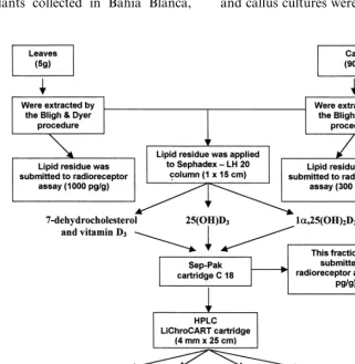
A scheme of the procedure used in the extrac-tion, purification and detection of
7-dehydro-cholesterol and vitamin D3metabolites is outlined
in Fig. 1. First, vitamin D3-related compounds
present inN.glaucacultured calli (first subculture)
and leaves were extracted following essentially the method of Bligh and Dyer [19]. Briefly, the leaves and callus cultures were homogenized with
chloro-Fig. 1. Flow diagram of the purification and detection of vitamin D3metabolites fromN.glaucaleaves and callus cultures. Weight
form-methanol (1:2, v/v; 3.6 ml/g FW) using an Ultraturrax homogenizer (Janke and Kunkel, Ger-many) for 2 min at maximum speed. The samples were further homogenized twice for 30 s: (1) after
the addition of chloroform (1.2 ml/g) and then (2)
after the addition of water (1.2 ml/g),
homogeniz-ing between additions. The final homogenate was centrifuged in a Sorvall SS-34 rotor (Du Pont,
Delaware, USA) at 4385×g for 10 min and the
lower lipid soluble phase was collected. When the
lipid extract was used for 1a,25(OH)2D3
radiore-ceptor assays, it was evaporated by flushing nitro-gen at 35°C and then dissolved in an appropriate volume of isopropanol. Prior to HPLC, the ex-tracts were passed through a Sephadex LH-20
column (1×15 cm) using
hexane-chloroform-methanol (9:1:1, v/v) as the solvent system. The
fractions which eluted with similar retention times to the authentic 7-dehydrocholesterol and vitamin
D3 (collected together), 25(OH)D3 and
1a,25(OH)2D3 were saved. Each fraction from the
Sephadex column was passed through a Sep-Pak C 18 cartridge eluted with 90% methanol for
7-de-hydrocholesterol and vitamin D3; 80 – 100%
methanol for 25(OH)D3 and 60 – 80% methanol
for 1a,25(OH)2D3, as solvent systems. Fractions
were collected which eluted with similar retention times to the authentic 7-dehydrocholesterol and
vitamin D3 (one fraction), 25(OH)D3 and
1a,25(OH)2D3. Each fraction was further resolved
by high performance liquid chromatography
(HPLC). HPLC was performed with an Isco Model 2360 gradient programmer equipped with a
Model Isco V4multiwavelength programmable
de-tector set at 265 nm and a pump Isco Model 2350 (Isco, Lincoln, NE, USA). A reverse phase
LiChroCART (4 mm×25 cm) cartridge eluted
with different solvent systems was used. Vitamin
D3 eluted with methanol-water (86:14, v/v); for
eluting 7-dehydrocholesterol, after the vitamin D3
peak, the solvent was changed to methanol-water
(90:10). Isocratic elutions of methanol-water
(75:25) and (86:14) were used to isolate 25(OH)D3
and 1a,25(OH)2D3, respectively.
2.4. Mass spectrometry
Mass spectra were obtained on a Micromass Model Autospec magnetic sector mass spectrome-ter, electron impact 70 eV (Micromass, UK). To that end, each HPLC fraction was evaporated and
redissolved in isopropanol, except that containing 7-dehydrocholesterol which was solubilized in ethanol. The mass spectra of samples were com-pared with those of authentic compounds from a database (Wiley 138.1).
2.5. 1a,25(OH)2D3 radioreceptor assays
The 1a,25(OH)2D3 content of Bligh and Dyer
[19] lipid extracts from N. glaucaleaves and callus
and the Sep-Pak chromatographic fraction of
1a,25(OH)2D3derived therefrom was measured by
a specific radioreceptor assay. The 1a,25(OH)2D3
receptor preparation was obtained by homoge-nization of mucosa from duodena of 4-week-old vitamin D-deficient chicks in TEKDP buffer (10 mM Tris – HCl, pH 7.4; 1.5 mM EDTA; 0.3 M KCl; 2 mM dithiothreitol; 0.3 mM phenylmethyl-sulfonylfluoride) followed by centrifugation at
100 000×g for 60 min. The supernatant was
col-lected and stored at −70°C until use. Binding
assays were performed incubating the samples
with 5.87×10⁻¹⁰ M [3H]1
a,25(OH)2D3 for
4 h at 0°C. Free radioactive steroid was separated from bound by the hydroxylapatite procedure [20].
3. Results
Radioreceptor assays of N. glauca leaf and
cal-lus lipid extracts revealed the presence of 1000 and
300 pg/g FW, respectively, of 1a,25(OH)2D3. The
metabolite was also detected in the corresponding fraction of the Sep-Pak purification step prior to HPLC (Fig. 1).
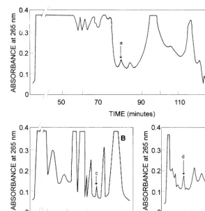
The presence of vitamin D3 and its related
metabolites (for chemical structures see Ref. [5]) in N. glauca was further investigated by high perfor-mance liquid chromatography. HPLC analysis on a LiChroCART cartridge of lipid extracts from calli and leaf, previously fractionated by Sephadex LH-20 and Sep-Pak chromatography, revealed the presence of peaks with elution times similar to
those of synthetic samples of vitamin D3 (peak a;
80.1 min), 7-dehydrocholesterol (peak b; 129.0
min), 25(OH)D3 (peak c; 34.7 min) and
1a,25(OH)2D3 (peak d; 8.9 min) (Fig. 2, A – C).
The above purification scheme has proved to be
satisfactory for purification of vitamin D3 and its
Fig. 2. Isolation of 7-dehydrocholesterol, vitamin D3 and hydroxylated derivatives from N. glauca by HPLC. Lipids were
extracted from calli according to Bligh and Dyer [13] followed by chromatography on a Sephadex LH-20 column and a Sep-Pak C 18 cartridge to obtain fractions containing 7-dehydrocholesterol and vitamin D3, 25(OH)D3and 1a,25(OH)2D3as indicated in
Section 2. Each fraction was further resolved by HPLC using a LiChroCART cartridge. (A) Vitamin D3and 7-dehydrocholesterol
(eluted with methanol/water, 86:14; after 90 min solvent changed to methanol/water, 90:10). (B) 25(OH)D3 was eluted
methanol/water (75:25). (C) 1a,25(OH)2D3was eluted methanol/water (86:14); the arrows indicate the elution of authentic samples
of: (a) vitamin D3; (b) 7-dehydrocholesterol; (c) 25(OH)D3; and (d) 1a,25(OH)2D3.
tissues [21], prior to their identification by mass spectrometry.
Mass spectrometry of the putative vitamin D3,
7-dehydrocholesterol, 25(OH)D3 and
1a,25(OH)2D3HPLC peaks was performed to
elu-cidate their chemical structures (Fig. 3, A – D). As shown in Fig. 3A, the electron impact mass spec-trum of HPLC peak a showed a molecular ion at
m/z 384. Loss of water and methyl group from the
parent peak gave m/z 351. The peak at m/z 325
was produced by loss of 59 atomic mass units (loss of water plus an isopropyl group) from the
molec-ular ion. The peak at m/z 271 was caused by loss
of the side chain from the parent ion. Loss of
water from peak m/z 271 gave m/z 253. The peak
at m/z 199 was produced by loss of C4H8O from
peak at m/z 271. The peak at m/z 136 results from
formal cleavage between carbons 7 and 8, whereas
the base peak at m/z 118 results from dehydration
of the peak at m/z 136. Peaks occurring at m/z 118
and 136 are characteristic of the vitamin D-triene system. These peaks and others of Fig. 3A
demon-strated that this compound is vitamin D3.
The mass spectrum of HPLC peak b (Fig. 3B)
has a molecular ion at m/z 384. Peaks at m/z 351,
325, 271, 253 and 199 also indicate that the steroid nucleus has remained unchanged. Collectively, from the above data, this metabolite could be 7-dehydrocholesterol.
Peak c of the HPLC run displayed a parent
molecular ion of m/z 400 (Fig. 3C) in agreement
with the molecular weight of 25(OH)D3. Peak at
m/z 382 represents loss of water from the
molecu-lar ion. Loss of water and methyl group from the
parent peak gave m/z 367. The peaks at m/z 271,
253, 136 and 118 arise in the manner discussed above for Fig. 3A. Altogether, these results indi-cate that the secoesteroid nucleus of their parent,
vitamin D3, has remained unchanged and that this
occur-ring only on the side chain. From the above data,
this metabolite can be identified as 25(OH)D3.
As shown in Fig. 3D, the mass spectrum of
HPLC peak d with a molecular ion at m/z 416
indicates that this metabolite contains one
addi-tional hydroxyl group when compared to
25(OH)D3, in accordance with the structure of
1a,25(OH)2D3. The peaks at m/z 398 and 380 are
characteristic of the parent peak minus one and two molecules of water, respectively. The peak at
m/z 287 is due to side chain cleavage from the
main steroid molecule (C-17/C-20 cleavage). Loss
of the side chain plus one and two molecules of
water of the parent ion gives the peaks at m/z 269
and 251, respectively. The peak at m/z 152 results
from cleavage between carbons 7 and 8 and repre-sents the A ring plus the carbon 6 and 7 fragment.
Loss of water from peak at m/z 152 gives rise to
the base peak at m/z 134. Collectively, these peaks
and others exhibited in the mass spectrum of Fig.
3D, demonstrate that this metabolite is
1a,25(OH)2D3.
4. Discussion
The data of this study demonstrate the existence
of 7-dehydrocholesterol, vitamin D3, 25(OH)D3
and 1a,25(OH)2D3 in N. glauca, a member of the
Solanaceae hitherto not known to contain these metabolically related steroids. The presence of
vi-tamin D3 compounds was established in the
lipophilic fraction of this plant species. Various
reports have previously shown that in the
Solanaceae, the concentration of the free form of
vitamin D3 metabolites generally exceeds that of
the corresponding glycosidic derivatives
[12,13,22 – 24]. Moreover, the possibility has been raised that formation of glycoconjugates of
vita-min D3 compounds in plants occurs to a great
extent during storage of collected tissues at the expense of the content of free sterols [12,13]. In
our study, N. glauca leaves or calli were
immedi-ately extracted after collection in the field or re-moval from the culture medium, respectively.
Regarding the identification of vitamin D3
metabolites, the high specificity of the
radiorecep-Fig. 3. Mass spectra of HPLC fractions from N. glauca containing vitamin D3, 7-dehydrocholesterol, 25(OH)D3 and
1a,25(OH)2D3. The fractions of vitamin D3-related compounds were obtained by chromatography of N.glaucalipid extracts on
tor assay, as revealed by its inability to detect
closely related analogs such as 24,25(OH)2D3,
25(OH)D3 and 1a(OH)D3, even when present in
large concentration [25], is clear evidence of the
presence of 1a,25(OH)2D3 in leaf and callus
cul-tures fromN.glauca. More conclusive evidence was
furnished by mass spectrometry of the HPLC peak
(d) coeluting with 1a,25(OH)2D3, which yielded a
molecular ion and a fragmentation profile corre-sponding to the structure of this metabolite. Also,
the presence of 7-dehydrocholesterol, vitamin D3
and 25(OH)D3 in leaf and callus cultures of this
plant was confirmed by chromatographic and spec-tral means, as discussed in Section 3.
This report adds one more example to the
grow-ing list of plants shown to contain vitamin D3
derivatives. Including N. glauca, there are so far
eight species belonging to the Solanaceae for which
biological and/or chemical evidence of the presence
of vitamin D3and related sterols has been provided.
These compounds are also found in varying con-centrations in species belonging to other taxonom-ical families, such as Gramineae, Leguminoseae and Cucurbitaceae [4,13,26].
There is little information about the formation of
and the subsequent hydroxylations of vitamin D3in
plants. Only low activities of vitamin D3
-25-hy-droxylase and 25(OH)D3-1a-hydroxylase have been
detected inSolanum glaucophyllumleaf extracts and
microsomal membranes using assays analogous to
those employed in studies on vitamin D3
hydroxy-lation in animals [27]. Much remains to be done to
elucidate the metabolism of vitamin D3 in plants
and its regulation.
The fact that the levels of 1a,25(OH)2D3in leaves
and cultured tissue from N. glauca are similar to
those found inS. glaucophyllum[15], a well-known
accumulator of vitamin D3 metabolites, but
geo-graphically restricted to certain areas within Ar-gentina and Brazil, indicates that the former species, which is widespread, may represent a more accessible source for investigations on this subject.
Finally, the finding that calli from N. glauca
pro-duce the hormonally active vitamin D3metabolite
may lead to the use of plant culture systems to produce this compound.
Acknowledgements
This research was supported by grants from the
Agencia Nacional de Promocio´n Cientı´fica y Tec-nolo´gica and Comisio´n de Investigaciones Cientı´-ficas de la Provincia de Buenos Aires (to Ricardo Boland), and the Universidad Nacional del Sur (to Mario Skliar), Argentina. Assistance in mass spec-trometry analysis by Drs Javier Sardina Lo´pez and Gabriel Tojo Sua´rez from the University of
San-tiago de Compostela (Spain) is gratefully
acknowledged.
References
[1] N.A. Worker, B.J. Carrillo, ‘Enteque seco’, calcification and wasting in grazing animals in the Argentine, Nature 215 (1967) 72 – 74.
[2] K.M.L. Morris, Plant induced calcinosis: a review, Vet. Hum. Toxicol. 24 (1982) 34 – 48.
[3] R.H. Wasserman, J.D. Henion, M.R. Haussler, T.A. McCaine, Calcinogenic factor in Solanum malacoxylon: evidence that it is 1,25-dihydroxyvitamin D3-glycoside,
Science 194 (1976) 853 – 854.
[4] R.L. Boland, Plants as a source of vitamin D3
metabo-lites, Nutr. Rev. 44 (1986) 1 – 8.
[5] M.I. Skliar, R.L. Boland, A. Mourin˜o, G. Tojo, Isola-tion and identificaIsola-tion of vitamin D3,
25-hydroxy-vita-min D3, 1,25-dihydroxy-vitamin D3 and
1,24,25-trihydroxy-vitamin D3inSolanum glaucophyllum
incubated with ruminal fluid, J. Steroid Biochem. Mol. Biol. 43 (1992) 677 – 682.
[6] A.J. Buchala, A. Schmid, Vitamin D and its analogues as a new class of plant growth substances affecting rhizoge-nesis, Nature 280 (1979) 230 – 231.
[7] B.C. Jarvis, A. Booth, Influence of indole-butyric acid, boron, myo-inositol, vitamin D2 and seedling age on
adventitious root development in cuttings of Phaseolus aureus, Physiol. Plant. 53 (1981) 213 – 218.
[8] M. Talmon, R. Vega, R. Boland, Cytohistological stud-ies on the action of vitamin D3 and stigmasterol on
Phaseolus 6ulgarisroots growing in vitro, Plant Sci. 59
(1989) 183 – 190.
[9] M. Vega, E. Santamarı´a, A. Morales, R. Boland, Vita-min D3 affects growth and Ca2+ uptake by Phaseolus
6ulgarisroots cultured in vitro, Physiol. Plant. 65 (1985)
423 – 426.
[10] M. Vega, R. Boland, Vitamin D3 induces de novo
syn-thesis of calmodulin in Phaseolus6ulgarisroot segments
growing in vitro, Biochim. Biophys. Acta 881 (1986) 364 – 374.
[11] M. Vega, L. Fernandez, R. Boland, Mediation of sterol-induced calmodulin synthesis inPhaseolus6ulgarisroots
by Ca2+ and its possible relationship to plant growth
regulators, Physiol. Plant. 75 (1988) 499 – 505.
[12] T.P. Prema, N. Raghuramulu, Vitamin D3 and its
metabolites in the tomato plant, Phytochemistry 42 (1996) 617 – 620.
[13] T. Aburjai, S. Al-Khalil, M. Abuirjeie, Vitamin D3and
[14] T. Aburjai, S. Bernasconi, L. Manzocchi, F. Pelizzoni, Effect of calcium and cell immobilization on the produc-tion of cholecalciferol and its derivatives by Solanum malacoxylon cell cultures, Phytochemistry 43 (1996) 773 – 776.
[15] A. Curino, M. Skliar, R. Boland, Identification of 7-de-hydrocholesterol, vitamin D3, 25(OH)-vitamin D3 and
1,25(OH)2-vitamin D3inSolanum glaucophyllumcultures
grown in absence of light, Biochim. Biophys. Acta 1425 (1998) 485 – 492.
[16] R. Milla´n, Las especies del ge´nero Nicotiana de la flora argentina, Rev. Fac. Agron. Vet. 6 (1928) 169 – 216. [17] J.G. Hawkes, R.N. Lester, A.D. Skelding, The biology
and taxonomy of the Solanaceae, Linn. Soc. Symp. Ser. 7 (1979) 1 – 738.
[18] T. Murashige, F. Skoog, A revised medium for rapid growth and bioassays with tobacco tissue cultures, Phys-iol. Plant. 15 (1962) 473 – 497.
[19] E.G. Bligh, W.J. Dyer, A rapid method of total lipid extraction and purification, Can. J. Biochem. Physiol. 37 (1959) 911 – 917.
[20] W.R. Wecksler, A.W. Norman, An hydroxylapatite batch assay for the quantitation of 1,25-dihydroxyvita-min D3-receptor complexes, Anal. Biochem. 92 (1979)
314 – 323.
[21] H. Schmidt-Gayk, F.P. Armbruster, R. Bouillon,
Cal-cium Regulating Hormones, Vitamin D Metabolites, and Cyclic AMP. Assays and Their Clinical Application, Springer, Berlin, 1990, pp. 247 – 279.
[22] T. Aburjai, S. Bernasconi, L. Manzocchi, F. Pelizzoni, Effect of calcium and cell immobilization on the produc-tion of cholecalciferol and its derivatives by Solanum malacoxylon cell cultures, Phytochemistry 46 (1997) 1015 – 1018.
[23] T.P. Prema, N. Raghuramulu, Free vitamin D3
metabo-lites in Cestrum diurnum leaves, Phytochemistry 37 (1994) 677 – 681.
[24] M. Weissenberg, A. Levy, R.H. Wasserman, Distribution of calcitriol activity inSolanum glaucophyllumplants and cell cultures, Phytochemistry 28 (1989) 795 – 798. [25] R. Boland, A. Norman, E. Ritz, W. Hasselbach,
Pres-ence of a 1,25-dihydroxy-vitamin D3 receptor in chick
skeletal muscle myoblasts, Biochem. Biophys. Res. Com-mun. 128 (1985) 305 – 311.
[26] R.L. Horst, T.A. Reihardt, J.R. Russell, J.L. Napoli, The isolation and identification of vitamin D2and
vita-min D3 from Medicago sati6a (alfalfa plant), Arch.
Biochem. Biophys. 231 (1984) 67 – 71.
[27] M. Esparza, M. Vega, R. Boland, Synthesis and compo-sition of vitamin D3 metabolites, Biochim. Biophys.
Acta 719 (1982) 633 – 640.