Original Contribution
Regulation of mitochondrial genome replication by hypoxia: The role
of DNA oxidation in D-loop region
Viktor M. Pastukh, Olena M. Gorodnya, Mark N. Gillespie, Mykhaylo V. Ruchko
nDepartment of Pharmacology and Center for Lung Biology, University of South Alabama College of Medicine, Mobile, AL 36688, USA
a r t i c l e
i n f o
Article history:
Received 21 November 2015 Received in revised form 18 March 2016 Accepted 14 April 2016
Keywords: Hypoxia
Reactive oxygen species Oxidative DNA modifications D-loop
Mitochondrial DNA replication TFAM binding
Ogg1 overexpression
a b s t r a c t
Mitochondria of mammalian cells contain multiple copies of mitochondrial (mt) DNA. Although mtDNA copy number canfluctuate dramatically depending on physiological and pathophysiologic conditions, the mechanisms regulating mitochondrial genome replication remain obscure. Hypoxia, like many other physiologic stimuli that promote growth, cell proliferation and mitochondrial biogenesis, uses reactive oxygen species as signaling molecules. Emerging evidence suggests that hypoxia-induced transcription of nuclear genes requires controlled DNA damage and repair in specific sequences in the promoter re-gions. Whether similar mechanisms are operative in mitochondria is unknown. Here we test the hy-pothesis that controlled oxidative DNA damage and repair in the D-loop region of the mitochondrial genome are required for mitochondrial DNA replication and transcription in hypoxia. We found that hypoxia had little impact on expression of mitochondrial proteins in pulmonary artery endothelial cells, but elevated mtDNA content. The increase in mtDNA copy number was accompanied by oxidative modifications in the D-loop region of the mitochondrial genome. To investigate the role of this sequence-specific oxidation of mitochondrial genome in mtDNA replication, we overexpressed mitochondria-tar-geted 8-oxoguanine glycosylase Ogg1 in rat pulmonary artery endothelial cells, enhancing the mtDNA repair capacity of transfected cells. Overexpression of Ogg1 resulted in suppression of hypoxia-induced mtDNA oxidation in the D-loop region and attenuation of hypoxia-induced mtDNA replication. Ogg1 overexpression also reduced binding of mitochondrial transcription factor A (TFAM) to both regulatory and coding regions of the mitochondrial genome without altering total abundance of TFAM in either control or hypoxic cells. These observations suggest that oxidative DNA modifications in the D-loop region during hypoxia are important for increased TFAM binding and ensuing replication of the mi-tochondrial genome.
&2016 Elsevier Inc. All rights reserved.
1. Introduction
Reactive oxygen species (ROS) from mitochondria and other endogenous and exogenous sources constantly generated in living cells have the potential to damage lipids, proteins, RNA and DNA
[1]. DNA damage, in particular, may trigger cell death pathways or lead to mutations[2]. Pathogenic oxidant stress is directed not only at the nuclear genome; oxidative damage and resulting mu-tations in the mitochondrial DNA also are linked to a range of human disorders, including aging, cancer, neurodegenerative and cardiovascular diseases[3,4].
Traditional concepts hold that maintenance of DNA integrity is indispensable to normal cellular physiology; damage must be re-paired, or cells die or harbor potentially deleterious mutations. However, there is emerging evidence that, at least for nuclear genes, controlled DNA damage and repair may be necessary for normal transcriptional regulation. For example,in vitrostudies on the p50 subunit of the NF-
κ
B transcription factor binding to the NF-κ
B promoter showed that oxidation of guanine at sites critical for protein recognition increases p50 binding affinity[5]. In MCF-7 cells, activation of estrogen responsive elements in selected genes leads to transient, oxygen radical-mediated DNA strand breaks that appear to be required for long-range changes in DNA topo-graphy and increased mRNA expression [6,7]. We have shown Contents lists available atScienceDirectjournal homepage:www.elsevier.com/locate/freeradbiomed
Free Radical Biology and Medicine
http://dx.doi.org/10.1016/j.freeradbiomed.2016.04.011 0891-5849/&2016 Elsevier Inc. All rights reserved.
Abbreviations:ANOVA, analysis of variance; ATP6, ATP synthase subunit 6; BER, base excision repair; ChIP, chromatin immunoprecipitation; Cox2, cytochrome c oxidase subunit 2; DIG, digoxigenin; Fpg, formamidopyrimidine DNA glycosylase; mtDNA, mitochondrial DNA; ND4, NADH dehydrogenase subunit 4; Ogg1, 8-ox-oguanine glycosylase; PAECs, pulmonary artery endothelial cells; PBS, phosphate-buffered saline; ROS, reactive oxygen species; RT-PCR, reverse transcription PCR; SE, standard error; TE, Tris-EDTA; TFAM, mitochondrial transcription factor A; VEGF, vascular endothelial growth factor
n
Correspondence to: University of South Alabama, Department of Pharmacology, 5851 USA Dr. N., MSB 3370, Mobile, AL 36688, USA.
E-mail addresses:[email protected](V.M. Pastukh),
[email protected](O.M. Gorodnya),
[email protected](M.N. Gillespie),
previously that a similar pathway is operative in pulmonary artery endothelial cells (PAECs) where hypoxia, like other physiologic signals using reactive oxygen species as second messengers[8,9], causes ROS-dependent modifications at specific bases within hy-poxia-response elements of hypoxia-inducible genes[10–12]. The oxidative lesions associated with hypoxic signaling are temporally related to mRNA accumulation[11]and restricted to hypoxia-re-sponse elements associated with transcriptionally-active nucleo-somes[13]. Mimicking the effect of hypoxia by introducing model oxidative base modifications in the hypoxia-response element of the VEGF promoter leads to enhanced sequenceflexibility, altered transcription factor binding and more robust reporter gene ex-pression[10,14]. Collectively, these findings support the concept that controlled, ROS-mediated nuclear DNA damage and repair are associated with normal physiologic signaling and function to alter the topology and flexibility of key promoter sequences thereby facilitating regulatory protein binding and productive transcrip-tion[7,15]. These observations raise an intriguing question relative to the mitochondrial genome: could a similar model for ROS-de-pendent transcriptional activation be operative in mitochondria?
In mammalian cells, each of the hundreds-to-thousands of mitochondria per cell harbors 2–10 copies of mitochondrial DNA
[16]. Increases in the cellular contents of mtDNA can be stimulated by a variety of metabolic and/or pathophysiologic stresses. For example, increased mtDNA content has been reported in aging tissues[17–19], some cancers[20], in cells treated with lipopoly-saccharide[21], and in cells treated with non-lethal concentrations of hydrogen peroxide[22]. In many cell types, including lung cells, both hypoxia and hyperoxia can also stimulate an increase in mtDNA content[23–27]. Importantly, a feature common to all of these diverse conditions is increased oxidant stress. However, the mechanism by which oxidant stress stimulates mtDNA replication remains unknown.
As discussed subsequently, multiple lines of indirect evidence suggest that“DNA damage and repair pathway”believed to govern nuclear gene expression also functions to regulate mtDNA re-plication. In many cell types, oxidant stress leads to upregulated expression of the key transcription factor driving mitochondrial gene expression and mtDNA replication, mitochondrial transcrip-tion factor A (TFAM)[28]. TFAM, being the main structural protein of the nucleoid, plays a significant role not only in mitochondrial genome transcription and replication, but also in packaging and repair of the mtDNA [29]. TFAM initiates mitochondrial tran-scription by binding to a non-coding regulatory sequence known as the D-loop region, which contains light-strand and heavy-strand promoter sequences. While thefine mechanism of mtDNA replication remains controversial, both existing models of this process hold that mtDNA synthesis requires extension of RNA primers, the generation of which is mediated by transcription machinery [30]. Thus, the TFAM-mediated processes of mtDNA transcription and replication are tightly and inextricably linked
[31,32].
The D-loop region is known to be exquisitely sensitive to oxi-dant stress. In this regard, while mtDNA is about 30-fold more sensitive to ROS-mediated damage than the nuclear genome[33], the few studies focusing on the D-loop region suggest that it is even more prone to oxidant attack than the coding portion of the mitochondrial genome[34,35]. It has also been reported that in-corporation of oxidative base damage products into model oligo-nucleotides enhances DNA binding affinity for TFAM[36].
Previously, we have successfully shown that hypoxia induces ROS production in rat pulmonary artery endothelial cell culture
[8,9]and that mitochondria are involved in this process[8,13,37]. Against this background, the present study tested the hypothesis that controlled oxidative DNA damage and repair in the D-loop region of the mitochondrial genome are required for mtDNA
replication and transcription in hypoxia. To address this issue and to decrease hypoxia-induced mtDNA oxidative damage, we used our previously published strategy–targeting human DNA glyco-sylase Ogg1 (hOgg1), an enzyme executing thefirst step in base excision repair, to mitochondria of rat pulmonary artery en-dothelial cells [38]. Oxidative stress can result in a variety of mtDNA lesions, including single-strand breaks, abasic sites and oxidized DNA bases, among which guanine is the base most sus-ceptible to oxidation [39]. Mitochondria employ multiple DNA repair mechanisms to repair ROS-induced damage, but base exci-sion repair (BER) is the primary pathway used to remove oxida-tively-modified DNA bases[39,40]. Several DNA glycosylases re-sponsible for recognition and removal of base lesions during the
first step in the BER pathway have been identified in mitochon-dria, including Ogg1, NEIL1 and NEIL2, MutY homolog MYH, Endo III homolog NTH1, and uracil DNA glycosylase UNG1 [40]. Ogg1 and NEIL enzymes have overlapping specificity to 8-oxoguanine; nevertheless, Ogg1 remains the primary enzyme for the repair of oxidized purines [40,41]. In our previous work we showed that cells deficient in Ogg1 demonstrated increased mtDNA damage and oxidant-mediated apoptosis, indicating that Ogg1 may be a rate-limiting enzyme in the mitochondrial BER[42]. On the other hand, overexpression of mitochondria-targeted human Ogg1 in rat PAECs and other cell types significantly enhanced DNA repair ca-pacity and protected mtDNA from oxidant-induced damage
[38,43–45]. In the present study we expected that overexpression of mitochondria-targeted hOgg1 would improve mitochondrial DNA repair and, thus, decrease hypoxia-induced oxidative damage to the mitochondrial genome. Then we analyzed replication and transcription of mtDNA in control cells and cells transfected with hOgg1 under hypoxic conditions. Abundance of TFAM, as a key mitochondrial transcription factor, and its binding to the mtDNA were also studied.
2. Materials and methods
2.1. Cell culture and treatment
Rat PAECs were harvested and cultured as described previously
[42]. Control (normoxic) cells were cultured in a water-jacketed incubator purged with airþ5% CO2, while hypoxic cells were
cul-tured for the indicated periods in an incubator purged with air, N2,
and CO2to create an environment consisting of 2% O2, 5% CO2, and
93% N2.
2.2. Ogg1 overexpression and its assessment
We stably transfected rat PAECs with lentivirus to overexpress hOgg1 in mitochondria. Empty lentiviral vector and vector con-taining hOgg1 gene were prepared as described previously [38]. Transfected cells were cultured in medium containing 10
μ
g/ml puromycin for 72 h to exclude cells without lentiviral construct. Transfection was confirmed by real time and conventional RT-PCR analyses, and Western blot analysis as described earlier [11,38]. Briefly, for quantitative RT-PCR analysis total RNA was isolated from rat PAECs using PrepEase RNA Spin Kit (Afflymetrix, Santa Clara, CA) and quantitative real-time RT-PCR was then performed using the USB VeriQuest SYBR Green One-Step qRT-PCR Master Kit with Fluorescein (Afflymetrix, Santa Clara, CA) according to the manufacturer's protocol using sets of primers for rat and human Ogg1 listed in Table 1. Immunoblotting analysis was performed using an antibody to Ogg1 (Abcam, Cambridge, UK).analyses for Ogg1 and TFAM
Subcellular fractions were prepared as described previously
[46]with minor modifications. In brief, PAECs were rinsed three times with PBS and two times with 0.25 M sucrose and 10 mM triethanolamine-acetic acid, pH 7.8, at room temperature and harvested in ice-cold 0.25 M sucrose, 1 mM EDTA, and 10 mM triethanolamine-acetic acid, pH 7.8. The following steps were carried out at 0–4°C. The cell suspension was transferred to a Dounce grinder and homogenized with 10 strokes. The homo-genate was centrifuged on a cushion (5 ml) containing 0.35 M sucrose, 20 mM HEPES-NaOH pH 7.4, and 1 mM EDTA at 700gfor 10 min at 4°C. The fraction around and above the interphase was collected as crude mitochondria and reserved for mitochondrial isolation. The nuclear pellet was suspended in 3 ml of nuclear isolation buffer (0.25 M sucrose, 20 mM HEPES-NaOH pH 7.4, 25 mM KCl, and 5 mM MgCl2) and purified on a 3-ml cushion
containing 0.8 M sucrose, 20 mM HEPES-NaOH pH 7.4, 25 mM KCl, and 5 mM MgCl2at 3000gfor 15 min at 4°C. The nuclear pellet so
obtained was washed with nuclear isolation buffer and cen-trifuged at 1000gfor 10 min. The pellet containing purified nuclei was suspended in 300
μ
l of RIPA buffer (Cell Signaling Technology, Danvers, MA), incubated for 30 min on ice, and centrifuged at 18,000gfor 15 min. The supernatant was designated as thenuclear fraction. The crude mitochondrial fraction, collected as described above, was centrifuged at 18,000gfor 20 min to pellet mitochon-dria, which were suspended in 2 ml of mitochondrial isolation buffer (0.2 M mannitol, 50 mM sucrose, 20 mM HEPES-NaOH pH 7.4, and 1 mM EDTA) and centrifuged under the same conditions. The pellet containing mitochondria was suspended in 300μ
l of RIPA buffer (Cell Signaling Technology, Danvers, MA), incubated for 30 min on ice, and centrifuged at 18,000gfor 15 min. This latter supernatant was designated as the mitochondrial fraction.Nuclear and mitochondrial fractions were subjected to Western immunoblot analysis as described previously[38]using antibody against Ogg1 (Abcam, Cambridge, UK) to determine subcellular distribution of the enzyme in transfected cells. To assess total TFAM abundance, cells were harvested after hypoxic exposure, lysed in 2% SDS electrophoresis loading buffer, and subjected to immunoblot analysis using TFAM antibody (Santa Cruz Bio-technology, Santa Cruz, CA). Antibodies against
β
-actin (Sigma, St. Louis, MO), ATP synthase (Complex V) subunit alpha (Thermo Fisher Scientific, Waltham, MA) and against lamin B (Santa Cruz Biotechnology, Santa Cruz, CA) were used as markers for total protein and mitochondrial and nuclear fractions, respectively. The peroxidase-conjugated secondary antibody were detected by chemiluminescence with SuperSignal West Dura substrate (Ther-mo Fisher Scientific, Waltham, MA) using Gel Logic 1500 Imaging System (Kodak, Rochester, NY).2.4. Assessment of mtDNA replication and transcription
The content of mtDNA in rat PAECs was determined by slot blot analysis as described previously[47]. In brief, isolated DNA was precisely quantified, adjusted to the same concentration with H2O,
and treated with 0.4 N NaOH for 10 min at room temperature to denature the DNA. The indicated amounts of total DNA were then blotted onto a nylon membrane (Roche Diagnostics, Sigma, St. Louis, MO) using a slot blot apparatus (Hoefer, Holliston, MA), membranes were hybridized with a DIG-labeled mtDNA-specific probe and processed as described below. Mitochondrial DNA content was normalized to the nuclear DNA, using a probe to the VEGF gene. Primers used for PCR-generation of mtDNA probe (coding region) and VEGF probe are listed inTable 2.
Transcription of mitochondrial genes ATP6, Cox2, and ND4 was assessed by real time RT-PCR analysis as described earlier in Methods, using sets of primers listed inTable 1.
2.5. Detection of mtDNA oxidative damage
Two different strategies were employed to detect damage to the mitochondrial genome. First, quantitative Southern blot ana-lysis was used to detect oxidative damage in large sequences of mtDNA. To measure oxidative mtDNA damage separately in two mtDNA regions, we cut cellular mtDNA with restriction enzymes into two fragments of interest: a smaller sequence containing the D-loop region and a larger fragment containing most of the coding sequences. Second, we used a PCR-based assay to detect base modifications in short sequences of selected mitochondrial re-gions. For both assays, total DNA was isolated immediately after treatment using the DNeasy Blood and Tissue Kit (Qiagen GmbH, Valencia, CA). Before isolation all buffers were purged with ni-trogen to prevent DNA oxidation.
Southern blot analysis was performed as published previously with minor modifications[42]. In brief, purified DNA was digested withPpuMI and AhdI restriction enzymes (New England Biolabs, Beverly, MA), 10 U/
μ
g DNA, overnight at 37°C. This resulted in cutting mtDNA into two fragments– a small (2.7 kb) sequence containing the D-loop region and a large (13.6 kb) coding se-quence. Digested DNA samples were precipitated, dissolved in TE buffer, and precisely quantified on the Bio-Rad Versa Fluorfl uo-rometer (Bio-Rad Laboratories, Hercules, CA) using Quant-iT Pi-coGreen dsDNA Assay Kit (Life Technologies, Carlsbad, CA). To re-veal oxidative base modifications, DNA was treated with for-mamidopyrimidine glycosylase (Fpg; New England Biolabs, Bev-erly, MA), a bacterial DNA repair enzyme that cleaves DNA at sites of oxidized purines. Samples containing 500 ng DNA were treated with 8 units of Fpg in 20μ
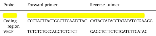
l of reaction volume at 37°C for 1 h. Subsequently, Fpg-treated and untreated samples were incubated with 0.1 N NaOH for 15 min at 37°C, and resolved in 0.6% agarose alkaline gel. After electrophoresis and DNA transfer to a nylon membrane (Roche Diagnostics, Sigma, St. Louis, MO), portions of the membrane with DNA sequences of interest were hybridized with PCR-generated probes to the corresponding regions of mtDNA. The mtDNA probes, labeled with a DIG-labeling kit (Roche Diagnostics, Sigma, St. Louis, MO), were generated with rat mtDNATable 1
Primer sequences used for PCR assessment of mRNA abundance, Fpg-sensitive oxidative base damage, and for mtDNA/protein cross-linking and im-munoprecipitation analysis.
Gene Forward primer Reverse primer
D-loop ATTTATCCTCATAGACAAAG TTTACCAATGCTAAGATTT
Primer sequences used for PCR-generation of probes for the Southern and slot-blot analyses.
Probe Forward primer Reverse primer
D-loop TATTTTCCCCAAGCATATAAGC CATTGAAGTTTCAGGTGTAGG Coding
region
CCCTACTTACTGGCTTCAATCTAC CATACCATACCTATATATCCGAAGG
sequences used as templates and the primers listed in Table 2. After cross-linking, the membranes were washed and processed according to the manufacturer’s suggestions. Hybridization bands were detected with Amersham Hyperfilm ECL (GE Healthcare, Piscataway, NJ) and a Gel Logic 1500 Imaging System (Kodak, Rochester, NY). Single-strand breaks formed at the sites of oxi-dized purines by Fpg treatment result in decrease of Southern blot hybridization band intensity that depends on the relative amount and integrity of DNA. Changes in hybridization band intensity between Fpg-treated and untreated DNA indicate the extension of oxidative base damage. These changes in the Fpg-sensitive lesion density were calculated as negative ln of the quotient of hy-bridization band intensities in Fpg-treated and untreated samples and normalized to 10 kb. This method of calculation is based on the application of a Poisson equation for the undamaged se-quences and has been traditionally used for the analyses of DNA damage by Southern blot and quantitative PCR[33,48].
As a complimentary method to detect mtDNA oxidative da-mage we employed Fpg-sensitive real time PCR analysis as de-scribed earlier[8], using the primers encompassing the sequences in the D-loop region and in the ND4 gene and listed inTable 1. The basis of the assay is treatment of mtDNA with Fpg that removes oxidized purines from DNA, thereby creating single-strand breaks and blocking PCR amplification at these sites. Differences in PCR amplification between Fpg-treated and untreated DNA are thus a specific indicator of the presence of oxidative base damage. The Fpg cleavage reaction was performed by incubating 250 ng of DNA with 8 units of Fpg in 1NEBuffer 1 (10 mM Bis-Tris propane-HCl, 10 mM MgCl2, 1 mM DTT, pH 7.0) and 100
μ
g/ml BSA in a volumeof 50
μ
l at 37°C for 1 h. Fpg was then inactivated by heating at 60°C for 5 min. An aliquot containing 10 ng DNA was then used for the PCR assay to detect Fpg-sensitive cleavage sites. Data are presented as the fraction of intact DNA, calculated as the quotient of signal intensities in Fpg-treated and untreated DNA.2.6. Analysis of TFAM binding to mtDNA
TFAM binding to the D-loop and coding regions of mtDNA was studied with mtDNA/protein cross-linking and immunoprecipita-tion analysis using a standard, commercially-available ChIP assay kit (Active Motif, Carlsbad, CA) and adapted ChIP protocol as de-scribed elsewhere[49]with modifications. Briefly, after exposure to hypoxia, 2107 PAECs were
fixed with 1% formaldehyde (Sigma, St. Louis, MO) for 10 min, washed with ice-cold 1 PBS, andfixation reaction terminated by addition of Glycine Stop Fix solution for 5 min. PAECs were washed and collected in Cell Scraping Solution supplemented with 0.5 mM PMSF. DNA was sheared to500 bp fragments by sonication for ten 20 s pulses at 25% amplitude with a Vibracell VCX 130PB (Sonics & Materials, Newtown, CT). Protein-mtDNA complexes were then im-munoprecipitated with TFAM antibody (Santa Cruz Biotechnology, Santa Cruz, CA) according to the manufacturer's instructions. The yield of target region DNA in each sample after precipitation was analyzed by real-time PCR using USB VeriQuest Fast SYBR Green qPCR Master Kit with Fluorescein (Afflymetrix, Santa Clara, CA). The primers used for the analysis of TFAM binding to the D-loop region and to the ATP6 site in coding region are listed inTable 1. Amplification of input DNA before immunoprecipitation at a di-lution of 1:10 was used as a positive control. A companion analysis without any antibody served as a negative control.
2.7. Statistical analysis
Reduced data are presented as the mean7standard error (SE). Depending on the experimental design, differences in mean values were assessed using unpairedt-test or one-way ANOVA combined
with Dunnett test. P values o0.05 were taken as evidence of statistical significance.
3. Results
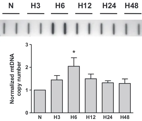
3.1. Hypoxia increases the abundance of mtDNA in rat PAECs
Our initial experiments with rat PAECs using slot-blot analysis showed that hypoxia caused a time-dependent increase in mtDNA abundance. The results of these studies are presented inFig. 1and show the time course of mtDNA content in the rat PAECs after cell exposure to hypoxia. Mitochondrial DNA copy number in PAECs peaked at 6 h of hypoxic exposure and remained elevated till 24 h under hypoxia. According to our hypothesis, this elevation in mtDNA content requires increased base oxidation in the D-loop region of mtDNA.
3.2. Hypoxia selectively damages the D-loop region of mtDNA in rat PAECs
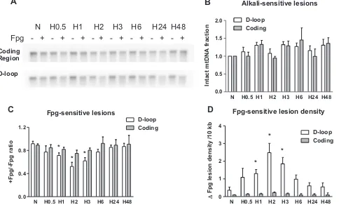
Southern blot analysis of mtDNA from PAECs for oxidative modifications in the coding and D-loop regions during hypoxia revealed significantly different levels of oxidative damage in these two sequences of the mitochondrial genome (Fig. 2A). To enable detection of oxidative modifications, we treated DNA with Fpg glycosylase, which cleaves DNA at sites with purine base oxidative damage. Application of alkaline gel electrophoresis and Southern hybridization to samples not treated with Fpg was used to reveal alkali-detectable lesions such as strand breaks and abasic sites. Treatment of the samples with Fpg along with alkali was em-ployed to detect oxidative damage to purine bases. In experiments with rat PAECs we found that the hypoxia did not induce alkali-detectable DNA damage in either the coding region or the D-loop region (Fig. 2B), but generated oxidative base modifications that were prominent in the mtDNA D-loop region after 1–3 h of hy-poxic exposure and were absent in the coding region of the mi-tochondrial genome (Fig. 2C). Quantitative data calculated as in-crease in purine oxidative lesion density are depicted inFig. 2D and show that hypoxia significantly elevated the density of purine
N H3 H6 H12 H24 H48
base oxidation products in the D-loop region of mitochondrial genome.
As an additional approach to confirm the detection of oxidative base modifications in sequences of the D-loop and coding regions of the mitochondrial genome, we used Fpg-sensitive conventional and real time PCR analyses. The results of experiments with both PCR techniques confirmedfindings obtained in the Southern blot studies; hypoxia-induced damage was restricted to the regulatory D-loop region in the mitochondrial genome (Fig. 3). Fpg-sensitive lesions were observed only in the D-loop region after culturing rat PAECs in hypoxic conditions for 2 and 3 h, whereas the coding region of mtDNA, specifically a sequence nested within the ND4 gene, did not show any oxidative damage after hypoxic exposure. It is important to note that the oxidative lesions to the D-loop region during hypoxia temporally preceded hypoxia-induced in-crease in mtDNA copy number.
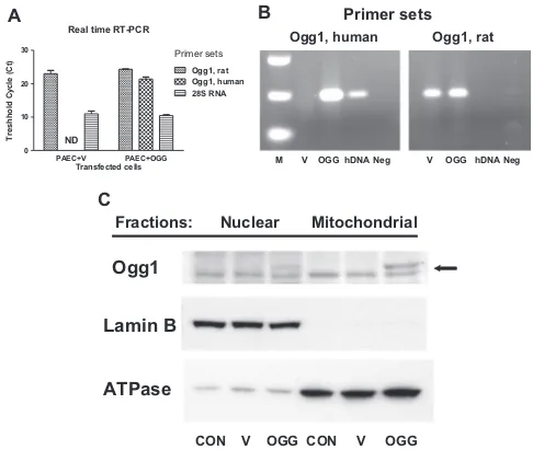
3.3. Transfection of rat PAECs with mitochondria-targeted hOgg1
To explore the role of oxidative DNA damage to the D-loop region in mtDNA replication, we overexpressed mitochondria-targeted human DNA repair glycosylase Ogg1 and studied the ef-fect of enhanced DNA repair on mitochondrial DNA replication and transcription during hypoxia. The results of real time and con-ventional RT-PCR analysis shown inFig. 4A–B confirmed successful overexpression of hOgg1 in rat PAECs. The results of Western blot analysis depicted in Fig. 4C show preferential mitochondrial lo-calization of hOgg1. Empty vector- and hOgg1-transfected cells were then exposed to hypoxia and analyzed for oxidative mtDNA damage, mtDNA copy number and transcription of mitochondrial
genes.
3.4. Overexpression of hOgg1 eliminates hypoxia-induced oxidative damage in D-loop region and attenuates hypoxia-induced mtDNA replication in rat PAECs
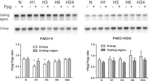
The quantitative Southern blot analysis shown in Fig. 5 in-dicates that overexpression of mitochondria-targeted hOgg1 in PAECs suppressed hypoxia-induced oxidative modifications to the
Alkali-sensitive lesions
Fpg lesion density /10 kb
B
Fig. 2.Hypoxia-induced oxidative base damage is localized to the D-loop region of the mitochondrial genome in rat pulmonary artery endothelial cells. (A) Representative quantitative Southern blot analysis of oxidative damage in coding and D-loop regions of mtDNA from normoxic (N) rat pulmonary artery endothelial cells and cells after hypoxia exposure for 0.5, 1, 2, 3, 6, 24, and 48 h (H0.5–H48). Samples were treated (þ) or not treated ( ) with Fpg to reveal oxidatively modified purines. (B) Calculated data showing absence of alkali-detectable lesions in hypoxic samples without Fpg treatment normalized to normoxic control. (C) Calculated data expressed as ratio between Fpg treated and not treated samples showing the fraction of mtDNA without oxidative damage in above mentioned experimental groups. (D) Calculated data showing change in Fpg-sensitive lesion density per 10 kb. Mean7SE, N¼3–9, *Po0.05, significantly different from normoxic controls.
N
H0.5
H1
H2
H3
H6
H24
regulatory D-loop region of the mitochondrial genome, but did not change the baseline level of oxidation in the coding region of the mtDNA. If our hypothesis is correct, then elimination of D-loop oxidation should prevent changes in mtDNA replication and transcription. Slot blot analysis was used to assess the mtDNA content in normoxic and hypoxic vector- and hOgg1-transfected cells. Results shown inFig. 6revealed that the increase in mtDNA copy number in PAECs exposed to hypoxia was completely atte-nuated in cells overexpressing hOgg1.
Since mechanisms of replication and transcription of the mtDNA are tightly connected, we analyzed the abundance of three mitochondria-encoded transcripts, ATP6, Cox2, and ND4, using real time RT-PCR. The results of these experiments are shown in
Fig. 7. Neither hypoxia, nor hOgg1 overexpression, had significant effects on the selected mitochondrial mRNAs, although there was some tendency toward decrease in abundance of some transcripts at 24 h of hypoxic exposure.
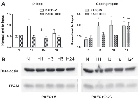
3.5. Overexpression of hOgg1 attenuates TFAM binding to both
D-loop and coding regions of mtDNA in rat PAECs
To explore a possible mechanism whereby mtDNA oxidation during hypoxia regulates mtDNA replication, we determined if mtDNA oxidative modifications impacted TFAM binding to the mtDNA. Since TFAM preferentially binds to oxidized DNA, we ex-pected that enhanced mtDNA repair in cells overexpressing hOgg1 should affect its binding. A modification of the ChIP assay –
mtDNA/protein cross-linking and immunoprecipitation analysis–
was performed on vector- and hOgg1-transfected rat PAECs after cell exposure to hypoxia for 1, 3, and 6 h using an antibody to TFAM. The results of the PCR analysis, using primers for coding and D-loop regions and precipitated DNA as a template, are depicted in theFig. 8A. Surprisingly, in vector-transfected PAECs, TFAM bind-ing to the D-loop region of mtDNA increased only transiently after 1 h of hypoxia, whereas its binding to the coding region of mtDNA was elevated at all times in hypoxic PAECs when compared to normoxic cells. Mitochondria-targeted hOgg1 overexpression at-tenuated TFAM binding to both the D-loop and coding regions of mtDNA. The results of Western blot analyses showed that hypoxia
Ogg1, human
Ogg1, rat
M V OGG hDNA Neg V OGG hDNA Neg
Primer sets
Real time RT-PCR
PAEC+V PAEC+OGG 0
10 20 30
Ogg1, rat Ogg1, human 28S RNA
ND
Primer sets
Transfected cells
T
resh
h
o
ld
C
y
cl
e (
C
t)
A
B
Ogg1
Lamin B
ATPase
Fractions: Nuclear Mitochondrial
CON V OGG CON V OGG
C
PAEC+V
N H1 H3 H6 H24
0.0 0.5 1.0 1.5
D-loop
Coding region
*
*
+Fpg/-Fpg ratio
PAEC+OGG
N H1 H3 H6 H24
0.0 0.5 1.0 1.5
D-loop
Coding region
+Fpg/-Fpg ratio
Fpg -
+ - + - + -
+ - + -
+ - + -
+ - + - +
Coding
region
D-loop
N H1 H3 H6 H24 N H1 H3 H6 H24
Fig. 5.Impact of Ogg1 overexpression on hypoxia-induced oxidative damage to mitochondrial genome in rat pulmonary artery endothelial cells (PAECs). Quantitative Southern blot analysis of oxidative damage in coding and D-loop regions of mtDNA in rat PAECs transfected either with empty vector (PAECþV) or with Ogg1 construct (PAECþOGG) in normoxic (N) conditions and after hypoxia exposure for 1, 3, 6, and 24 h (H1–H24).Top:Representative Southern blot analysis of Fpg-treated (þ) or untreated ( ) samples to reveal oxidatively modified purines.Bottom:Calculated data expressed as ratio between Fpg treated and not treated samples showing the fraction of mtDNA without oxidative damage in above mentioned experimental groups. Mean7SE, N¼4–5, *Po0.05, significantly different from normoxic controls.
N H1 H3 H6 H24
PAEC+V
PAEC+OGG
N H1 H3 H6 H24
0.0 0.5 1.0 1.5 2.0 2.5
PAEC+V
PAEC+OGG
*
*
Normalized mtDNA
copy number
Fig. 6.Impact of Ogg1 overexpression on hypoxia-induced mtDNA replication in rat pulmonary artery endothelial cells (PAECs).Left:Representative slot-blot analysis of mtDNA from rat PAECs transfected either with empty vector (PAECþV) or with Ogg1 construct (PAECþOGG) in normoxic (N) conditions and after hypoxia exposure for 1, 3, 6, and 24 h (H1–H24).Right:Reduced results of hybridization band intensities normalized to nuclear DNA. Mean7SE, N¼5–6, *Po0.05, significantly different from nor-moxic controls.
ATP6 mRNA
N H1 H3 H6 H24 0.0
0.5 1.0 1.5
PAEC+V PAEC+OGG
Normalized Fold Expression
Cox2 mRNA
N H1 H3 H6 H24 0.0
0.5 1.0 1.5
PAEC+V PAEC+OGG
Normalized Fold Expression
ND4 mRNA
N H1 H3 H6 H24 0.0
0.5 1.0 1.5
PAEC+V PAEC+OGG
Normalized Fold Expression
A
B
C
Fig. 7.Transcription of mitochondrial genes in rat pulmonary artery endothelial cells (PAECs) transfected either with empty vector (PAECþV) or with Ogg1 construct
did not change TFAM abundance in total cell lysates (Fig. 8B).
4. Discussion
Hypoxia, as a fundamental stimulus, which complicates many cardiopulmonary, infectious and neoplastic disorders, displays complex and sometimes controversial action on cells and mi-tochondria. Indeed, the cell's reaction to hypoxia depends on many factors: cell origin, nutrient conditions, duration and severity of hypoxia[50]. In some cases hypoxia appears as a damaging factor, inducing autophagy, inflammation, cell injury, and apoptosis[50–
52]. But in many tissues, especially in the vasculature, hypoxia, by up-regulating VEGF expression, acts as a powerful stimulus for cell proliferation, vasculogenesis and angiogenesis[53–56]. The effect of hypoxia on mtDNA replication and mitochondrial biogenesis is also complex and may depend on many factors, including sex specificity[57]. Although some cell types respond to hypoxia by reducing their mitochondrial biogenesis and mtDNA content[58–
60], mtDNA copy number is reported to increase under hypoxic conditions in many other tissues, including brain, liver, heart, placenta, sperm and blood cells[23–25,61–67]. Hypoxia, like many other factors that affect mitochondrial biogenesis, utilizes mi-tochondria-generated ROS as second messengers[68–71]. In the context of ROS-mediated nuclear gene expression, a surprising target of free radicals is specific DNA sequences in gene promoters,
and emerging evidence suggests that a process of “controlled oxidative DNA damage and repair” may be necessary for normal transcriptional regulation[6,7,10,11,15,72–76]. In this model, ROS generated during signaling lead to oxidative DNA “damage” and activation of base excision repair in key promoter sequences. Complete execution of the repair entails the formation and sub-sequent re-ligation of DNA strand breaks; such breaks have pro-found effects on DNA topology, flexibility, and conformation, which may contribute to regulation of promoter function and gene expression.
Could a similar model for ROS-dependent transcriptional acti-vation be operative in the mitochondrial genome, triggering mtDNA replication and transcription via formation and repair of oxidative base lesions in the mtDNA transcriptional regulatory region–the D-loop? Such a mechanism could be involved in ei-ther mtDNA replication or transcription, since initiation of both processes is mediated by the same transcription machinery[30]. Results of the present study support this intriguing idea. Whereas in our earlier research we focused mostly on the“common dele-tion”region of the mtDNA[9], in the present work we searched for oxidative damage in two discrete, functionally relevant mtDNA regions: the D-loop region withflanking sequences, and the cod-ing region of the mitochondrial genome.
We were particularly interested in the D-loop region of the mtDNA because of its exquisite sensitivity to oxidative damage
[34,35]. In addition, it has been reported that base oxidation in the
D-loop
N
H1
H3
H6
0.0
0.5
1.0
1.5
PAEC+V
PAEC+OGG
*
Normalized to Input
Coding region
N
H1
H3
H6
0.0
0.5
1.0
1.5
PAEC+V
PAEC+OGG
*
*
* **
Normalized to Input
A
Beta-actin
TFAM
PAEC+V
PAEC+OGG
B
model oligonucleotides enhance DNA binding affinity for TFAM, the main factor for regulation of the transcription and replication of the mtDNA[36]. Our experiments on PAECs showed that hy-poxia-induced transient oxidative damage to mtDNA was localized in the D-loop region and was absent in the coding region of the molecule. This mtDNA oxidation temporarily coincided with a transient increase in TFAM binding and an elevation in mtDNA copy number, suggesting the potential role of oxidative modifi ca-tions in the D-loop region for the TFAM binding and mtDNA replication.
Being part of the High Mobility Group domain family proteins, TFAM is capable of introducing specific structural alterations in DNA; in the mtDNA D-loop region, binding of TFAM is believed to unwind[77,78]and bend the promoter sequences[79]. The en-hanced bending of the promoter DNA imparted by the C-terminal tail is considered as a critical component of the ability of TFAM to initiate transcription and subsequent mtDNA replication machin-ery[29,79].
The requirement for a TFAM-induced conformational change in the D-loop is central to our model. For TFAM binding to initiate conformational changes in the D-loop, it must overcome the in-trinsic stiffness of the sequence. In mammalian cells, the circular mtDNA is covalently closed and adopts a supercoiled configuration
[80]. Replication of mtDNA, like any other closed-circular DNA, requires cycles of nicking and closing of strands in parental mtDNA to relieve tensional stress caused by supercoiling[81]. Recently it was shown that TFAM binding to mtDNA increases itsflexibility which can be explained by local denaturation of the DNA[82]. We have observed hypoxia-induced formation of oxidative mtDNA base modifications in the D-loop region in a very narrow time interval (1–3 h of hypoxia) that might be sufficient for the tran-sient increased binding of TFAM to this region. We suggest that this increased TFAM binding temporarily decreases stiffness in the regulatory sequence, increases itsflexibility, and changes its con-formation. This, in turn, may reduce the steric obstruction and facilitate assembly of other components of the replication ma-chinery, which results in increased mtDNA replication. We can also suggest involvement of DNA repair enzymes in the regulation of mtDNA replication, since D-loop oxidative lesions under hypoxic exposure were transient in character and were eventually re-paired. Such a possibility is supported by some existing data on potential involvement of DNA repair enzymes in mitochondrial and nuclear transcription[7,12,83].
To explore the importance of sequence-specific oxidation of the mtDNA for TFAM binding and initiation of mtDNA replication, we overexpressed the mitochondria-targeted human DNA repair gly-cosylase Ogg1 in PAECs as described previously. Increased Ogg1 expression should enhance DNA repair capacity, eliminate oxi-dized guanines in the DNA, and thereby decrease TFAM binding to the sequence. As predicted, hOgg1 overexpression eliminated hy-poxia-induced oxidative damage in the D-loop region of the mi-tochondrial genome in rat PAECs and attenuated TFAM binding to both D-loop and coding regions of mtDNA, which resulted in prevention of hypoxia-induced mtDNA replication. Interestingly, Ogg1 overexpression suppressed TFAM binding not only to the oxidized D-loop sequence, but also to the mtDNA coding region, which, unlike the D-loop, was not oxidatively modified by hypoxia. These results may indicate that transient oxidative modifications in the regulatory sequence not only increase TFAM binding to the D-loop region, but also facilitate further TFAM binding to the rest of the mtDNA molecule. Elimination of such transient site-specific mtDNA oxidation decreases the amount of TFAM bound to the entire mtDNA molecule and prevents its replication. Interestingly, TFAM has the unique ability to bind to the promoter region in a sequence-specific manner and to non-promoter DNA in a non sequence-specific manner [84,85]. The mechanisms of TFAM
binding to promoter and non-promoter regions are not under-stood, but according to our results, preferential oxidation of the D-loop region in response to stimuli could be the key factor in orchestrating TFAM interaction with other regions of the mtDNA that leads to its replication.
It is important to emphasize that changes in TFAM binding and mtDNA replication occur without increase in TFAM expression, suggesting that binding of TFAM to the promoter region of the mtDNA, but not its abundance, is a key event in initiation of mtDNA replication. Although it has been reported that mtDNA copy number is directly proportional to total TFAM levels[86], several lines of evidence show that the link between TFAM abundance and mtDNA replication might be more complicated. For example, Noack et al. showed that mild oxidative stress led to proliferation of the mtDNA, but did not change TFAM protein le-vels[87]. Another study with transient overexpression of TFAM demonstrated that increase in TFAM abundance in the cell is
suf-ficient to stimulate mtDNA transcription, but not sufficient to in-crease mtDNA copy number[88].
Interestingly, despite the fact that hypoxia increased mtDNA copy number, changes in mRNA transcripts of the examined mi-tochondrial proteins did not occur. It has been reported that mtDNA copy number is regulated by cellular proliferation[89]and, though not studied here, previous reports show that prolonged hypoxia induces proliferation of pulmonary endothelial cells[53–
55]. We consider the observed increase in mtDNA copy number only as an early stage of mitochondrial biogenesis on the threshold of cell proliferation. Initiation of mitochondrial DNA replication uses the same pathways as employed for transcription initiation; therefore the processes of replication and transcription must be highly coordinated. Recently it has been proposed that replication and transcription are mutually exclusive processes in human mi-tochondria that allow the corresponding machineries to avoid the detrimental consequences of a head-on collision[32]. It was sug-gested that human transcription elongation factor serves as a molecular switch that allows the organelles either to replicate the mtDNA and regulate its copy number or to elevate its transcription rates. The modest effect of hypoxia on mtDNA transcription ob-served in our experiments may indicate that, prior to hypoxia-induced cell proliferation, PAECs undergo mtDNA replication without altering the transcription rate.
In summary, our observations suggest that hypoxia causes oxidative modifications that are prominent in the D-loop region of the mitochondrial genome of the PAECs and that such DNA modifications may be important for TFAM binding and subsequent mtDNA replication. These data support our hypothesis that con-trolled oxidative DNA damage and repair in the D-loop region of the mitochondrial genome are required for mitochondrial DNA replication in hypoxia. The concept that oxidative mtDNA damage might be important for mitochondrial proliferation is supported indirectly by other recentfindings. For example, the robust mi-tochondrial biogenesis that occurs during the postnatal cardiac development in rodents is accompanied by oxidative mtDNA da-mage and repair[83]. Here too, it was suggested that up-regula-tion of DNA damage response genes may be involved in control of mitochondrial biogenesis.
The present results are significant because they extend the emerging concept that controlled DNA damage and repair also contribute to regulation of mtDNA replication. In addition, our
Acknowledgments
We acknowledge Dr. Mikhail Alexeyev and Mrs. Viktoriya Pas-tukh from Gene Delivery Core for their work on generation and production of lentiviral vectors. We also appreciate the assistance of Mrs. Gina Capley Bardwell and Ms. Mita Patel. This study was funded by National Heart, Lung, and Blood Institute Grants R21 HL102789 (PI M. Ruchko), R01 HL113614 (PI M. Gillespie), and R01 HL058234 (PI M. Gillespie).
References
[1]B.A. Freeman, J.D. Crapo, Biology of disease: free radicals and tissue injury, Lab.
Invest. 47 (1982) 412–426.
[2]S. Dimauro, G. Davidzon, Mitochondrial DNA and disease, Ann. Med. 37 (2005)
222–232.
[3]D.C. Wallace, A mitochondrial paradigm of metabolic and degenerative
dis-eases, aging, and cancer: a dawn for evolutionary medicine, Annu. Rev. Genet.
39 (2005) 359–407.
[4]O.A. Sedelnikova, C.E. Redon, J.S. Dickey, A.J. Nakamura, A.G. Georgakilas, W.
M. Bonner, Role of oxidatively induced DNA lesions in human pathogenesis,
Mutat. Res. 704 (2010) 152–159.
[5]M.K. Hailer-Morrison, J.M. Kotler, B.D. Martin, K.D. Sugden, Oxidized guanine
lesions as modulators of gene transcription. Altered p50 binding affinity and
repair shielding by 7,8-dihydro-8-oxo-2’-deoxyguanosine lesions in the
NF-kappaB promoter element, Biochemistry 42 (2003) 9761–9770.
[6]B.G. Ju, V.V. Lunyak, V. Perissi, I. Garcia-Bassets, D.W. Rose, C.K. Glass, M.
G. Rosenfeld, A topoisomerase IIbeta-mediated dsDNA break required for
regulated transcription, Science 312 (2006) 1798–1802.
[7]B. Perillo, M.N. Ombra, A. Bertoni, C. Cuozzo, S. Sacchetti, A. Sasso, L. Chiariotti,
A. Malorni, C. Abbondanza, E.V. Avvedimento, DNA oxidation as triggered by H3K9me2 demethylation drives estrogen-induced gene expression, Science
319 (2008) 202–206.
[8]A.B. Al-Mehdi, V.M. Pastukh, B.M. Swiger, D.J. Reed, M.R. Patel, G.C. Bardwell,
V.V. Pastukh, M.F. Alexeyev, M.N. Gillespie, Perinuclear mitochondrial clus-tering creates an oxidant-rich nuclear domain required for hypoxia-induced
transcription, Sci. Signal. 5 (2012) ra47.
[9]V. Grishko, M. Solomon, J.F. Breit, D.W. Killilea, S.P. Ledoux, G.L. Wilson, M.
N. Gillespie, Hypoxia promotes oxidative base modifications in the pulmonary
artery endothelial cell VEGF gene, FASEB J. 15 (2001) 1267–1269.
[10]K.A. Ziel, V. Grishko, C.C. Campbell, J.F. Breit, G.L. Wilson, M.N. Gillespie,
Oxi-dants in signal transduction: impact on DNA integrity and gene expression,
FASEB J. 19 (2005) 387–394.
[11]V. Pastukh, M. Ruchko, O. Gorodnya, G.L. Wilson, M.N. Gillespie,
Sequence-specific oxidative base modifications in hypoxia-inducible genes, Free Radic.
Biol. Med. 43 (2007) 1616–1626.
[12]V. Pastukh, J.T. Roberts, D.W. Clark, G.C. Bardwell, M. Patel, A.B. Al-Mehdi, G.
M. Borchert, M.N. Gillespie, An oxidative DNA“damage”and repair mechanism
localized in the VEGF promoter is important for hypoxia-induced VEGF mRNA
expression, Am. J. Physiol. Lung Cell. Mol. Physiol. 309 (2015) L1367–L1375.
[13]M.V. Ruchko, O.M. Gorodnya, V.M. Pastukh, B.M. Swiger, N.S. Middleton, G.
L. Wilson, M.N. Gillespie, Hypoxia-induced oxidative base modifications in the
VEGF hypoxia-response element are associated with transcriptionally active
nucleosomes, Free Radic. Biol. Med. 46 (2009) 352–359.
[14]J.F. Breit, K. Ault-Ziel, A.B. Al-Mehdi, M.N. Gillespie, Nuclear protein-induced
bending andflexing of the hypoxic response element of the rat vascular
en-dothelial growth factor promoter, FASEB J. 22 (2008) 19–29.
[15]M.N. Gillespie, G.L. Wilson, Bending and breaking the code: dynamic changes
in promoter integrity may underlie a new mechanism regulating gene
ex-pression, Am. J. Physiol. Lung Cell. Mol. Physiol. 292 (2007) L1–L3.
[16]E.D. Robin, R. Wong, Mitochondrial DNA molecules and virtual number of
mitochondria per cell in mammalian cells, J. Cell. Physiol. 136 (1988) 507–513.
[17]H.C. Lee, C.Y. Lu, H.J. Fahn, Y.H. Wei, Aging- and smoking-associated alteration
in the relative content of mitochondrial DNA in human lung, FEBS Lett. 441
(1998) 292–296.
[18]V. Pesce, A. Cormio, F. Fracasso, J. Vecchiet, G. Felzani, A.M. Lezza, P. Cantatore,
M.N. Gadaleta, Age-related mitochondrial genotypic and phenotypic
altera-tions in human skeletal muscle, Free Radic. Biol. Med. 30 (2001) 1223–1233.
[19]M. Masuyama, R. Iida, H. Takatsuka, T. Yasuda, T. Matsuki, Quantitative change
in mitochondrial DNA content in various mouse tissues during aging, Biochim.
Biophys. Acta 1723 (2005) 302–308.
[20]M. Yu, Generation, function and diagnostic value of mitochondrial DNA copy
number alterations in human cancers, Life Sci. 89 (2011) 65–71.
[21]H.B. Suliman, M.S. Carraway, K.E. Welty-Wolf, A.R. Whorton, C.A. Piantadosi,
Lipopolysaccharide stimulates mitochondrial biogenesis via activation of
nu-clear respiratory factor-1, J. Biol. Chem. 278 (2003) 41510–41518.
[22]H.C. Lee, P.H. Yin, C.Y. Lu, C.W. Chi, Y.H. Wei, Increase of mitochondria and
mitochondrial DNA in response to oxidative stress in human cells, Biochem. J.
348 (Pt 2) (2000) 425–432.
[23]L.E. Costa, A. Boveris, O.R. Koch, A.C. Taquini, Liver and heart mitochondria in
rats submitted to chronic hypobaric hypoxia, Am. J. Physiol. 255 (1988)
C123–C129.
[24] D.R. Gutsaeva, M.S. Carraway, H.B. Suliman, I.T. Demchenko, H. Shitara,
H. Yonekawa, C.A. Piantadosi, Transient hypoxia stimulates mitochondrial biogenesis in brain subcortex by a neuronal nitric oxide synthase-dependent
mechanism, J. Neurosci. 28 (2008) 2015–2024.
[25] H.M. Lee, G.H. Greeley Jr., E.W. Englander, Sustained hypoxia modulates
mi-tochondrial DNA content in the neonatal rat brain, Free Radic. Biol. Med. 44
(2008) 807–814.
[26] M.S. Carraway, H.B. Suliman, C. Kliment, K.E. Welty-Wolf, T.D. Oury, C.
A. Piantadosi, Mitochondrial biogenesis in the pulmonary vasculature during
inhalational lung injury andfibrosis, Antioxid. Redox Signal. 10 (2008)
269–275.
[27] E. Samper, L. Morgado, J.C. Estrada, A. Bernad, A. Hubbard, S. Cadenas,
S. Melov, Increase in mitochondrial biogenesis, oxidative stress, and glycolysis
in murine lymphomas, Free Radic. Biol. Med. 46 (2009) 387–396.
[28] J.V. Virbasius, R.C. Scarpulla, Activation of the human mitochondrial
tran-scription factor A gene by nuclear respiratory factors: a potential regulatory link between nuclear and mitochondrial gene expression in organelle
bio-genesis, Proc. Natl. Acad. Sci. USA 91 (1994) 1309–1313.
[29] C.T. Campbell, J.E. Kolesar, B.A. Kaufman, Mitochondrial transcription factor A
regulates mitochondrial transcription initiation, DNA packaging, and genome
copy number, Biochim. Biophys. Acta 1819 (2012) 921–929.
[30] R. Kasiviswanathan, T.R. Collins, W.C. Copeland, The interface of transcription
and DNA replication in the mitochondria, Biochim. Biophys. Acta 1819 (2012)
970–978.
[31]G.S. Shadel, D.A. Clayton, Mitochondrial DNA maintenance in vertebrates,
Annu. Rev. Biochem. 66 (1997) 409–435.
[32] K. Agaronyan, Y.I. Morozov, M. Anikin, D. Temiakov, Mitochondrial biology.
Replication-transcription switch in human mitochondria, Science 347 (2015)
548–551.
[33] F.M. Yakes, B. Van Houten, Mitochondrial DNA damage is more extensive and
persists longer than nuclear DNA damage in human cells following oxidative
stress, Proc. Natl. Acad. Sci. USA 94 (1997) 514–519.
[34] E. Mambo, X. Gao, Y. Cohen, Z. Guo, P. Talalay, D. Sidransky, Electrophile and
oxidant damage of mitochondrial DNA leading to rapid evolution of
homo-plasmic mutations, Proc. Natl. Acad. Sci. USA 100 (2003) 1838–1843.
[35] O. Rothfuss, T. Gasser, N. Patenge, Analysis of differential DNA damage in the
mitochondrial genome employing a semi-long run real-time PCR approach,
Nucleic Acids Res. 38 (2009) e24.
[36] Y. Yoshida, H. Izumi, T. Ise, H. Uramoto, T. Torigoe, H. Ishiguchi, T. Murakami,
M. Tanabe, Y. Nakayama, H. Itoh, H. Kasai, K. Kohno, Human mitochondrial transcription factor A binds preferentially to oxidatively damaged DNA,
Bio-chem. Biophys. Res. Commun. 295 (2002) 945–951.
[37]D.W. Clark, T. Phang, M.G. Edwards, M.W. Geraci, M.N. Gillespie, Promoter
G-quadruplex sequences are targets for base oxidation and strand cleavage
during hypoxia-induced transcription, Free Radic. Biol. Med. 53 (2012) 51–59.
[38] M. Ruchko, O. Gorodnya, S.P. LeDoux, M.F. Alexeyev, A.B. Al-Mehdi, M.
N. Gillespie, Mitochondrial DNA damage triggers mitochondrial dysfunction and apoptosis in oxidant-challenged lung endothelial cells, Am. J. Physiol.
Lung Cell. Mol. Physiol. 288 (2005) L530–L535.
[39] R.X. Santos, S.C. Correia, X. Zhu, M.A. Smith, P.I. Moreira, R.J. Castellani,
A. Nunomura, G. Perry, Mitochondrial DNA oxidative damage and repair in
aging and Alzheimer’s disease, Antioxid. Redox Signal. 18 (2013) 2444–2457.
[40] S.D. Cline, Mitochondrial DNA damage and its consequences for mitochondrial
gene expression, Biochim. Biophys. Acta 1819 (2012) 979–991.
[41]M. Muftuoglu, M.P. Mori, N.C. de Souza-Pinto, Formation and repair of
oxi-dative damage in the mitochondrial DNA, Mitochondrion 17 (2014) 164–181.
[42] M.V. Ruchko, O.M. Gorodnya, A. Zuleta, V.M. Pastukh, M.N. Gillespie, The DNA
glycosylase Ogg1 defends against oxidant-induced mtDNA damage and apoptosis in pulmonary artery endothelial cells, Free Radic. Biol. Med. 50
(2011) 1107–1113.
[43] A.W. Dobson, V. Grishko, S.P. LeDoux, M.R. Kelley, G.L. Wilson, M.N. Gillespie,
Enhanced mtDNA repair capacity protects pulmonary artery endothelial cells from oxidant-mediated death, Am. J. Physiol. Lung Cell. Mol. Physiol. 283
(2002) L205–L210.
[44]L.I. Rachek, V.I. Grishko, S.I. Musiyenko, M.R. Kelley, S.P. LeDoux, G.L. Wilson,
Conditional targeting of the DNA repair enzyme hOGG1 into mitochondria, J.
Biol. Chem. 277 (2002) 44932–44937.
[45] N.M. Druzhyna, S.B. Hollensworth, M.R. Kelley, G.L. Wilson, S.P. Ledoux,
Tar-geting human 8-oxoguanine glycosylase to mitochondria of oligodendrocytes
protects against menadione-induced oxidative stress, Glia 42 (2003) 370–378.
[46] J.M. Chouteau, B. Obiako, O.M. Gorodnya, V.M. Pastukh, M.V. Ruchko, A.
J. Wright, G.L. Wilson, M.N. Gillespie, Mitochondrial DNA integrity may be a determinant of endothelial barrier properties in oxidant-challenged rat lungs,
Am. J. Physiol. Lung Cell. Mol. Physiol. 301 (2011) L892–L898.
[47]V.M. Pastukh, L. Zhang, M.V. Ruchko, O. Gorodnya, G.C. Bardwell, R.M. Tuder,
M.N. Gillespie, Oxidative DNA damage in lung tissue from patients with COPD
is clustered in functionally significant sequences, Int. J. Chron. Obstruct.
Pul-mon. Dis. 6 (2011) 209–217.
[48] V.A. Bohr, C.A. Smith, D.S. Okumoto, P.C. Hanawalt, DNA repair in an active
gene: removal of pyrimidine dimers from the DHFR gene of CHO cells is much
more efficient than in the genome overall, Cell 40 (1985) 359–369.
[49] H. Ryu, J. Lee, S. Impey, R.R. Ratan, R.J. Ferrante, Antioxidants modulate
mi-tochondrial PKA and increase CREB binding to D-loop DNA of the
mitochon-drial genome in neurons, Proc. Natl. Acad. Sci. USA 102 (2005) 13915–13920.
survival? Curr. Opin. Cell Biol. 22 (2010) 177–180.
[51]K.M. Rouschop, C.H. Ramaekers, M.B. Schaaf, T.G. Keulers, K.G. Savelkouls,
P. Lambin, M. Koritzinsky, B.G. Wouters, Autophagy is required during cycling hypoxia to lower production of reactive oxygen species, Radiother. Oncol. 92
(2009) 411–416.
[52]R.M. Gurevich, K.M. Regula, L.A. Kirshenbaum, Serpin protein CrmA
sup-presses hypoxia-mediated apoptosis of ventricular myocytes, Circulation 103
(2001) 1984–1991.
[53]M. Tucci, S.I. Hammerman, S. Furfaro, J.J. Saukonnen, T.J. Conca, H.W. Farber,
Distinct effect of hypoxia on endothelial cell proliferation and cycling, Am. J.
Physiol. 272 (1997) C1700–C1708.
[54]I.T. Toby, L.G. Chicoine, H. Cui, B. Chen, L.D. Nelin, Hypoxia-induced
pro-liferation of human pulmonary microvascular endothelial cells depends on epidermal growth factor receptor tyrosine kinase activation, Am. J. Physiol.
Lung Cell. Mol. Physiol. 298 (2010) L600–L606.
[55]K.M. Porter, B.Y. Kang, S.E. Adesina, T.C. Murphy, C.M. Hart, R.L. Sutliff, Chronic
hypoxia promotes pulmonary artery endothelial cell proliferation through
H2O2-induced 5-lipoxygenase, PLoS One 9 (2014) e98532.
[56]E.M. Conway, D. Collen, P. Carmeliet, Molecular mechanisms of blood vessel
growth, Cardiovasc. Res. 49 (2001) 507–521.
[57]J. Sharma, M.V. Johnston, M.A. Hossain, Sex differences in mitochondrial
bio-genesis determine neuronal death and survival in response to oxygen glucose
deprivation and reoxygenation, BMC Neurosci. 15 (2014) 9.
[58]P.H. Oliveira, J.S. Boura, M.M. Abecasis, J.M. Gimble, C.L. da Silva, J.M. Cabral,
Impact of hypoxia and long-term cultivation on the genomic stability and mitochondrial performance of ex vivo expanded human stem/stromal cells,
Stem Cell. Res. 9 (2012) 225–236.
[59]H. Hoppeler, M. Vogt, E.R. Weibel, M. Fluck, Response of skeletal muscle
mi-tochondria to hypoxia, Exp. Physiol. 88 (2003) 109–119.
[60]H. Zhang, P. Gao, R. Fukuda, G. Kumar, B. Krishnamachary, K.I. Zeller, C.
V. Dang, G.L. Semenza, HIF-1 inhibits mitochondrial biogenesis and cellular
respiration in VHL-deficient renal cell carcinoma by repression of C-MYC
ac-tivity, Cancer Cell 11 (2007) 407–420.
[61]J. Carabelli, A.L. Burgueno, M.S. Rosselli, T.F. Gianotti, N.R. Lago, C.J. Pirola,
S. Sookoian, High fat diet-induced liver steatosis promotes an increase in liver mitochondrial biogenesis in response to hypoxia, J. Cell. Mol. Med. 15 (2011)
1329–1338.
[62]W. Yin, A.P. Signore, M. Iwai, G. Cao, Y. Gao, J. Chen, Rapidly increased neuronal
mitochondrial biogenesis after hypoxic-ischemic brain injury, Stroke 39
(2008) 3057–3063.
[63]Y. Luo, W. Liao, Y. Chen, J. Cui, F. Liu, C. Jiang, W. Gao, Y. Gao, Altitude can alter
the mtDNA copy number and nDNA integrity in sperm, J. Assist. Reprod.
Genet. 28 (2011) 951–956.
[64]Y. Luo, G. Lu, Y. Chen, F. Liu, G. Xu, J. Yin, Y. Gao, Long-term cycles of hypoxia
and normoxia increase the contents of liver mitochondrial DNA in rats, Eur. J.
Appl. Physiol. 113 (2013) 223–232.
[65]M. Zungu, M.P. Alcolea, F.J. Garcia-Palmer, M.E. Young, M.F. Essop, Genomic
modulation of mitochondrial respiratory genes in the hypertrophied heart
reflects adaptive changes in mitochondrial and contractile function, Am. J.
Physiol. Heart Circ. Physiol. 293 (2007) H2819–H2825.
[66]D. Lattuada, F. Colleoni, A. Martinelli, A. Garretto, R. Magni, T. Radaelli, I. Cetin,
Higher mitochondrial DNA content in human IUGR placenta, Placenta 29
(2008) 1029–1033.
[67]D. Lacedonia, G.E. Carpagnano, E. Crisetti, G. Cotugno, G.P. Palladino,
G. Patricelli, R. Sabato, M.P. Foschino Barbaro, Mitochondrial DNA alteration in
obstructive sleep apnea, Respir. Res. 16 (2015) 47.
[68]I. Ishida, H. Kubo, S. Suzuki, T. Suzuki, S. Akashi, K. Inoue, S. Maeda, H. Kikuchi,
H. Sasaki, T. Kondo, Hypoxia diminishes toll-like receptor 4 expression through reactive oxygen species generated by mitochondria in endothelial
cells, J. Immunol. 169 (2002) 2069–2075.
[69]R. Paddenberg, B. Ishaq, A. Goldenberg, P. Faulhammer, F. Rose, N. Weissmann,
R.C. Braun-Dullaeus, W. Kummer, Essential role of complex II of the re-spiratory chain in hypoxia-induced ROS generation in the pulmonary
vasculature, Am. J. Physiol. Lung Cell. Mol. Physiol. 284 (2003) L710–L719.
[70]R.D. Guzy, P.T. Schumacker, Oxygen sensing by mitochondria at complex III:
the paradox of increased reactive oxygen species during hypoxia, Exp. Physiol.
91 (2006) 807–819.
[71]E.D. Yoboue, A. Devin, Reactive oxygen species-mediated control of
mi-tochondrial biogenesis, Int. J. Cell Biol. 2012 (2012) 403870.
[72]J.F. Haince, M. Rouleau, G.G. Poirier, Transcription. Gene expression needs a
break to unwind before carrying on, Science 312 (2006) 1752–1753.
[73]M.N. Gillespie, V. Pastukh, M.V. Ruchko, Oxidative DNA modifications in
hy-poxic signaling, Ann. NY Acad. Sci. 1177 (2009) 140–150.
[74]M.N. Gillespie, V.M. Pastukh, M.V. Ruchko, Controlled DNA "damage" and
re-pair in hypoxic signaling, Respir. Physiol. Neurobiol. 174 (2010) 244–251.
[75]S. Amente, A. Bertoni, A. Morano, L. Lania, E.V. Avvedimento, B. Majello,
LSD1-mediated demethylation of histone H3 lysine 4 triggers Myc-induced
tran-scription, Oncogene 29 (2010) 3691–3702.
[76]S. Amente, L. Lania, E.V. Avvedimento, B. Majello, DNA oxidation drives Myc
mediated transcription, Cell Cycle 9 (2010) 3002–3004.
[77]R.P. Fisher, T. Lisowsky, M.A. Parisi, D.A. Clayton, DNA wrapping and bending
by a mitochondrial high mobility group-like transcriptional activator protein,
J. Biol. Chem. 267 (1992) 3358–3367.
[78]M. Falkenberg, N.G. Larsson, C.M. Gustafsson, DNA replication and
transcrip-tion in mammalian mitochondria, Annu. Rev. Biochem. 76 (2007) 679–699.
[79]C.S. Malarkey, M. Bestwick, J.E. Kuhlwilm, G.S. Shadel, M.E. Churchill,
Tran-scriptional activation by mitochondrial transcription factor A involves
pre-ferential distortion of promoter DNA, Nucleic Acids Res. 40 (2012) 614–624.
[80]J. Chen, F.F. Kadlubar, J.Z. Chen, DNA supercoiling suppresses real-time PCR: a
new approach to the quantification of mitochondrial DNA damage and repair,
Nucleic Acids Res. 35 (2007) 1377–1388.
[81]D.L. Robberson, D.A. Clayton, Replication of mitochondrial DNA in mouse L
cells and their thymidine kinase–derivatives: displacement replication on a
covalently-closed circular template, Proc. Natl. Acad. Sci. USA 69 (1972)
3810–3814.
[82]G. Farge, N. Laurens, O.D. Broekmans, S.M. van den Wildenberg, L.C. Dekker,
M. Gaspari, C.M. Gustafsson, E.J. Peterman, M. Falkenberg, G.J. Wuite, Protein sliding and DNA denaturation are essential for DNA organization by human
mitochondrial transcription factor A, Nat. Commun. 3 (2012) 1013.
[83]J.L. Pohjoismaki, T. Boettger, Z. Liu, S. Goffart, M. Szibor, T. Braun, Oxidative
stress during mitochondrial biogenesis compromises mtDNA integrity in growing hearts and induces a global DNA repair response, Nucleic Acids Res.
40 (2012) 6595–6607.
[84]B.A. Kaufman, N. Durisic, J.M. Mativetsky, S. Costantino, M.A. Hancock,
P. Grutter, E.A. Shoubridge, The mitochondrial transcription factor TFAM co-ordinates the assembly of multiple DNA molecules into nucleoid-like
struc-tures, Mol. Biol. Cell. 18 (2007) 3225–3236.
[85]T.I. Alam, T. Kanki, T. Muta, K. Ukaji, Y. Abe, H. Nakayama, K. Takio,
N. Hamasaki, D. Kang, Human mitochondrial DNA is packaged with TFAM,
Nucleic Acids Res. 31 (2003) 1640–1645.
[86]M.I. Ekstrand, M. Falkenberg, A. Rantanen, C.B. Park, M. Gaspari, K. Hultenby,
P. Rustin, C.M. Gustafsson, N.G. Larsson, Mitochondrial transcription factor A regulates mtDNA copy number in mammals, Hum. Mol. Genet. 13 (2004)
935–944.
[87]H. Noack, T. Bednarek, J. Heidler, R. Ladig, J. Holtz, M. Szibor, TFAM-dependent
and independent dynamics of mtDNA levels in C2C12 myoblasts caused by
redox stress, Biochim. Biophys. Acta 1760 (2006) 141–150.
[88]K. Maniura-Weber, S. Goffart, H.L. Garstka, J. Montoya, R.J. Wiesner, Transient
overexpression of mitochondrial transcription factor A (TFAM) is sufficient to
stimulate mitochondrial DNA transcription, but not sufficient to increase
mtDNA copy number in cultured cells, Nucleic Acids Res. 32 (2004)
6015–6027.
[89]M. Trinei, I. Berniakovich, P.G. Pelicci, M. Giorgio, Mitochondrial DNA copy
number is regulated by cellular proliferation: a role for Ras and p66(Shc),