ZOOTAXA
ISSN 1175-5326 (print edition)ISSN1175-5334(online edition) Copyright © 2010 · Magnolia Press
Zootaxa 2405: 1–47 (2010)
www.mapress.com/zootaxa/
Article
North West Pacific deep-sea barnacles (Cirripedia, Thoracica) collected by the
TAIWAN expeditions, with descriptions of two new species
BENNY K.K. CHAN1 , ROMANUS EDY PRABOWO2 & KWEN-SHEN LEE 3,* 1Biodiversity Research Center, Academia Sinica, Taipei 115, Taiwan 2Faculty of Biology, Jenderal Soedirman University, Purwokerto, Indonesia 3 National Museum of Natural Science, Taichung, Taiwan* = corresponding author Table of contents Abstract ... 2Introduction ... 2
Material and methods ... 2
Taxonomy ... 3
Order Lepadiformes Buckeridge & Newman, 2006 ... 3
Suborder Lepadomorpha Pilsbry, 1916 ... 3
Family Poecilasmatidae Annandale, 1909 ... 3
Genus Glyptelasma Pilsbry, 1907 ... 3
Glyptelasma gigas (Annandale, 1916) ... 3
Order Scalpelliformes Buckeridge & Newman, 2006 ... 3
Family Calanticidae Zevina, 1978 ... 3
Genus Euscalpellum Hoek, 1907 ... 3
Euscalpellum rostratum (Darwin, 1851) ... 3
Family Scalpellidae Pilsbry, 1916 ... 5
Sub-family Arcoscalpellinae Zevina, 1978... 5
Genus Arcoscalpellum Hoek, 1907 ... 5
Arcoscalpellum truncatum (Hoek, 1883) ... 5
Genus Tarasovium Zevina, 1978... 8
Tarasovium orientale Ren, 1983 ... 8
Genus Trianguloscalpellum Zevina, 1982 ... 9
Trianguloscalpellum diota (Hoek, 1907) ... 9
Trianguloscalpellum hirsutum (Hoek, 1883) ... 13
Trianguloscalpellum regium (Thomson, 1878) ... 13
Trianguloscalpellum weltnerianum (Pilsbry, 1911) ... 16
Genus Verum Zevina, 1981 ... 17
Verum novaezelandiae (Hoek, 1907) ... 17
Sub-family Meroscalpellinae Zevina, 1978 ... 17
Genus Annandaleum Newman & Ross, 1971 ... 17
Annandaleum japonicum biramosum (Pilsbry, 1911) ... 17
Genus Litoscalpellum Newman & Ross, 1971 ... 23
Litoscalpellum spinosus sp. nov. ... 23
Subfamily Scalpellinae Pilsbry, 1907 ... 24
Genus Scalpellum Leach, 1817 ... 24
Scalpellum stearnsii Pilsbry, 1890... 24
Order Sessilia Lamarck, 1818 ... 28
Suborder Verrucomorpha Pilsbry, 1916 ... 28
Family Verrucidae Darwin, 1854 ... 28
Altiverruca Pilsbry, 1916... 28
Altiverruca longimandible sp. nov. ... 28
Genus Metaverruca Pilsbry, 1916 ... 31
Metaverruca defayeae Buckeridge, 1994 ... 31
Metaverruca recta (Aurivillius, 1898)... 36
Genus Rostratoverruca Broch, 1922 ... 36
Rostratoverruca krugeri (Broch, 1922) ... 36
Suborder Balanomorpha Pilsbry, 1916 ... 41
Superfamily Pachylasmatoidea Buckeridge, 1983... 41
Family Pachylasmatidae Utinomi, 1968 (emend.) ... 41
Genus Hexelasma Hoek, 1913 ... 41
Hexelasma velutinum Hoek, 1913 ... 41
Discussion ... 44
Acknowledgement ... 44
References ... 45
Abstract
Taiwan is a large island in north western Pacific waters with the sea floor connecting to two major deep-sea basins, the eastern waters facing the Pacific Ocean (to 4000 m depth) and linking to the Philippine Basin, whilst south western waters are associated with the South China Sea Basin (up to 1000 m). Previously, the biodiversity of Taiwanese deep-sea barnacles had not been studied extensively, due to a lack of deep-sea expeditions and sampling. Recently, several TAIWAN deep-sea cruises investigated the biodiversity of the deep-sea fauna of Taiwan and sampling was conducted to depths of 4000 m. The present study reports on the biodiversity of the deep-sea barnacles of Taiwan, a total of 18 species. One species was previously recorded from Taiwanese waters and 17 are new records, including two new species belong to the genera Litoscalpellum and Altiverruca.
Key words:
Introduction
Taiwan is a large island located in the north western Pacific, supporting a high diversity of shallow water and deep-sea habitats. The waters to the south west of Taiwan connect to the South China Sea Basin (depth to 1000 m). Eastern coasts face the Pacific Ocean, linking to the West Philippine Basin (to 4000 m) and north eastern waters open to the Okinawa Trough (Wang, 1991). The biodiversity of the deep-sea barnacles of Taiwan have received scant attention and few deep-sea species have been reported. Previous records in Taiwan were often focused on intertidal and shallow water species (e.g. Hiro, 1939a, b). In recent years, with support from the National Science Council, Taiwan, several cruises were conducted to survey the deep-sea fauna of Taiwanese waters. Sampling by the TAIWAN expeditions was conducted using benthic French beam trawls in depths to 4000 m (Tsai et al., 2009). The present study reports on 18 deep-sea species collected by the recent TAIWAN expeditions plus collections from deep-sea fishing markets. One species had been previously recorded from Taiwanese waters, two were new species and 15 new records for Taiwanese waters.
Material and methods
Taxonomy
Poecilasma gigas Annandale, 1916: 299, pl. 4 fig. 4, pl. 5 figs 10–14, pl. 6 figs 7, 8.
Megalasma (Glyptelasma) gigas. — Calman, 1919: 364. — Nilsson-Cantell, 1928: 20. — Zevina, 1982: 85, fig. 75. Glyptelasma gigas. — Broch, 1931: 32, fig. 12.
Material examined. NMNS 005087-00078, 8 specimens, Stn. CP132 (22°20.98’N, 120°6.73’E, 21 Nov.
2001, depth: 690–700 m), CL 7.67–15.41 mm, CW 4.46–9.28 mm, PL 3.72–9.79 mm.
Diagnosis. Largest known species of Glyptelasma; peduncle long; carinal base laterally expanded; scutum
with basal and occludent margins forming right angle.
Description. Capitulum large, 5 smooth, white, opaque plates, separated by narrow, chitinous inter-spaces
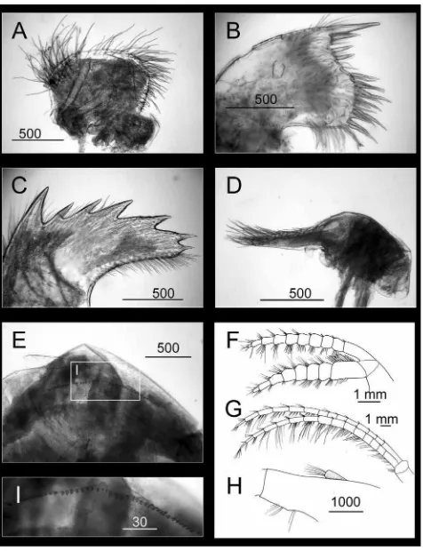
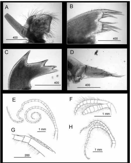
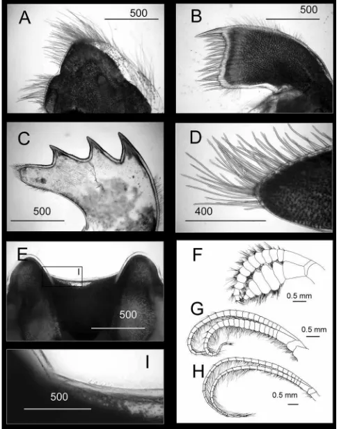
(Fig. 1A); tergum quadrangular, all margins straight; scutum ovoid, occludent margin convex, basal and tergal margins straight, carinal margin strongly convex, basal and occludent margins forming right angle (Fig. 1A); carina bowed, basal laterally expanded (Fig. 1A). Peduncle long, yellow, surface concentrically wrinkled (Fig. 1A). Maxilla bilobed, setae in 2 main clusters (Fig. 3A); maxillule notched, 2 large, long, cuspidate setae above notch, short spines in notch, 10 cuspidate setae below notch, region below notch expanded outwards (Fig. 3B); mandible with 6 teeth excluding inferior angle, first tooth separated from remaining teeth, lower margin very short, smooth, inferior angle terminating in 1 sharp seta (Fig. 3C); mandibular palp narrow, elongated, setae on superior margin (Fig. 3D); labrum concave, without deep notch, cutting edge with dense, fine teeth (Fig. 3E, I). Cirrus I with rami subequal; outer ramus 10-segmented, inner ramus 8-segmented (Fig. 3F); cirrus II with outer ramus 14-segmented, inner ramus 15-segmented (Fig. 3G); caudal appendage short, 1-segmented, length < height of basal segment of pedicle of cirrus VI (Fig. 3H).
Distribution. Malaysia, South China Sea, Taiwan. Remarks. This is a new record for Taiwanese waters.
Order Scalpelliformes Buckeridge & Newman, 2006
Family Calanticidae Zevina, 1978
Genus Euscalpellum Hoek, 1907
Euscalpellum rostratum (Darwin, 1851)
Figures 1B, 4
Scalpellum rostratum Darwin, 1851: 259, pl. 6 fig. 7.
Scalpellum (Euscalpellum) rostratum. — Hoek, 1907: 65, pl. 5 fig. 13. — Stubbings, 1936: 19 fig. 7.
Euscalpellum rostratum. — Pilsbry, 1908: 107, figs 1e–f. — Utinomi, 1968: 162, fig. 1. — Zevina, 1978: 1001. — Liu &
Ren, 1985: 190, fig. 5, pl. 3 figs 1–2. — Liu & Ren, 2007: 214, fig. 90.
Scalpellum (Smilium) rostratum. — Annandale, 1914: 274.
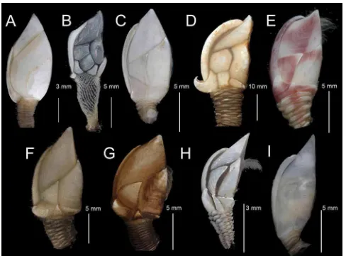
FIGURE 1. Taiwanese deep-sea barnacles (right side view). A. Glyptelasma gigas, B. Euscalpellum rostratum, C.
Arcoscalpellum truncatum, D. Tarasovium orientale, E. Trianguloscalpellum diota, F. Trianguloscalpellum hirsutum, G. Trianguloscalpellum regium, H. Trianguloscalpellum weltnerianum, I. Verum novazelandiae.
Material examined. ASIZCR000225, 1 specimen, Stn. CP164 (22°15.57’N, 120°35.56’E, 25 May 2002,
depth: 60–90 m), CL 9.76 mm, CW 6.44 mm, PL 7.6 mm.
Diagnosis. Capitulum higher than wide; 15 white plates; subcarina present; rostrum large, well developed;
inframedian latus diamond-shaped; rostrolatus quadrangular, umbo apical.
Description. Capitulum higher than wide, 15 fully calcified, white plates; tergum truncated, quadrangular,
Distribution. South China Sea, Taiwan, the Philippines, Malay Archipelago, Arabian Sea. Remarks. This is a new record for Taiwanese waters.
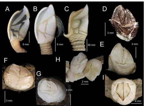
FIGURE 2. Taiwanese deep-sea barnacles (right side view on A–C, top view for D–I). A. Annandaleum japonicum
biramosum, B. Litoscalpellum spinosus sp. nov., C. Scalpellum stearnsii, D. Altiverruca longimandible sp. nov., E. Altiverruca navicula, F. Metaverruca defayeae, G. Metaverruca recta, H. Rostratoverruca krugeri, I. Hexelasma velutinum.
Family Scalpellidae Pilsbry, 1916
Sub-family Arcoscalpellinae Zevina, 1978
Genus Arcoscalpellum Hoek, 1907
Arcoscalpellum truncatum (Hoek, 1883)
Figures 1C, 5
Scalpellum truncatum Hoek, 1883: 92, pl. 6 fig. 13. — Zevina, 1973: 846. Arcoscalpellum truncatum. — Zevina, 1981: 334, fig. 254.
Material examined. NMNS 005087-00073, 1 specimen, Stn. CP185 (22º0.54’N, 119º 27.94’E, 26 Aug.
Diagnosis. 14 fully calcified capitular plates, white, surfaces striated with radiating lines; tergum
pentagonal, bluntly truncated; inframedian latus narrow, triangular, touching upper latus; carinolatus with umbo at basicarinal angle, located in middle of carinal margin; carina bowed, umbo apical.
FIGURE 3. Glyptelasma gigas. A. Maxilla, B. Maxillule, C. Mandible, D. Mandibular palp, E. Labrum, F. Cirrus I, G.
FIGURE 4. Euscalpellum rostratum. A. Maxilla, B. Maxillule, C. Mandible, D. Mandibular palp, E. Caudal appendage,
Description. 14 fully calcified, white plates, surfaces with prominent ridges (Fig. 1C); tergum pentagonal,
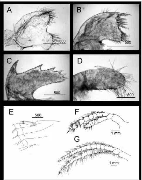
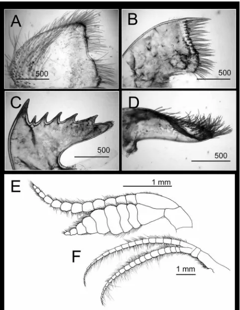
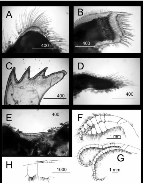
bluntly truncated, occludent and basal margins straight, carinal margin slightly convex (Fig. 1C); scutum with umbo apical, apex projecting over tergum, occludent and basal margins straight, tergal margin slightly concave, occludent and basal margins forming right angle (Fig. 1C); upper latus trapeziform, carinal margin short, straight (Fig. 1C); scutal margin long, concave, umbo apical, apex projecting over scutum (Fig. 1C); rostrolatus quadrangular, scutal and basal margins parallel, umbo at occludent margin of scutum (Fig. 1C); carinolatus irregular shape, subquadrangular, carinal margin concave, other margins straight, umbo at basicarinal angle, in middle portion of carinal margin, angle not extending beyond carina (Fig. 1C); rostrum narrow, small (Fig. 1C). Maxilla triangular, with setae on all margins, maxillary lobe long, naked (Fig. 5A); cutting edge of maxillule notched, 4 cuspidate setae above notch, notch naked, >7 cuspidate setae below notch (Fig. 5B); mandible tri-dentate, teeth sharply pointed (Fig. 5C), lower margin short, with sparse setae, inferior angle setose (Fig. 5C); mandibular palp elongated triangle, superior margin with sparse setae, denser distally (Fig. 5D); cirrus I with rami subequal (Fig. 5F), separated from cirri II–VI; outer ramus longer, slender (10-segmented), inner ramus flattened (8-segmented); cirrus II with outer ramus longer (19-segmented) than inner ramus (14-segmented; Fig. 5H); cirri III–VI similar, rami long, slender, 28–30 segmented (Fig. 5E); caudal appendages short, slender, slightly longer than first segment of basal pedicle of cirrus VI, 7-segmented (Fig. 5G).
Distribution. New Zealand, Australia and Taiwan. Remarks. This is a new record for Taiwanese waters.
Genus Tarasovium Zevina, 1978
Tarasovium orientale Ren, 1983
Figures 1D, 6
Tarasovium orientale Ren, 1983: 76, fig. 1 (13–23). — Liu & Ren, 1985: 201, pl. 2 (15–17). — Liu & Ren, 2007:
246–247, fig. 106.
Material examined. NMNS 005734-00009, 1 specimen, benthic trawl, Tainan County, south west Taiwan (15
Aug. 2005, depth unknown), CL 32.05 mm, CW 21.07 mm, PL 18.66 mm.
Diagnosis. 14 capitular plates, fully calcified; upper latus pentagonal, umbo sub-apical; inframedian latus
quadrilateral, umbo close to basal margin; carinolatus horn-shaped, umbo at basicarinal angle, angle extending beyond carina.
Description. Capitulum pale yellow (Fig. 1D), subtriangular, with 14 fully calcified plates, covered by
membrane, with sparse setae (Fig. 1D); tergum triangular, umbo apical, occludent and basal margins slightly convex, carinal margin straight; scutum quadrangular, small apex extending beyond margin of tergum, umbo apical, occludent margin convex, basal and upper latus margins concave (Fig. 1D); upper latus pentagonal, umbo subapical (Fig. 1D); rostrolatus narrow quadrangular, wider than high (Fig. 1D); inframedian latus broad, rectangular, umbo close to basal margin (Fig. 1D); carinolatus horn-shaped, umbo at basi-carinal angle, angle extending beyond carina; carina convex, umbo apical (Fig. 1D). Maxilla subtriangular, dense setae along entire margin (Fig. 6A); maxillule not notched, cutting edge with numerous setae (Fig. 6B); mandible with 6 teeth excluding inferior angle (Fig. 6C), lower margin straight, smooth, without setae, inferior angle setose (Fig. 6C); mandibular palp elongated, dense setae along superior margin (Fig. 6D), inferior margin naked. Cirrus I with rami unequal (Fig. 6E), separated from cirri II–VI, outer ramus slender, longer (13-segmented), inner ramus broader, shorter (9-segmented); cirri II–VI similar in morphology (Fig. 6F), outer and inner rami similar lengths, 18–20 segmented; caudal appendage short, 1-segmented, length < height of basal segment of pedicle of cirrus VI.
FIGURE 5. Arcoscalpellum truncatum. A. Maxilla, B. Maxillule, C. Mandible, D. Mandibular palp. Line drawing of E.
Cirrus VI, F, Cirrus I, G. Caudal appendages, H. Cirrus II. Scale bars in µm, except E, F, H in mm.
Genus Trianguloscalpellum Zevina, 1982
Trianguloscalpellum diota (Hoek, 1907)
Figures 1E, 7, 8
FIGURE 6. Tarasovium orientale. A. Maxilla, B. Maxillule, C. Mandible, D. Mandibular palp, E. Cirrus I, F. Cirrus II.
Material examined. NMNS 003636-00002, 1 specimen, benthic trawl at Donggang, S. Taiwan (26 Apr.
2001, depth: 250 m), on spine of sea urchin Stylocidaris renei, CL 5.01 mm, CW 2.92 mm, PL 2.5 mm.
Diagnosis. Capitulum with 14 fully calcified plates, occludent margin nearly straight, carina margin
strongly convex, surfaces with diffused pink patches (colour not faded out after preservation in ethanol); apex of carinolatus strongly recurved, umbo apical.
FIGURE 7. Trianguloscalpellum diota. A. Maxilla, B. Maxillule, C. Mandible, D. Mandibular palp, E. Lower margin of
FIGURE 8. Trianguloscalpellum diota. Line drawings of A. Cirrus I, B. Cirrus II, C. Cirrus VI, showing the caudal
appendage. Scale bars in mm.
Description. Capitulum elongated, diffused light pink color (color not faded out after preservation in
(6-segmented); cirri II–VI long, slender, similar in length (Fig. 8B, C); cirrus II with outer ramus 12-segmented; inner ramus 13-12-segmented; cirrus VI with outer ramus 15-segmented, inner ramus 17-12-segmented; caudal appendages short, length ~ 1/3 inner ramus of cirrus VI, 10-segmented, setae distally (Fig. 8C).
Distribution. Indonesia, the Philippines and Taiwan. Remarks. This is a new record for Taiwanese waters.
Trianguloscalpellum hirsutum (Hoek, 1883)
Figures 1F, 9
Scalpellum hirsutum Hoek, 1883: 88, pl. 4 fig. 19. — Newman & Ross, 1971: 63, fig. 28. Trianguloscalpellum hirsutum. — Zevina, 1981: 309, fig. 233.
Material examined. NMNS 005087-00082, 1 specimen, Stn. CP300 (22º17.156’N, 119º59.963’E, 11 Aug.
2005, depth: 960–972 m), CL 20.03 mm, CW 12.76 mm, PL 8.54 mm; CEL-BB-26B, 1 specimen, Stn. CP375 (24º16.240’N, 122º11.720’E, 27 Aug. 2006, depth: 2216–2497 m), CL 21.38 mm, CW 19.42 mm, PL 13.78 mm; CEL-BB-63, 1 specimen, Stn. CP371 (24º28.521’N, 122º12.821’E, 26 Aug. 2006, depth: 582–613 m), CL 21.5 mm, CW 13.2 mm, PL 12.03 mm.
Diagnosis. Capitulum with 14 fully calcified plates; surface of capitulum and peduncle covered by dense
setae; tergum with long, sharp apex, upper latus triangular
Description. Capitulum with 14 fully calcified plates, surfaces covered by long setae (Fig. 1F); tergum
large, elongated rhomboid, higher than wide, apex apical, produced, acute, umbo apical, occludent margin straight; scutum quadrangular, occludent and tergal margins convex, lateral and basal margin straight, apex produced at tip of occludent margin, umbo apical (Fig. 1F); upper latus triangular, carinal margin straight; inframedian latus triangular, subequilateral, umbo apical; rostrolatus flattened, width twice height (Fig. 1F); carinolatus horn-shaped, apex apical, produced, touching carina. Peduncle short, scales in longitudinal rows. Maxilla bilobed, setae in 3 main clusters (Fig. 9A); maxillule not notched, cutting edge with ~ 19 cuspidate setae on cutting edge (Fig. 9B); mandible with 3 equally spaced teeth, lower margin straight, smooth, inferior angle blunt with dense setae (Fig. 9C); mandibular palp elongated, setae on superior margin and apically (Fig. 9D); labrum concave, cutting edge smooth, with fine setae, teeth absent (Fig. 9E, I). Cirrus I separated from cirri II-VI, rami unequal, outer ramus longer, slender, (13-segmented), inner ramus shorter, broad (7-segmented) (Fig. 9F); cirrus II with rami subequal, outer ramus 23-segmented, inner ramus 25-segmented (Fig. 9G); cirri III–VI similar length, longer than cirri I and II, cirrus IV with outer ramus 26-segmented, inner ramus 25-segmented (Fig. 9H); caudal appendage 1-segmented, appendage length < height of basal segment of pedicle of cirrus VI.
Distribution. Borneo, Antarctica and Taiwan.
Remarks. This is a new record for Taiwanese waters. Trianguloscalpellum hirsutum is morphologically
close to Arcoscalpellum foresti Rosell, 1989, in that both species have setae on the whole body, and in the shape of the tergum. Trianguloscalpellum hirsutum has a triangular upper latus, whilst that of A. foresti is quadrangular. The umbo of the carinolatus of A. foresti is located in the middle of the carinal margin, whilst that of T. hirsutum is located apically.
Trianguloscalpellum regium (Thomson, 1878)
Figures 1G, 10
Scalpellum regium Wyville Thomson, 1878: 11, figs 2–3. — Hoek, 1883: 106: pl. 4 figs 3–5. — Pilsbry, 1907: 28, pl. 3
figs 4, 5. — Gruvel, 1920: 30, pl. 1 fig. 7.
Trianguloscalpellum regium regium. — Zevina, 1981: 309, fig. 234.
Material examined. CEL-BB-46C, 1 specimen, Stn. CP375 (24º16.240’N, 122º11.720’E, 27 Aug. 2006,
FIGURE 9. Trianguloscalpellum hirsutum. A. Maxilla, B. Maxillule, C. Mandible, D. Mandibular palp, E. Labrum, F.
FIGURE 10. Trianguloscalpellum regium. A. Maxilla, B. Maxillule, C. Mandible, D. Mandibular palp, E. Labrum, F.
Cirrus I, G. Cirrus II, H. Caudal appendage. Scale bars in µm, except F, G in mm.
Diagnosis. Capitulum quadrangular, covered by smooth membrane, 14 fully calcified plates; tergum
than wide, apex produced at junction of occludent and tergal margins, umbo apical; carinolatus higher than wide, curved, apex produced, umbo apical.
Description. Capitulum quadrangular, 14 plates, covered by smooth membrane (Fig. 1G); tergum large,
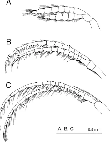
triangular, carinal margin convex, occludent margin straight, umbo apical; scutum quadrangular, higher than wide, apex produced at junction of occludent and tergal margins, umbo apical (Fig. 1G); upper latus triangular, tergal and lateral margins convex, scutal margin concave (Fig. 1G); inframedian latus equilateral triangle, umbo apical; rostrolatus very narrow, wider than high; carinolatus higher than wide, curved, apex produced, umbo apical (Fig. 1G); carina bowed, roof flat; peduncle with densely packed, concentric scales (Fig. 1G). Maxilla densely setose (Fig. 10A); cutting edge of maxillule not notched, >22 large cuspidate setae along cutting edge (Fig. 10B); mandible with 3 evenly separated teeth excluding inferior angle, lower margin long, naked, inferior angle covered by dense setae (Fig. 10C); mandibular palp narrow, elongated, setae along superior margin, inferior margin naked (Fig. 10D); labrum concave, cutting edge smooth, teeth absent (Fig. 10E). Cirrus I separated from remaining cirri, rami unequal, outer ramus longer, slender, 10-segmented, inner ramus broader, shorter, 6-segmented (Fig. 10F); cirrus II with rami equal, both rami 22-segmented (Fig. 10G); cirrus VI with rami equal, outer and inner rami 33-segmented; caudal appendage short, 4-segmented, length < height of basal segment of pedicle of cirrus VI (Fig. 10H).
Distribution. Cosmopolitan: Atlantic and Pacific Oceans, including Taiwan.
Remarks. This is a new record for Taiwanese waters. This species was named by the chief scientist,
Professor Sir Charles Wyville Thomson (see Hoek, 1883: 106), but no detailed species description was provided. Hoek (1883) fully described the species and retained Thomson’s name, Scalpellum regium.
Trianguloscalpellum weltnerianum (Pilsbry, 1911)
Figures 1H, 11
Scalpellum weltnerianum Pilsbry, 1911: 64, pl. 9 figs 5–7.
Trianguloscalpellum weltnerianum. — Zevina, 1981: 306, fig. 230.
Material examined. CEL-BB-88, 1 specimen, Stn. CP320 (20º50.090’N, 117º27.170’E, 19 Aug. 2005,
depth: 720–730 m), CL 8.31 mm, CW 3.57 mm, PL 3.56 mm.
Diagnosis. Capitulum elongated, length twice width; 14 capitular plates, white, surfaces sculptured with
radial fine riblets, setae absent; rostrum oval, large; inframedian latus narrow, triangular; peduncle covered with strongly imbricating, laterally interlocking, white scales.
Description. Capitulum elongated, length twice width; 14 white capitular plates, surfaces striated with
radial riblets (Fig. 1H); tergum higher than wide, quadrangular (Fig. 1H); scutum quadrilateral, umbo apical, occludent and basal margins straight, intersecting at right angles (Fig. 1H); upper latus quadrangular, higher than wide, scutal margin concave, basal margin short (Fig. 1H); inframedian latus triangular, height twice width, umbo apical; carinolatus upwardly curved, umbo apical (Fig. 1H); rostrolatus quadrangular; rostrum large, oval; carina bowed, roof flat (Fig. 1H). Peduncle covered with strongly imbricating, laterally interlocking, white scales. Maxilla subquadrangular, bilobed, setae clustered into 3 regions (Fig. 11A); maxillule with cutting edge narrow, notched, 4 large cuspidate setae above notch, notch naked, 5 cuspidate setae below notch (Fig. 11B); mandible tridentate, first tooth separated from second and third, lower margin very short, inferior angle pectinated with cuspidate setae (Fig. 11C); mandibular palp with setae apically and along superior margin, inferior margin naked (Fig. 11D). Cirrus I with rami unequal, separated from cirri II–VI, outer ramus longer, slender, 10-segmented, inner ramus slightly broader, 6-segmented (Fig. 11E); cirri II–VI long, slender; cirrus II with outer ramus 12-segmented, inner ramus 12-segmented (Fig. 11F); cirrus III with outer ramus 12-segmented, inner ramus 11-segmented; cirrus VI with outer ramus 14-segmented, inner ramus 15-segmented (Fig. 11G); caudal appendage very short, 1-segmented, length < basal segment of pedicle of cirrus VI (Fig. 11H).
Remarks. This record of T. weltnerianum is a new record for Taiwanese waters. Pilsbry (1911) noted that
T. weltnerianum is morphologically close to T. album (Hoek, 1883) collected from the Malay Archipelago. Trianguloscalpellum weltnerianum differs from T. album in having the surfaces of the capitular plates striated, whilst those of T. album are smooth (Hoek, 1883; Pilsbry, 1911). At present, this species is only reported to live on crinoids, suggesting an association relationship.
Genus Verum Zevina, 1981
Verum novaezelandiae (Hoek, 1907)
Figures 1I, 12, 13
Scalpellum novaezelandiae Hoek, 1883: 124, pl. 5 figs 7, 8. Scalpellum poculum Hoek, 1907: 100, pl. 8 figs 4, 4a. Verum novaezelandiae. — Zevina, 1981: 288, fig. 165.
Material examined. CEL-BB-76, 2 specimens, Stn. CP364 (22º06.335’N, 121º08.224’E, 24 Aug. 2006,
depth: 1260–1275 m), on sunken wood, CL 4.22–6.86 mm, CW 1.86–3.15 mm, PL 0.94–0.96 mm.
Diagnosis. Capitulum elongated, higher than wide, 14 fully calcified, white, plates; inframedian latus
pentagonal or vase-shaped, umbo sub-basal; carinolatus with basicarinal angle not extending beyond carina.
Description. Capitulum elongated, flattened, 14 fully calcified, closely packed, smooth, white plates (Fig.
1I); tergum triangular, umbo apical, occludent and scutal margins straight, intersecting at right angles; scutum quadrangular, apex produced at tergal margin, umbo apical, occludent margin straight, lateral margin slightly convex; upper latus pentagonal, apex produced at tergal-scutal angle, umbo apical (Fig. 1I); inframedian margin shortest; inframedian latus pentagonal or vase-shaped, umbo submedial; rostrolatus quadrangular, wider than high (Fig. lI); carinolatus higher than wide, umbo at basicarinal angle, angle not extending beyond scutum; carina bowed, roof flat; rostrum small, oval-shaped; peduncle covered by dense scales (Fig. 1I). Maxilla bilobed, setae clumped into 2 main clusters, sparse setae on superior margin (Fig. 12A); cutting edge of maxillule notched, 4 cuspidate setae above notch, notch naked, > 6 cuspidate setae below notch (Fig. 12B); mandible with 3 large teeth, first separated from second and third (Figs 12C, G); lower margin short, pectinated, inferior angle pectinated by >3 large, cuspidate setae (Fig. 12E); mandibular palp triangular, setae along superior margin and apically, inferior margin naked (Fig. 12D); labrum with fine, sharp teeth on cutting edge (Figs 12F, H). Cirrus I separated from remaining cirri, rami unequal; outer ramus slender, longer, 9-segmented, inner ramus broader, short, 6-segmented (Fig. 13A); cirri II–VI with rami subequal; cirrus II with outer ramus 13-segmented, inner ramus 14-segmented (Fig. 13B); cirrus VI with outer ramus 15-segmented, inner ramus 17-segmented (Fig. 13C); caudal appendage very short,1-segmented, length < height of basal segment of pedicle of cirrus VI (Fig. 13D).
Distribution. Pacific Ocean, including Taiwan. Remarks. This is a new record for Taiwanese waters.
Sub-family Meroscalpellinae Zevina, 1978
Genus Annandaleum Newman & Ross, 1971
Annandaleum japonicum biramosum (Pilsbry, 1911)
Figures 2A, 14, 15
Scalpellum japonicum Hoek, 1883: 67, pl. 3 figs 9, 10.
FIGURE 11. Trianguloscalpellum weltnerianum. A. Maxilla, B. Maxillule, C. Mandible, D. Mandibular palp, E. Cirrus
FIGURE 12. Verum novaezelandiae. A. Maxilla, B. Maxillule, C. Mandible, D. Mandibular palp, E. Lower margin of
FIGURE 13. Verum novaezelandiae. A. Cirrus I, B. Cirrus II, C. Cirrus VI, D. Caudal appendage.
Material examined. NMNS 003636-00062, 1 specimen, Stn. CP130 (22º18.77’N, 120º6.99’E, 22 Aug. 2001,
FIGURE 14. Annanadaleum japonicum biramosum. A. Maxilla, B. Maxillule, C. Mandible, D. Mandibular palp, E.
FIGURE 15. Annanadaleum japonica biramosum. A. Cirrus I, B. Cirrus II, C. Cirrus VI with caudal appendage.
Diagnosis. Opercular plates not completely calcified; tergum inverted V-shaped; scutum with apicolateral
margin, bifid, simple on carinal side; inframedian latus higher than wide, vase-shaped, umbo submedial; caudal appendage relatively long, 10-segmented.
Description. Capitulum covered by semi-transparent, pale yellow membrane (Fig. 2A); 14 partially
calcified capitular plates, separated by broad, chitinous interspaces (Fig. 2A); tergum inverted V-shaped, umbo apical, branch at carinal margin longer than occludent margin branch (Fig. 2A); scutum long, narrow, umbo apical, slightly overlapping tergal margin, lateral margin with apicolateral arm ~ 1/3 length of tergal margin; upper latus irregular, subtriangular, branch at scutal margin bifid, branch at carinal margin simple (Fig. 2A); rostrum very small, narrow; rostrolatus quadrangular, wider than high (Fig. 2A); inframedian latus higher than wide, vase-shaped, umbo submedial (Fig. 2A); carinolatus V-shaped, lateral margin bifid; carina bowed, roof flat, width greater in basal region. Maxilla subtriangular, margin covered with dense setae in 3 clusters (Fig. 14A); maxillule slightly notched, 3 long and 1 short cuspidate setae above notch, notch naked, 9 cuspidate setae below notch (Fig. 14B); mandible with 3 teeth (Fig. 14C), first tooth separated from second and third (Fig. 14E, G), lower margin very short, setae absent, inferior angle blunt with 5 setae (Fig. 14E); mandibular palp subtriangular, elongated, setae distally and along superior margin, inferior margin naked (Fig. 14D); cutting edge of labrum straight, with very fine teeth (Fig. 14F, H). Cirrus I separated from cirri II–VI; rami of cirrus I subequal, outer ramus longest, 12 segmented (Fig. 15A), inner ramus broader, shorter, 11 segmented; cirri II–VI with rami subequal; cirrus II with outer ramus 25-segmented, inner ramus 21-segmented (Fig. 15B), cirrus VI with outer ramus 29-21-segmented, inner ramus 26-21-segmented (Fig. 15C); caudal appendage ~ 1/3 length of cirrus VI, 10-segmented (Fig. 15C).
Distribution. Pacific waters in Japan and Taiwan.
Remarks. Pilsbry (1911) identified the sub-species biramosum of Annandaleum japonicum. Compared to
Annandaleum japonicum, A. j. biramosum has the umbo of the carina closer to the upper end of the plate with the branch of the upper latus extending along the scutal margin bifid and simple on the carinal side. The inframedian latus is less excavated along its upper border in A. j. biramosum (Pilsbry, 1911). However, Pilsbry (1911) doubted that such difference in plate morphology represented intra-specific variation. In the present study, the maxillule and mandible of A. j. biramosum are similar to Pilsbry’s illustration of the maxillule and mandible of A. japonicum (Pilsbry, 1911). The taxonomic status and morphological variation of A. japonicum and A. j. biramosum should be evaluated using molecular techniques. The present record of A. j. biramosum is a new record for Taiwanese waters.
Genus Litoscalpellum Newman & Ross, 1971
Litoscalpellum spinosus sp. nov.
Figures 2B, 16, 17
Material examined. Holotype: NMNS 6058-001, 1 specimen, Stn. CP238 (25º12.28’N, 123º1.85’E, 23 Jul.
2004, depth: 1650–1689 m), CL 27.75 mm, CW 17.54 mm, PL 11.85 mm.
Diagnosis. Capitulum with 14 partially calcified plates; plates white, surfaces covered by dense, short
setae; inframedial latus not touching upper latus; somatic body with dorsal thoracic, sharp processes, located at bases of cirri III and IV.
Description. Capitulum with 14 partially calcified plates, plates white, surfaces covered by dense, short
bowed, roof flat, umbo apical (Figs 2B, 17A). Peduncle with white, concentric scales (Fig. 2B). Maxilla quadrangular, setae divided into 3 clusters (Fig. 16A); cutting edge of maxillule short, notched, 3 large, long cuspidate setae and 2 short, cuspidate seta above notch, several large cuspidate setae below notch (Fig. 16B); mandible with 3 teeth, first widely separated from second and third, lower margin smooth, inferior angle sharp with dense setae (Figs 16C, E, G); mandibular palp elongated, narrow, setae distally and along superior margin, inferior margin naked (Fig. 16D); labrum strongly concave, cutting edge with fine, sharp teeth (Figs 16F, H). Cirrus I separated from cirri II–VI, rami subequal, outer ramus slender, longer, 13-segmented, inner ramus broader, shorter, 8-segmented (Fig. 17C); cirri II–VI with rami subequal; cirrus II with outer ramus 19-segmented, inner ramus 30-segmented (Fig. 17E); cirrus III with outer ramus 18-segmented; inner ramus 21-segmented; cirrus IV with outer ramus 25-segmented, inner ramus 7-21-segmented; cirrus V with outer ramus 25-segmented, inner ramus 25-segmented; cirrus VI with outer ramus 29-segmented, inner ramus 32-segmented; caudal appendage very short, 1-segmented, length < height of basal segment of cirrus VI (Figs 17B, D); somatic body with dorsal thoracic, sharp processes, located at bases of cirri III and IV (Fig. 17B).
Etymology. The name spinosus is to denote the dorsal thoracic, sharp processes, located at the bases of
cirri III and IV, a diagnostic character of this species.
Distribution. Presently known only from the type locality.
Remarks. The capitular morphology of Litoscalpellum spinosus sp. nov. is similar to L. affbricatum
Foster, 1978. However, the capitulum of L. affbricatum is covered by a naked, smooth integument (Foster, 1978) whilst that of L. spinosus sp. nov. is covered by dense, short hairs. The rostrum of L. affbricatum is large, wide and triangular (Fig. 17G; Foster, 1978), whilst the rostrum of L. spinosus sp. nov. is narrow (Fig. 17F). The somatic body of L. affbricatum is not reported to have dorsal thoracic, sharp processes at the bases of cirri III and IV. Previously, no Litoscalpellum species has been reported to possess dorsal thoracic, sharp processes.
Subfamily Scalpellinae Pilsbry, 1907
Genus Scalpellum Leach, 1817
Scalpellum stearnsii Pilsbry, 1890
Figures 2C, 18
Scalpellum stearnsii Pilsbry, 1890: 441. — Broch, 1922: 235, fig. 6. — 1931: 16. — Nilsson-Cantell, 1934: 33. — Hiro,
1939c: 237. — Rosell, 1981: 279, pl. Ie.
Scalpellum stearnsi. — Gruvel, 1905: 44, fig. 46.
Material examined. CEL-BB-56, 1 specimen, Nan Fang Ao Fishing market (27 May 2004), CL 34.6 mm,
CW 22.84 mm, PL 26.27 mm.
Diagnosis. Carina strongly bowed; tergum triangular; scutum with umbo at tergo-occludent angle;
inframedian latus pentagonal, umbo sub-basal; carinolateral horn-shaped, umbo at basicarinal angle, angle extending beyond carina.
Description. Capitulum almost square-shaped; 14 perfectly calcified plates, covered by yellow membrane
V-shaped, with fine teeth on cutting edge. Cirrus I with rami unequal, inner ramus oval (12-segmented), outer ramus long, slender (18-segmented) (Fig. 18E); cirri II–VI similar in morphology, rami almost equal length, outer and inner rami 25–36-segmented (Fig. 18F); caudal appendages short, 5–6 segmented (Fig. 18G).
FIGURE 16. Litoscalpellum spinosus sp. nov. A. Maxilla, B. Maxillule, C. Mandible, D. Mandibular palp, E. Lower
FIGURE 17. Litoscalpellum spinosus sp. nov. A. Side view of the capitulum, B. Somatic body showing the dorsal
Distribution. Indo-Pacific waters including Taiwan.
Remarks. The Nan Fang Ao fishing market is a local deep-sea fishery market in Taiwan, selling fishery
catches collected daily by the fishermen operating in the deep-sea in the eastern Taiwanese waters. The sample collected in the Nan Fang Ao fish market was found among the local catches from the fishermen.
FIGURE 18. Scalpellum stearnsii. A. Maxilla, B. Maxillule, C. Mandible, D. Mandibular palp, E. Cirrus I, F. Cirrus VI,
Order Sessilia Lamarck, 1818
Material examined. Holotype: ASIZCR 000224, 1 specimen, Stn. CP375 (24º16.240’N, 122º11.720’E, 27
Aug. 2006, depth: 2216–2497 m), BD 9.78 mm. Paratype: NMNS-6006-003, 2 specimens, locality same as holotype, BD 8.2–9.8 mm.
Diagnosis. Altiverruca with single interlock between rostrum and carina, movable tergum interlocking
with movable scutum by 3 main ribs; lower margin of mandible long, smooth, naked; caudal appendage long, 1/2 length of cirrus VI.
Description. Shell white, some individuals covered by dark brown coating (Figs 2D, 20A); apices of
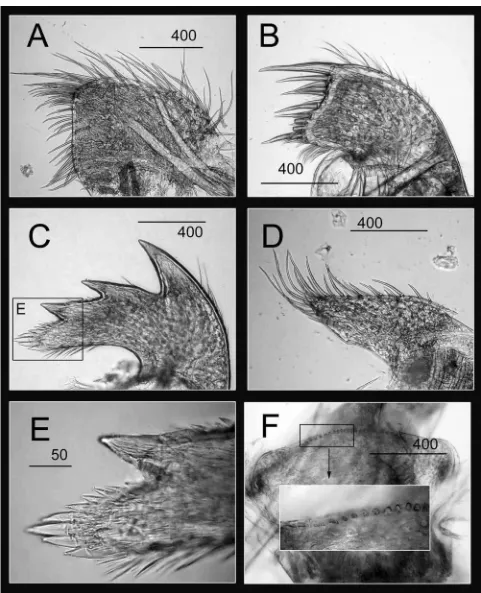
rostrum and carina marginal, produced, curved outwards (Figs 2D, 20A); rostrum and carina interlocking by 1 major rib; rostrum without secondary ridges directed towards tergal base (Figs 2D, 20A); movable tergum quadrangular, interlocking with movable scutum by 3 main ribs; movable scutum triangular (Figs 2D, 20A). Maxilla bilobed, dense, shorter setae clustered on lobes, long setae along superior margin (Fig. 19A); maxillule with wide notch, basal region of cutting edge expanded (Fig. 19B); 2 large, robust, cuspidate setae above notch, ~ 10 fine, cuspidate setae in notch, 8 large, cuspidate setae below notch (Fig. 19B); mandible elongated, with 3 teeth, equally spaced, lower margin very long, smooth, naked, inferior angle blunt, minutely pectinated (Figs 19C, E); mandibular palp elongated, setae along superior margin (Fig. 19D); labrum not concave, cutting edge straight, with numerous fine teeth (Fig. 19F). Cirrus I with rami unequal, outer ramus antenniform, 24-segmented, inner ramus short, 10-segmented (Fig. 20B); cirrus II with rami unequal, outer ramus antenniform, 21-segmented, inner ramus short, 10-segmented, with dense setae (Fig. 20C); cirri III–VI similar, rami subequal, long, slender; cirrus III with outer ramus 28-segmented, inner ramus 22-segmented; cirrus IV with outer ramus 32-segmented, inner ramus 34-segmented; cirrus V with outer ramus 27-segmented, inner ramus 30-segmented; cirrus VI with outer ramus 33-27-segmented, inner ramus 33-segmented (Figs 20D, E, F, G); caudal appendage long, 1/2 length of cirrus VI, 32-segmented (Fig. 20G).
Etymology. The species is named with respect to the long, lower margin of the mandible. The name of
this species is treated as a noun in apposition.
Distribution. Presently known only from the type locality.
Remarks. In the present study, Altiverruca longimandible sp. nov. was recorded in the eastern waters of
Taiwan. From external shell morphology, Altiverruca longimandible sp. nov. is similar to A. navicula (Hoek, 1913) in having the apices of the rostrum and carina produced, the rostrum and carina interlocking with 1 rib and the movable scutum and tergum interlocking by several strong ribs. The mandible of A. longimandible sp.
nov., however, has a very long, smooth, fragile lower margin that is devoid of setae, terminating at the blunt
inferior angle, whilst that of A. navicula has a pectinated lower margin. Buckeridge (1994, 1997) reported that variation in mandible morphology may occur in A. navicula. In the present study, dissection of three specimens of A. longimandible sp. nov. revealed similar mandible morphology, suggesting that morphological variation of the mandible is not obvious in this species.
FIGURE 19. Altiverruca longimandible sp. nov. A. Maxilla, B. Maxillule, C. Mandible, D. Mandibular palp, E. Lower
FIGURE 20. Altiverruca longimandible sp. nov. A. External view of the whole specimen, B. Cirrus I, C. Cirrus II, D.
Altiverruca navicula (Hoek, 1913)
Figures 2E, 21, 22
Verruca navicula Hoek, 1913: 134, figs 4–6. — Nilsson-Cantell, 1927: 778, figs a–f. Altiverruca navicula. — Buckeridge, 1994: 100, fig. 5.
Material examined. NMNS 005087-00077, 1 specimen, Stn. CP285 (24º16.09’N, 122º11.52’E, 16 Jun.
2005, depth 2268–2426 m), BD 11.37 mm.
Diagnosis. Rostral and carinal apices marginal; myophore absent from fixed scutum; operculum nearly
vertical to base; movable scutum and movable tergum interlocking with 4 ribs, caudal appendages long.
Description. Shell white, rostral and carinal apices marginal, carina and rostrum interlocking in a single
rib; movable tergum quadrangular, scutal margin with 5 ribs, apex curved; movable scutum triangular, tergal margin with 4 ribs, apex curved (Fig. 2E). Maxilla bilobed, with dense setae, sparse setae along superior margin (Fig. 21A); maxillule with strong, wide notch on cutting edge, 2 large cuspidate setae above notch, dense setae in notch, 7 setae below notch (Fig. 21B); mandible with 3 teeth (Fig. 21C), lower margin long, with > 8 small, cuspidate setae, inferior angle pectinate, with cuspidate setae at angle (Fig. 21E); mandibular palp elongated, dense setae along superior margin (Fig. 21D); labrum slightly concave, fine, bi-dentate to quadridentate teeth on cutting margin (Fig. 21H). Cirrus I with rami unequal, outer ramus elongated, segmented, inner ramus shorter, 15-segmented (Fig. 22A); cirrus II with rami unequal, outer ramus segmented, inner ramus 11-segmented (Fig. 22B); cirrus III with outer ramus 30-segmented, inner ramus 27-segmented (Fig. 22C); cirrus VI with outer ramus 36-27-segmented, inner ramus 37-27-segmented (Fig. 22D); caudal appendage long, 34-segmented (Fig. 22D).
Distribution. Pacific Ocean
Remarks. This is a new record for Taiwanese waters.
Genus Metaverruca Pilsbry, 1916
Metaverruca defayeae Buckeridge, 1994
Figures 2F, 23, 24
Metaverruca defayeae Buckeridge, 1994: 109, fig. 9. — Buckeridge, 1997: 138.
Material examined. CEL-BB-100, 4 specimens, Stn. CP371 (24º28.521’N, 122º12.821’E, 26 Aug. 2006,
depth: 582–613 m), BD 10.22–13.10 mm.
Diagnosis. External shell smooth, with dense growth lines; movable tergum and scutum with 4 articular
ribs; mandible with 5 teeth; caudal appendage short, < length of basal segment of pedicle of cirrus VI.
Description. Shell white, low conic, sides steep, surface smooth, with dense growth lines; basis
FIGURE 21. Altiverruca navicula. A. Maxilla, B. Maxillule, C. Mandible, D. Mandibular palp, E. Lower margin of
FIGURE 22. Altiverruca navicula. A. Cirrus I, B. Cirrus II, C. Cirrus III, D. Cirrus VI, showing caudal appendage. Scale
FIGURE 23. Metaverruca defayeae. A. Maxilla, B. Maxillule, C. Mandible, D. Mandibular palp, E. Lower margin of
Remarks. This is a new record for Taiwanese waters. Buckeridge (1994) concluded Metaverruca
defayaea to be morphologically close to M. recta (Aurivillius, 1898). The major difference between these two species is the number of interlocking ribs on the movable scutum and tergum; M. defayaea has 4 whilst M. recta has 3. In the present study, the fifth tooth of the mandible of M. defayaea is represented by 2 small teeth, whilst the fifth of M. defayaea in Buckeridge (1994) is represented by a single small tooth. The taxonomic status of M. defayaea in Taiwanese waters will need to be further evaluated from larger samples sizes and with support from a molecular approach.
FIGURE 24. Metaverruca defayeae. A. Cirrus I, B. Cirrus II, C. Cirrus III, D. Cirrus IV, E. Cirrus VI, showing caudal
Metaverruca recta (Aurivillius, 1898)
Figures 2G, 25, 26
Verruca recta Aurivillius, 1898: 195. Verruca sculpta Aurivillius, 1898: 197.
Verruca halotheca Pilsbry, 1916: 188, pl. 12 figs 9, 10.
Verruca cookei. — Rosell, 1989: 299, pl. 11 figs r, s, u, v (non Verruca cookei Pilsbry, 1927). — Rosell, 1991: 33 (non Verruca cookei Pilsbry, 1927).
Metaverruca recta. — Buckeridge, 1994: 116, figs 13a–f. — Young, 1998: 52.
Material examined. NMNS 20050614, 1 specimen, Stn. CP278 (24º23.63’N, 122º14.13’E, 14 Jun. 2005,
depth: 1222–1239 m), BD 9.44 mm.
Diagnosis. External shell smooth, growth lines widely spaced; orifice D-shaped; movable tergum and
scutum with 3 articular ribs; fixed scutum with myophore.
Description. Shell white, smooth, low conic, with operculum sub-parallel to base, growth lines widely
spaced, orifice D-shaped; rostral and carinal apices marginal, movable tergum and scutum with 3 articular ribs (Fig. 2G). Maxilla bilobed, dense setae in 3 main clusters (Fig. 25A); maxillule notched, 2 large cuspidate setae above notch, 7 fine setae in notch, 7 large setae below notch on expanded margin (Fig. 25B); mandible with 3 teeth (Fig. 25C, G), lower margin with several small teeth or large pectinations, inferior angle terminating in several cuspidate setae (Fig. 25E); labrum cutting edge with sharp, fine teeth (Fig. 25F, H). Cirrus I with rami subequal, outer ramus 13-segmented, inner ramus 12-segmented (Fig. 26A); cirrus II with rami subequal, outer ramus 11-segmented, inner ramus 8-segmented (Fig. 26B); caudal appendage short, 7-segmented, length within basal length of basal segment of cirrus VI (Fig. 26C).
Distribution. Caledonia, the Philippines, Taiwan, Atlantic Ocean,
Remarks. Metaverruca recta is one of the largest verrucids. This is a new record for Taiwanese waters.
Genus Rostratoverruca Broch, 1922
Rostratoverruca krugeri (Broch, 1922)
Figures 2H, 27, 28
Verruca krügeri Broch, 1922: 295, figs 43–44. Verruca (Rostratoverruca) krügeri Broch, 1931: 46.
Rostratoverruca koehleri. — Liu & Ren, 2007: 273, fig. 119 (non R. koehleri (Gruvel, 1907)).
Material examined. NMNS 0003328-00076, 55 specimens, benthic trawl at Donggang, on spines of the sea
urchin Stylocidaris renei (17 Jan. 2000), BD 2.25–6.45 mm.
Diagnosis. Operculum parallel to basis; rostrum patelliform, apex of rostrum removed from opercular
margin; fixed scutum without myophore; movable tergum and scutum with 4 articular ribs; rostrum and carina with prominent rounded ribbing.
Description. Shell white, basis membranous; rostrum patelliform, apex removed from opercular margin;
10-segmented (Fig. 28A); cirrus II with rami subequal, outer ramus 15-10-segmented, inner ramus 11-10-segmented (Fig. 28B); caudal appendage long, 1/2 length of cirrus VI, 18-segmented (Fig. 28C).
FIGURE 25. Metaverruca recta. A. Maxilla, B. Maxillule, C. Mandible, D. Mandibular palp, E. Lower margin of
FIGURE 27. Rostratoverruca krugeri. A. Maxilla, B. Maxillule, C. Mandible, D. Mandibular palp, E. Lower margin of
FIGURE 28. Rostratoverruca krugeri. A. Cirrus I, B. Cirrus II, C. Cirrus VI, showing caudal appendage. For clarity,
Distribution. East China Sea, South China Sea, Taiwan, western Pacific.
Remarks. This is a new record for Taiwanese waters. The species has often been reported attached to the
spines of sea urchins, suggesting a commensal relationship. Buckeridge (1994) concluded Rostratoverruca koehleri (Gruvel, 1907) was morphologically close to R. krugeri (Broch, 1922). When compared to R. koehleri, the fixed tergum of R. krugeri has a more prominent apex and there is more ribbing on the movable plates. Liu & Ren (2007), however, consider R. krugeri to be a subjective synonym of R. koehleri. In the present study, we consider R. krugeri and R. koehleri to be separate species and the illustration in Liu & Ren (2007) suggested their specimen is R. krugeri.
Suborder Balanomorpha Pilsbry, 1916
Hexelasma velutinum Hoek, 1913: 246, pl. 26 figs 1–6. — Broch, 1931: 53. — Hiro, 1933: 70, pl. 3 fig. 2. — Utinomi,
1968: 30. — Newman & Ross, 1971:155. — Newman & Ross, 1976: 46. — Jones, 2000: 273, fig. 64, tabs 28–31. — Liu & Ren, 2007: 328, fig. 146.
Material examined. CEL-BB-102, 1 specimen, Stn. CP371 (24º28.521’N, 122º12.821’E, 26 Aug. 2006,
depth: 582–613 m), BD 13.88 mm.
Diagnosis. Shell with 6 compartmental plates; carina, carinolateral and lateral plates with alae, radii
absent; rostrum with radii and alae absent; parietes solid, longitudinal ribs on inner surface absent; basis membranous; intermediate segments of cirri IV–VI bearing 2 pairs of long setae.
Description. Shell white, conical or cylindrical, operculum large; shell surface with parallel growth lines,
covered by membrane with fine, short setae (Fig. 2I); 6 compartmental plates; carina, carinolateral and lateral plates with alae, radii absent; rostrum with radii and alae absent (Fig. 2I); parietes solid, longitudinal ribs on inner surface absent; basis membranous; scutum triangular, occludent margin straight, with large teeth, tergal margin straight; tergum narrow, scutal margin long, straight, basiscutal angle oblique, spur pronounced. Maxilla subtriangular (Fig. 29A), inferior and superior margins setose; maxillule with cutting edge notched, 2 long, cuspidate setae above notch, 3 fine short setae in notch, 12 fine setae below notch (Fig. 29B); mandible with 4 teeth, first separated from remainder (Figs 29C, G), lower margin smooth (Fig. 29E), inferior angle sharp (Fig. 29E); mandibular palp rectangular, apex blunt, dense setae on superior margin and distally (Fig. 29D); labrum concave, deep notch absent, cutting edge with fine bidentate and tridentate teeth (Fig. 29H). Cirrus I with rami unequal, outer ramus shorter, 11 segmented, inner ramus longer 11-segmented (Fig. 30A); cirrus II with rami subequal, outer ramus 14-segmented, inner ramus 13-segmented (Fig. 30B); cirrus III with rami subequal, outer ramus 37-segmented, inner ramus 43-segmented (Fig. 30C); intermediate segments of cirri IV–VI with 2 pairs of long setae (Fig. 30D).
FIGURE 29. Hexelasma velutinum. A. Maxilla, B. Maxillule, C. Mandible, D. Mandibular palp, E. Lower margin of
FIGURE 30. Hexelasma velutinum. A. Cirrus I, B. Cirrus II, C. Cirrus III, D. intermediate segments of inner ramus of
Discussion
The present study reports a total of 18 species, with 17 new records including two new species of deep-sea barnacles from Taiwanese waters, suggesting that the diversity of Taiwanese deep-sea barnacles is greater than has been previously reported. In the present study, Annandaleum japonicum, Trianguloscalpellum weltnerianum and Metaverruca defayaea were collected from the eastern waters of Taiwan. These three species have also been recorded from the Philippine Basin (Hoek, 1883; Buckeridge, 1994), suggesting that the distribution of some deep-sea species may be associated with the topography of the ocean floor, being confined within ocean basins. Previously, there has only been one record of Tarasovium orientale from the East China Sea (Liu & Ren, 2007). In the present study, T. orientale was recorded in the Taiwan Strait. The distribution of T. orientale may be affected by the China Coastal Current, which passes from the East China Sea into the Taiwan Strait in the winter months (Jan et al., 2002). Some of the species collected in the present study, including Euscalpellum rostratum, Scalpellum stearnsii, Trianguloscalpellum regium and Metaverruca recta, have wide geographical distributions, being distributed in several major oceans.
The distribution of deep-sea barnacles may be attributed to the length of time taken for larval development, the pattern of deep-sea currents and the phylogeography of the deep-sea floor. Deep-sea barnacles exhibit a longer developmental time than shallow water species, for example, Neoverruca sp. completed the larval stages in 1.0–1.5 month at a temperature of 4°C (Watanabe et al., 2004) compared to shallow water species, for example Tetraclita squamosa Bruguiére, 1789 and T. japonica (Pilsbry, 1916) completed their larval stages at about 14 days (Chan, 2003). Such long developmental times can allow larvae to be dispersed over a great geographical distance via deep-sea currents. Dispersal can also be affected by larval behaviour. Newmaniverruca albatrossiana Young, 1998, an obligate on the spines of deep-sea sea urchins, has the early development of a cyprid-like antennule in the late naupliar stage for securing such specific habitats (Watanabe et al., 2008). Such an adaptation could reduce the length of time available for larval dispersal.
Research on the biogeography, biodiversity and ecology of deep-sea barnacles is still limited due to a number of difficulties, such as obtaining adequate sample sizes, scattered sample locations and the fact that most old museum collections were fixed in formalin, which makes extraction of DNA impossible. Previous studies on the biogeography of deep-sea species have been based on morphological identification and may have overlooked the presence of cryptic species. With advancement in deep-sea sampling practices, including deep-sea submersibles and more efficient trawling devices (Fujikura et al., 2008; Tsai et al., 2009), samples can now be obtained over finer spatial scales in larger quantities and can be preserved in ethanol for potential molecular studies. Such advancements will allow the development of research in genetic differentiation, population genetics and phylogeography of deep-sea barnacles, using a combination of morphological and molecular approaches.
Acknowledgement
References
Annandale, N. (1909) An account of the Indian cirripedia Pedunculata Part 1. Family Lepadidae (sensu stricto). Records
of the Indian Museum, 2, 61–137.
Annandale, N. (1914) New and interesting pedunculate cirripedes from Indian Seas. Records of the Indian Museum, 10, 227–236.
Annandale, N. (1916) Three plates to illustrate the Scalpellidae and Iblidae of Indian seas with synonymy and notes.
Memoirs of the Indian Museum, 6, 127–131.
Aurivillius, C.W.C. (1898) Cirripèdes noveaux provenant des Campagnes de S. A. S. le Prince de Monaco. Bulletin de la
Société Zoologique de France, 23, 189–198.
Broch, H. (1922) Studies on Pacific Cirripedes. Papers from Dr Th. Mortensen's Pacific Expedition 1914–1916 (10).
Videnskabelige Meddelelser fra Dansk Naturhistorisk Forening i Kobenhavn, 73, 215–358.
Broch, H. (1931) Papers from Dr. Th. Mortensen’s Pacific Expedition 1914–1916, LVI. Indomalayan Cirripedia.
Videnskabelige Meddelelser fra Dansk Naturhistorisk Forening i Kobenhavn, 91, 1–146.
Bruguiére, M. (1789) Encyclopedie methodique: Historie naturelle des Vers, 1:158–173.
Buckeridge, J.S. (1983) Fossil barnacles (Cirripedia: Thoracica) of New Zealand and Australia. New Zealand Geological
Survey Palaeontological Bulletin, 50: 1–151, 14 pls.
Buckeridge, J.S. (1994) Cirripedia Thoracica: Verrucomorpha of New Caledonia, Indonesia, Wallis and Futuna Islands.
In: Crosnier, A. (ed.) Résultats des Campagnes MUSORSTOM, Volume 12. Mémoires du Muséum national d’Histoire naturelle, Paris, 161, 87–125.
Buckeridge, J.S. (1997) Cirripedia Thoracica: new ranges and species of Verrucomorpha from the Indian and Southwest Pacific Oceans. In: A Crosnier (ed.) Résultats des Campagnes MUSORSTOM, Volume 18. Mémoires du Muséum
national d’Histoire naturelle, Paris, 176, 125–149.
Buckeridge, J.S. & Newman, W.A. (2006) A revision of the Iblidae and the pedunculate barnacles (Crustacea: Cirripedia: Thoracica), including new ordinal, familial and generic taxa, and two new species from New Zealand and Tasmanian waters. Zootaxa, 1136, 1–38.
Calman, W.T. (1919) On barnacles of the genus Megalasma from deep-sea telegraph cables. Annals and Magazines of
the Natural History Museum, Series 9, 4, 361–374.
Chan, B.K.K. (2003) Studies on Tetraclita squamosa and Tetraclita japonica (Cirripedia: Thoracica) II: larval morphology and development. Journal of Crustacean Biology, 23, 522–547.
Darwin, C. (1851) A monograph on the sub-class Cirripedia with figures of all species. The Lepadidae: or pedunculated
barnacles. Ray Society, London. 400 pp.
Darwin, C. (1854) A monograph on the sub-class Cirripedia with figures of all species. The Balanidae, Verrucidae, etc. Ray Society, London. 684 pp.
Foster, B.A. (1978) The marine fauna of New Zealand: Barnacles (Cirripedia: Thoracica). Memoir of the New Zealand
Oceanographic Institute, 69, 1–143.
Fujikura, K., Okutan, K. & Maruyama, T. (2008) Deep-sea life – biological observations using research submersibles. Tokai University Press, Japan. 512 pp.
Gruvel, J.A. (1905) Monographic des Cirrhipèdes ou thecostraces. Masson et Cie. Paris. 472 pp.
Gruvel, J.A. (1907) Cirripèdes operculés de l’Indian Muséum de Calcutta. Memoirs of the Asiatic Society of Bengal, 2, 1–10.
Gruvel, J.A. (1920) Cirrhipèdes provenant des campagnes scientifiques de S. A. S. le Prince de Monaco (1885–1913).
Résultats des campagnes scientifiques decompiles sur son Yacht par Albert 1er Prince Souve rain de Monaco,
Monaco, 53, 1–88.
Hiro, F. (1933) Report on the Cirripedia collected by the surveying ships of the Imperial Fisheries Experimental Station on the continental shelf bordering Japan. Records of the Oceanographic Works in Japan, 5, 11–84.
Hiro, F. (1939a) Distribution of littoral barnacles in Formosa. Zoological Magazine Tokyo, 51, 128.
Hiro, F. (1939b) Studies on the cirripedian fauna of Japan IV. Cirripeds of Formosa (Taiwan), with some geographical and ecological remarks on the littoral forms. Memoirs of the College of Science, Kyoto Imperial University, Series B, 15, 245–284.
Hiro, F. (1939c) Studies on the Cirripedian fauna of Japan. III. Supplementary notes on the Cirripeds found in the Vicinity of Seto. Memoirs of the College of Science, Kyoto University, Series B, 15: 237–244.
Hoek, P.P.C. (1883) Report on the Cirripedia collected by H.M.S. Challenger during the years 1873–76. Report of the
Scientific Results from the Exploratory Voyages of H.M.S. Challenger, Zoology, 8, 1–169.
Hoek, P.P.C. (1907) Pedunculata. The Cirripedia of the Siboga Expedition. A. Cirripedia Pedunculata. Siboga-Expeditie
Monographe, 31a, 1–127.
Jan, S., Wang, J., Chern, C.S. & Chao, S.Y. (2002) Seasonal variation of the circulation in the Taiwan Strait. Journal of
Marine Systems, 35, 249–268.
Jones, D.S. (2000) Crustacea Cirripedia Thoracica: Chionelasmatoidea and Pachylasmatoidea (Balanomorpha) of New Caledonia, Vanuatu and Wallis and Futuna Islands, with a review of all currently assigned taxa. In: A. Crosner (ed.), Résultats des Campagnes MUSORSTOM, Volume 21. Mémoires de Muséum national d’Histoire naturelle, 184, 141–283.
Lamarck, J.B.P.A. de. (1818) Histoire naturelle des animaux sans vertèbres, présentant les caractères généraux et
particuliers de ces animaux, leur distribution, leurs classes, leurs familles, leurs genres, et la citation des principales espèces qui s'y rapportent; precédée d'une Introduction offrant la Détermination des caractères essentiels de l'Animal, sa distinction du végétal et des autres corps naturels, enfin, l'Exposition des Principes fondamentaux de la Zoologie. Tome 5. Deuxième édition: 1–612.
Leach, W.E. (1817) Distribution, systématique de la class Cirripèdes: par la même. Journal de Physique de Chimie et
d’Histoire naturelle, Paris, 85, 67–69.
Liu, R.Y. & Ren, X.Q. (1985) Studies on Chinese Cirripedia (Crustacea). VI. Suborder Lepadomorpha. Studia Marina
Sinica, 25, 179–281.
Liu, R.Y. & Ren, X.Q. (2007) Fauna Sinica. Invertebrata. Vol. 42 Crustacea Cirripedia Thoracica. Science Press, Beijing, China. 633 pp.
Newman, W. A. & Ross, A. (1971) Antarctic Cirripedia. Monographic account based on specimens collected chiefly under the United States Antarctic Research Program, 1962–1965. Antarctic Research Series, 14, 1–257.
Newman, W.A. & Ross, A. (1976) Revision of the balanomorph barnacles; including a catalog of the species. Memoirs of
the San Diego Society of Natural History, 9, 1–108.
Nilsson-Cantell, C.A. (1927) Some barnacles in the British Museum (Natural History). Proceedings of the Zoological
Society of London, 1927, 743–790.
Nilsson-Cantell, C.A. (1928) Studies on cirripeds in the British Museum. Annals and Magazine of the Natural History
Museum, 10, 1–39.
Nilsson-Cantell, C.A. (1934) Indo-Malayan cirripeds in the Raffles Museum, Singapore. Bulletin of the Ruffles Museum, 9, 42–73.
Nilsson-Cantell, C.A. (1938) Cirripeds from the Indian Ocean in the collection of the Indian Museum, Calcutta. Memoirs
of the Indian Museum, 13, 1–81.
Pilsbry, H.A. (1890) Description of a new Japanese Scalpellum. Proceedings of the Academy of the Natural Sciences,
Philadelphia, 42, 441–443.
Pilsbry, H.A. (1907) The barnacles (Cirripedia) contained in the collections of the U. S. National Museum. Bulletin of the
United States National Museum, 60, 1–122.
Pilsbry, H.A. (1908) On the classification of scalpelliform barnacles. Proceedings of the Academy of Natural Science of
Philadelphia, 15, 104–111.
Pilsbry, H.A. (191l) Barnacles of Japan and Bering Sea. Bulletin of the Bureau of Fisheries, Washington, 29, 61–84. Pilsbry, H.A. (1916) The sessile barnacles (Cirripedia) contained in the collections of the U.S. National Museum;
including a monograph of the American species. Bulletin of the United States National Museum, 93, 1–366.
Pilsbry, H.A. (1927) Littoral barnacles of the Hawaiian islands and Japan. Proceedings of the Academy of Natural
Science of Philadelphia, 79, 305–317.
Ren, X.Q. (1983) Five new species of Suborder Lepadomorpha (Cirripedia: Thoracica) from Chinese waters.
Oceanologia et Limnologia Sinica, 14, 74–87.
Ren, X.Q. (1984) Studies on Chinese Cirripedia (Crustacea) IV. Family Verrucidae. Studia Marina Sinica, 23, 165–179. Rosell, N.C. (1981) Crustacea: Cirripedia. Résultats des campagnes MUSORSTOM I PHILIPPINES (18–28 MARS
1976). Éditions de l’Office de la Récherche Scientifique et Technique Outre-Mer avec le concours du Muséum National d.Histoire Naturelle Collection Mémoires ORSTOM, nº 91 Paris. pp. 277–307.
Rosell, N.C. (1989) Thoracic cirripeds from the MUSORSTOM 2 Expedition. In: Forest, J. (ed.) Résultats des Campagnes MUSORSTOM, Volume 5. Mémoire du Muséum national d’Histoire naturelle (A), 144, 9–35.
Rosell, N.C. (1991) Crustacea Cirripedia Thoracica: MUSORSTOM 3 Philippine collection. In: Crosnier, A. (ed.) Résultats des Campagnes MUSORSTOM. Volume 9. Mémoire du Muséum national d’Histoire naturelle, (A), 152, 9–61.
Stubbings, H.G. (1936) Cirripedia. John Murray Expedition 1933–34 Scientific Report, 4(1), 1–70.
Tsai, P.-C., Yeh, H.-M., Chan, B.K.K. & Chan, T.-Y. (2009) Comparison between the catch composition of the French and ORE type beam trawl on deep-sea decapod crustaceans: implications for quantitative sampling of the deep-sea decapod biodiversity. Crustaceana, 82, 565–591.
Thomson, C.W. (1878) The voyage of the “Challenger”. The Atlantic. A preliminary account of the general results of the
exploring voyage of the H.M.S. “Challenger” during the year 1873 and the early part of the year 1876 in two volumes. Vol II. New York: Harpers and Brothers publishers.
classification of the Chthamalidae (Cirripedia, Thoracica). Publications of the Seto Marine Biological Laboratory, 16, 21–39.
Wang, Y.H. (1991) Marine Atlas of Bohai Sea, Yellow Sea, East China Sea, Chemistry. China Ocean Press, Beijing, pp. 257.
Watanabe, H., Kado, R., Tsuchida, S., Miyake, H., Kyo, M. & Kojima, S. (2004) Larval development and intermoult period of the hydrothermal vent barnacle Neoverruca sp. Journal of the Marine Biological Association of United
Kingdom, 84, 743–745.
Watanabe, H., Høeg, J.T., Chan, B.K.K., Kado, R., Kojima, S. & Sari, A. (2008) First report of antennular attachment organs in a barnacle nauplius larva. Journal of Zoology (London), 274, 284–291.
Young, P.S. (1998) Cirripedia (Crustacea) from the ’Campagne Biaçores’ in the Azores region, including a generic revision of the Verrucidae. Zoosystema, 20, 31–92.
Young, P.S. (2002) Revision of the Verrucidae (Crustacea, Cirripedia) from the Atlantic Ocean studied by Abel Gruvel (Travailleur and Talisman scientific expeditions). Zoosystema, 24, 771–797.
Zevina, G.B. (1973) Scalpellidae from the Indian Ocean. 1. Species of subgenera Scalpellum and Arcoscalpellum of the genus Scalpellum. Zoologicheskii Zhurnal, 52, 842–848. (in Russian).
Zevina, G.B. (1978) A new classification of the family Scalpellidae Pilsbry (Cirripedia, Thoracica). 1. Sub-families Lithotryinae, Calanticinae, Pollicipinae, Scalpellinae, Brochiinae and Scalpellipsinae. Zoologicheskii Zhurnal, 57, 998–1007. (in Russian).
Zevina, G.B. (1981) Barnacles of the suborder Lepadomorpha (Cirripedia, Thoracica) of the world oceans. I: Family Scalpellidae. Opredeliteli Faune SSSR, 127, 1–406. (in Russian).
Zevina, G.B. (1982) Barnacles of the suborder Lepadomorpha (Cirripedia, Thoracica) of the world oceans II. Opredeliteli
Faune SSSR, 133, 1–221. (in Russian).
Zevina, G.B. (1987) Deep-sea Verrucomorpha (Cirripedia, Thoracica) of the Pacific. 1. The north Pacific. Zoologicheskii
Zhurnal, 66, 1812–1821. (in Russian).
Zevina, G.B. (1988) Deep-sea Verrucomorpha (Cirripedia, Thoracica) of the Pacific. 2. The south Pacific. Zoologicheskii
Zhurnal, 67, 31–40. (in Russian).