Corresponding author: Emy Khoironi, Department of Dental Radiology Faculty of Dentistry Univ Hang Tuah, Indonesia P-ISSN 1979-0201, e-ISSN 2549-6212 Available from:http://jurnal.unpad.ac.id/pjd/index
DOI:
Submission: July 2017 Publishing: Nov 2017
Determination of pulp necrosis based on periapical digital
radiography histogram and pulp histopathology
Emy Khoironi*, Ria Noerianingsih Firman**, Azhari**, Fahmi Oscandar **
*Department of Dental Radiology Faculty of Dentistry Universitas Hang Tuah, Indonesia **Department of Dentomaxillofacial Radiology Faculty of Dentistry Universitas Padjadjaran,
Indonesia
ABSTRACT
Introduction: Radiographic examination is needed to determine the diagnosis of pulp necrosis in addition to a clinical examination. Visual observation was limited in seeing the colour change degree and hence an
efort taken by assessing the histogram value. The purpose of this study was to obtain the pulp chamber histogram pattern which reveals its grey scale value, trend, intensity average, histogram variation, and
histograms maximum regional of interest (ROI) through digital periapical radiograph. Methods: This study
was a descriptive study of the total of nine pulp chamber periapical radiograph data samples. The sam
-ples were divided into three groups, the 1st group was the data taken prior to the tooth extraction, the 2nd
group was the data collected after the teeth extraction, and the 3rd group was the data of priorly pulpless teeth. Results: There was a tendency of histogram graphic shifting to the left side, likely towards the
radiolucent area on ROI of the pulp at the apical region, whilst histopathologically, a massive iniltration of a round PMN cells was found in the area. This inding supporteded the determination of pulp necrosis
diagnose. Conclusion: The tooth with a pulp necrosis showed a tendency that led to radiolucency on periapical radiograph histogram, and histopathologic examination showed massive iniltration of a round PMN cells, thus supported the pulp necrosis diagnose.
Keywords: Pulp necrosis, periapical digital radiography, histogram graphic, histopathology
INTRODUCTION
Dental health problems in Indonesia are still an important public health problem because the prevalence of caries and periodontal disease
reaches 80% of the population. Eforts to overcome
them have not given tangible results when
measured with a community dental indicator.1 Caries occur due to the demineralization
process occurring on the tooth surface, as well
as periodontal disease occurring in periodontal
through deep caries, blood low or through non-sterile dentistry2
The pulp is soft tissue with various cell
components and extracellular matrix with the
largest component being ibroblasts. The process of inlammation in the pulp tissue if allowed to
cause necrosis.3 Pulp necrosis is the death of the pulp can be part or all of the pulp involved.
Clinically marked discolouration, absence of pulp
response to stimulation.3
The use of x-rays is an important part of dentistry. Radiography is often used as a
reference tool to support the diagnosis of various abnormalities of teeth and jaw. In assessing the
condition of the pulp, radiographic examination is
needed to support clinical examination.4 The result
of a radiograph produced by X-rays penetrating an organ has a diferent rate, depending on anatomical density. The inal picture on the ilm is due to the diference in X-ray absorption by body parts exposed to X-rays. This absorption depends on the volume and thickness of the tissue it passes, thus afecting the density of the image on the radiograph. Certain bone thickness will absorb more X-rays than blood or soft tissue, and they absorb more of the fat. This distinction opens the rough boundary of anatomy and pathological
processes.5
Interpretation of radiograph is the
process of inding or inding all information on a radiograph by giving a black, white and grey image. The objective is to identify the disease,
to complete the information of the nature and
to know the development of a disease, and to
enable the diagnosis to be obtained.6 The results of the periapical radiograph examination have
limitations that are only conjectural and subjective since they are two-dimensional images with three-dimensional objects. Digital radiography can produce images by numerical processing8 and quantitative analysis of the average value of greyscale pixels using digital radiographic histograms is observed by the scale of grey scale
from the examination results will be obtained an objective assessment of the results of radiograph
examination. The purpose of this research is to know the description of the histogram on pulp necrosis with periapical radiography and to know histopathology picture of pulp necrosis.
METHODS
The research design is descriptive research with
the results obtained in the form of quantitative
data. The population in this study were all patients who visited the RSGM Radiology clinic of Unpad during September-November 2013. The sample in this study was periapical radiograph taken
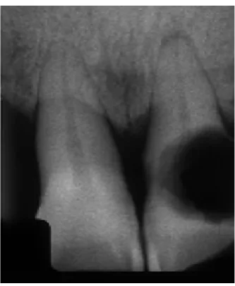
with the parallel technique from patients with a diagnosis of pulp necrosis as seen in Figure 1.
The samples were taken based on accidental
sampling method with inclusion criteria ie
teeth with complete and complete maxillary and maxillary pulp necrosis from crown to root, expressed from clinical examination, taken by parallel technique, pulp chamber studied no
other lesions such as radioopaque lesion so as
not disturbing judgment, the result of the photo is said to be of good quality, if the result of the photograph is no error on the result of photo like too dark, too bright, less contrast, less sharp, foreign body appearance or wrong position placement, stated by dentist radiology specialist in radiology. Exclusion criteria are age, gender, race, the oral condition of patient hygiene and systemic disease sufered by the patient.
The measurement scale in this study is the histogram obtained by determining the ROI of 30x30, along with the pulp chamber from the apical
direction. ROI obtained in one room pulp as much as
6 pieces, then in the input into the Matlab software
version R2012a to then processed with output data histogram graphics, ROI intensity, variance histogram ROI, and maximum histogram ROI.
The workings of the research through several stages are stage 1 (tooth not yet extracted), the
teeth diagnosed with pulp necrosis are done
periapical photographs of parallel technique, scanned on digital x-ray reader A4 size/scanner transparent resolution 1200DPI (Dot Per Inci).
Radiographs in the crop with a box shape on the
area to be studied as ROI, each tooth is divided into
6 segments from the bottom of the pulp duct near
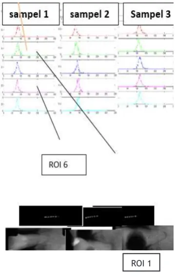
the apical, up to the top of the pulp, to determine the diference in pulp necrosis conditions (Figure 2), then open Matlab Software version of R2012a,
and performed data processing (Figure 3).
Stage 2 (tooth after being extracted with pulp), extracted maxillary anterior teeth per
-formed periapical radiography of parallel tech
-nique with conventional aircraft, then Scanning
on digital X-Ray reader A4 / transparent scanner resolution 1200 DPI (Dot Per Inch), then perform
-ing the examination as in step 1.
Stage 3 (tooth after extraction without pulp), anterior teeth of the extracted jaw in the drill using issure drill and pulp tissue removed are placed in glass preparation, ixation with formalin solution (formaldehyde dissolved in 1: 9 aquadest),
then perform histopathological examination with
eosin hematoxylin painting and photographed mi
-croscope of anterior neck mandibular prep against
the upper and lower anterior teeth.
Stage 4 (tooth after extraction without
pulp), maxillary anterior tooth element without
pulp tissue is done a periapical radiographic ex-amination of parallel technique with conventional
aircraft, then scans on digital X-Ray reader A4 / scanner transparent 1200 DPI (Dot Per Inci) reso
-lution. Then do the check as in stage 1.
Tools and research materials used by con
-ventional x-ray aircraft with speciications of RSGM Unpad radiology installations, Digital X-Ray Reader A4 / Transparent scanner with 1200 DPI (dot per inch) resolution, Toshiba Portege Intel Core 13 Laptop Windows 7, Matlab Software ver
-sion of R2012a. The research materials used are conventional radiography periapical parallel tech -nique and pulp of necrotic teeth.
RESULTS
The results of histopathological examination of
necrotic tooth pulp tissue from samples consist
of three teeth, two cannot be checked because the sample is not representative/preparation
Figure 2. Cropped Image of the necrotic pulp
is too small, Conducted Hematoxylin-eosin, the result is no colour diference from fat and protein
composition because the sample preparation is too small. On macroscopic examination with small
pulp tissue samples, brownish white. Microscopic examination there is massive iniltration of PMN
round cell.
The histogram pattern, the maximum value of the ROI histogram of samples 1, 2 and 3 on the tooth before it was pulled, shows the cervical pattern, the maximum peak of the histogram getting to the left of l, which means it is closer to
the lowest value of the radiolucent.
The histogram pattern of the maximum value of the histogram of samples 1, 2 and 3 on the tooth after being pulp removed, the pulp chamber shows an increasingly cervical histogram pattern, the maximum peak of the histogram getting to the left of each sample, which means closer to the
lowest value of the radiolucent.
The average intensity pattern of each ROI In the tooth before it is pulled, the more coronal radiolucent seen in table 1 (from point 0.45 to point 0.3), after revoked the pattern looks down then on the third ROI up to the 5th ROI, from radio-intermediate in the irst ROI position (apical) to radiolucency (approached coronal) After unplugged
without a pulp seen pattern from apical ROI to coronal down means from radio-intermediate to more radiolucent (from point 0.6 to point 0.4).
The variance histogram of the sample ROI of 1 tooth before it was removed ranged from 0.2335 to 0.2272, the tooth after being removed with pulps ranged from 0.2724 to 0.2136 and the tooth after being removed without pulp between 0.2479 to 0.1723. Obtained histogram variance data is
not much varied (If the width of the histogram
Figure 4. The histopathology of necrotic pulp of the 2nd tooth
ROI 1
Figure 5. Tooth histogram prior to extraction
Table 1. The average of intensity, histogram variance, and maximum histogram prior to the tooth extraction
ROI 1 ROI 2 ROI 3 ROI 4 ROI 5 ROI 6
Intensity average Sample 1 0.4249 0.3875 0.3789 0.3719 0.3642 0.3760 Sample 2 0.3942 0.3601 0.3759 0.3922 0.4069 0.3991 Sample 3 0.4740 0.4820 0.4715 0.4949 0.4886 0.4605
Histogram variance Sample 1 0.2335 0.2183 0.2339 0.2060 0.2095 0.2272
Sample 2 0.2120 0.2166 0.2413 0.1969 0.2393 0.1818 Sample 3 0.2291 0.2117 0.2111 0.2487 0.2182 0.2235 Histogram maximum Sample 1 0.3804 0.3490 0.3333 0.3098 0.3098 0.3451
variance the more variants, the more unpolluted
the pulp content.
Maximum histogram of dental ROI before
revoked on the irst ROI 0.3804 graph decreased until the 5th ROI 0.3098, visible pattern of numbers decreased, meaning the irst ROI of the apical part
to the coronal of the pulp getting radiolucent.
teeth after being pulled out with pulp from 0.2724 down until the fourth ROI of 0.1931, the ifth ROI pattern rises then the sixth ROI down 0.2136 and
the tooth after unplugged without pulp between
0.2479 to 0.1723. pulp obtained ranged from 0.0274 to 0.2136. histogram variance data does not vary much.
DISCUSSION
Pulp inlammation causes the tissue to undergo
central disintegration thus forming a liquefaction
necrosis region due to a lack of collateral circulation and a rigid dentin wall. This condition interferes with inlammatory drainage and increased tissue pressure increases, resulting in uncontrolled
destruction until all pulp tissues are necrotic.
The rate of progression of liquefaction
necrosis varies. Speed can be related to network
capability. The necrotizing region contains irritants from tissue and microbial destruction,
both anaerobic and aerobic.
Results of histopathological examination
of pulp tissue from three tooth samples, two
cannot be examined because the sample is too
small, marked with no visible colour diference
of fat and protein structure when given eosin
hematoxylin staining. characterized by no visible colour diference from fat and protein structures when stained with hematoxylin-eosin.
Carnerio et al in his study said the average grey scale can be used to look at caries remineralization, a smaller value for demineralization. this study mentions the minimum histogram value lies on the grey scale 50 and the highest histogram at number 200.9
Merete et al in his study stated that the accumulated
increase in mineral deposits occurred in teeth with smaller pulp cavities compared with larger pulp cavities in addition to composition factors of
pulps with diferent densities. As well as a barrier factor from absorption of x-rays in the oral cavity than outside the oral cavity.10
The average of tooth pulp intensity before it is pulled increasingly to the apical closer to
zero. Grossman said that the end result of pulp
decomposition is hydrogen sulphide, ammonia, fatty substance, ptomaine, water and carbon dioxide and further results are indol, skatol, putrescine, and cadaverin.3 Yoon et al, states that
dry pulp tissue consists of 5.7 % calcium, 3.5% phosphorus, and 11.7% are nitrogen, increasingly to the more radiolucent apical, where in the
coronal portion contains more air so that the
density is lower than the apical region containing
the end result of pulp decomposition.11
The average intensity of each ROI on a single tooth sample before being unplugged, unplugged with pulp and unplugged without pulp, shows an average pattern of intensity patterns increasingly coronal to radiolucent. This means
the demineralisation process runs from coronal to apical or the process of necrosis goes from coronal
to apical. The average ROI intensity in all tooth
samples before removal compared to the teeth that have been removed and the teeth removed and pulp is removed on each ROI as measured from the apical part of the pulp approaching to
the coronal down, meaning the coronal density further decreases so that more radiolucent, pulp density in the region approximates the maximum apical so that more radiopaque in this study is shown by the average number of ROI intensity on
the apical pulps higher approximation value.
The variance of the histogram of ROI in
one tooth sample was compared before it was
revoked after it was removed with pulp and tooth after unplugged without pulp, the data of histogram variance did not vary much. Similarly, the comparison of the three samples, namely the teeth have not been revoked, pulled out with
pulp and unplugged without pulp not too varied.
The maximum ROI histogram is the highest value in the one ROI that most often appears or how many of the most frequent intensities appear, from the histogram graph, the higher the maximum value of the histogram of the high value. These varying values indicate the diference in the rate
of the outcome of the pulp necrosis condition
occurring on the pulp. This is related to the pathophysiological process of pulp necrosis.
Maximum comparison of the histogram of
((0.3804) shows highest peak pattern of histogram chart decreased until ROI to ive (0.3098), meaning the irst ROI of the apical part towards coronal of pulp increasingly to left that is more radiolucent. Teeth after being pulled out with pulp (0.2724) down to the fourth ROI (0.1931), meaning the irst
ROI of the apical part to the coronal of the pulp
(ROI four) the radiolucent, meaning the density progressively decreases to the coronal gear after being unplugged without pulp from the irst ROI (0.6118) decreases until the ROI of six (0.3294)
means the top of the graph (maximum value) of the histogram gascic the more the coronal to
the radiolucent Graph The maximum histogram of ROI of all tooth samples prior to repeal,
after being pulverized and after unplug pulse
measured from irst ROI until ROI six shows the pattern approaching zero means the density is
decreasing in the coronal section the comparison
of all samples before removal, after unplug and after pulling, the pattern shows more trend to the right ie more radiopak on the teeth after unplug without pulp, this is due to formalin immersion or the presence of formalin luid on the tooth after it is unplugged without pulp formalin luid). Water has a larger density of 1.0 compared to the air 0.000129, which means here that water or liquids have more density or mass than gas or air. In accordance with the nature of absorption, the
lower the atomic number of an object the greater
the penetration of the X-ray and the greater the atomic number, the greater its absorption.12,13 So that radiographs result more radiopaque on the
teeth after soaking formalin.
Diferences in these variations of value are associated with an X-ray interaction when it comes to a material, in which case the material used is a
pulp with a diagnosis of pulp necrosis. Each atom
number (Z) afects the thickness of the resulting image, the greater the atomic number, the number
of electrons attached to each electron shell will
be greater. This greatly afects the radiodensity and contrast of the resulting image. The lower the atomic number of an object or matter, the greater the penetration of the X-ray, the greater the atomic number, the greater its absorption.12,13
The pulp decomposition of each compound has diferent atomic number. Consecutively for the number of hydrogen atoms, carbon, nitrogen, and oxygen are 1, 6, 7, and 8. The atomic number
(commonly denoted by Z) is the number of protons present in the atomic nucleus. The number of
protons is equal to the number of electrons in the
compound. The atomic number and the number
of electrons in the compounds are related to the
interaction of light on the object, in this study is a pulp that has been necrosis with diferent
decomposition results.
The density is afected by the material or material, the physical characteristics of the contrast material from the water of the efective atomic number (z) 7.42, density 1.0 gr/cm3; fat
atomic number efective (z) 5.92, density 0.91; air of efective atomic number (z) 7.64, density 0.00129; calcium efective atomic number (z) 20.0, density 1.55; efective atomic number (z) 7.46, density 1.0 gr/cm3; barium efective atomic
number (z) 56.0, density 3.5 gr/cm3; iodine
efective atomic number (z) 56.0, density 3.5 gr/
cm3. According to the nature of the absorbs of
x-rays when the mass of the material is greater, the absorption power of the x-rays will also be greater,14 so that grayscale pulp necrosis is more radiolucent.
CONCLUSION
The tooth with a pulp necrosis showed a tendency that led to radiolucency on periapical radiograph histogram, and histopathologic examination showed massive iniltration of a round PMN cells,
thus supported the pulp necrosis diagnose.
REFERENCES
1. National Institute of Health Research and Development (NIHRD). Indonesia Basic Health Research (RISKESDAS) 2007-2008. Jakarta: Ministry of Health Republic of Indonesia; 2008. 2. Rajendran R. Shafer’s Texbook of Oral
Pathology. 6th ed. New Delhi: Reed Elsevier
India Pvt. Ltd.; 2009. p. 682-3.
3. Grossman LI, Seymour O, Del Rio CE. Endodontic Practice, 11th ed. Philadelphia:
Lea & Febiger; 1988. p. 40-83.
4. Frommer HH, Stabulas-Savage JJ. Radiology for The Dental Professional, 9th ed. St. Louis:
Mosby-Elsevier; 2011.
Pathology: Its Etiology and Prevention. In: Ingle JI, Bakland LK. Endodontics, Volume 1.
4th ed. Hamilton: BC Decker Inc.; 2002. p. 95-174.
6. Novelline RA. Squire’s Fundamentals of
Radiology. 6th ed. Cambridge: President &
Fellows of Harvard College; 2004.
7. Attwood D, Sakdinawat A. Soft X-rays and
extreme ultraviolet radiation: Principles and
Application. 2nd ed. Cambridge: Cambridge
University Press; 2016. p. 2.
8. Nelson SJ. Wheeler’s Dental Anatomy,
Phisiology, and Occlusion. 9th ed. St. Louis:
Saunders-Elsevier; 2010. p. 333-5.
9. Carneiro LS, Nunes CA, Silva MA, Leles CR, Mendonca EF. In vivo study of pixel grey measurement in digital subtraction radiography
for monitoring caries remineralization.
Dentomaxillofac Radiol. Feb 2009;38(2): p. 73-8. DOI: 10.1259/dmfr/15857365.
10. Carneiro LS, Nunes CA, Silva MA, Leles CR, and Mendonc EF. 2009. In vivo study of pixel grey measurement in digital subtraction radiography
for monitoring caries remineralization.
Dentomaxillofacial Radiology Journal, 38, 73-78. http://dmfr.birjournals.org (diakses April 2012).(already cited in citation no. 8 above) 11. Markvart M, Bjørndal L, Darvann TA, Larsen
P, Dalstra M, Kreiborg S. Three-dimensional Analysis of The Pulp Cavity on Surface Models of Molar Teeth, Using X-ray Micro-Computed Tomography. Acta Odontol Scand. Mar 2012;70(2): p. 133-9. DOI: 10.3109/00016357.2011.600707.
12. Yoon SH, Brudevold F, Smith FA, Gardner DE. Flouride, Calcium, Phosphate, Ash, and Water Content of Human Dental Pulps. J Dent Res. Jul 1965;44: p. 696-700. DOI: 10.1177/00220345650440041601.
13. Whaites E. Essentials of Dental Radiography and Radiology. 4th ed. London: Churchill
Livingstone; 2007.
14. White SC, Oral Radiology: Principles and
Interpretation. 7th ed. St.Louis: Mosby-Elsevier; 2014.
15. Sprawls P Jr. The Physical Principles of Medical
Imaging, 2nd ed. Madison: Sprawls Educational