ORIGINAL ARTICLE
Cytotoxic and antioxidant activities of catechins in inhibiting
the malignancy of breast cancer
Endang Evacuasiany
1, Hana Ratnawati
1, Laella K. Liana
1, Wahyu Widowati
1,
Maesaroh Maesaroh
2, Tjandrawati Mozef
3, Chandra Risdian
31
Faculty of Medicine, Maranatha Christian University;
2
Biomolecular and Biomedical Research Center, Aretha Medika Utama ;
3
Research Center for Chemistry, Indonesian Institute of Sciences; Bandung, West Java, Indonesia
Received February 21, 2014 Jl. Prof drg. Suria Sumantri No.65, Bandung, West Java, 40164, Indonesia.
Objective: Breast cancer is a malignant disease of women most often found after cervical cancer in Indonesia. Increased levels of free radicals can cause DNA damage, which could lead to malignancy; this can play role in breast cancer etiopathogenesis. The present research was conducted to determine the activity of catechins as antioxidants and their potential efficacy in inhibiting breast cancer malignancy.
Methods: The research was done by examining the antioxidant and free radical scavenging activity including 1,1-diphenyl-2-picryl-hydrazyl (DPPH), the value of superoxide dismutase (SOD), and assays in breast cancer cell lines (T47D, MCF7). The cytotoxic potency was determined by the MTS (3-(4,5-dimethylthiazol-2-yl)-5-(3-carboxymethoxyphenyl)-2-(4-sulfophenyl)-2H-tetrazolium) assay.
Results: The highest DPPH scavenging activity is presented by (-)-epigallocatechin (EGC) and the lowest by gallo catechins (GC). The highest SOD value were reached with EGC at 500 µg/ml, followed by (-)-epicatechin gallate (ECG) at 125 µg/ml, and GC at 31.25 µg/ml concentrations. The highest cytotoxic activity in T47D cell line for 24 and 48 h incubation was exhibited by (-)-gallocatechin gallate (GCG). The greatest cytotoxic activity in MCF7 cell line for 24 h was presented by (-)-epigallocatechin gallate (EGCG), and for 48 h incubation by (+)-catechin (C). Conclusion: Catechins have high antioxidant activities proven by both DPPH scavenging and SOD activities. They possess higher anticancer action on T47D than on MCF7 cell line.
© 2014 GESDAV
INTRODUCTION
Breast cancer is a malignant disease of women most often found in Indonesia after cervical cancer. The high number of breast cancer caused by a hereditary gene mutation, improper diet and unhealthy lifestyle and environmental factors include air pollution, radiation, cigarette smoking, alcohol and carcinogenic substances. These factors may increase the free radicals level which is one of the factors causing DNA damage, consequently leading to malignancy [1]. Oxidative stress is triggered by imbalance between free radicals and antioxidant and can link to many diseases including cancer [2].
Cancer is the second leading cause of death; the most cancer drugs are synthetic agents with relatively high prices and side effects, but lower cure rates [3-5]. Cancer treatment combined with natural or herbal medicine can inhibit or lower tumor proliferation. Many herbal medicines have antioxidant activities which play role in curing many diseases [6]. Since having antioxidant, antiangiogenic and cytotoxic activities, the tea plant (or green tea; Camellia sinensis) is one possible herbal remedy which can be used for
cancer or related issues; this plant contains polyphenolic compounds [7, 8], including catechins, which have antioxidant activities that can protect cells from free radical attack [7-9]. Several of tea catechins or flavan-3-ols including (+)-catechin (C), epicatechin (EC), epigallocatechin (EGC), (-)-epicatechin gallate (ECG), (-)-epigallocatechin gallate (EGCG), catechin gallate (CG), gallo catechins (GC)
CG, EGC, GCG and GC (Biopurify Phytochemicals), Dulbecco’s modified Eagle’s medium (DMEM; Sigma-Aldrich), fetal bovine serum (FBS; Sigma-Sigma-Aldrich), penicillin Aldrich), streptomycin (Sigma-Aldrich), CellTiter Aqueous One Solution Cell Proliferation Assay (MTS; Promega). trypsin-EDTA (Sigma-Aldrich).
DPPH scavenging activity assay
Fifty microliters of catechins at various concentrations were introduced in microplates and 0.077 mmol DPPH in 200 µl DMSO was added. The mixture was shaken vigorously, incubated at room temperature for 30 min and then the absorbance was measured at 517 nm using a microplate reader. For negative controls 250 µl DPPH and for blank 250 µl methanol was used. The radical scavenging activity of each sample was expressed by the ratio of the lowering absorption rate of DPPH (%) relative to the absorption of DPPH solution in the absence of test sample (negative control). Here the formula for DPPH scavenging antioxidant activity (%):
Superoxide dismutase (SOD) assay
The SOD assay was performed using a commercial kit comprising assay buffer, sample buffer, radical detector, SOD standard and xanthine oxidase. SOD standards were prepared by introducing 200 μl diluted radical detector and 10 μl SOD standard (7-level standard) per well [15]. Catechins were dissolved in DMSO at concentrations of 500, 125 and 31.25 µg/ml. The sample well contained 200 µl diluted radical detector and 10 µl sample. To all wells 20 µl diluted xanthine oxidase was added. The mixtures were shaken carefully for few seconds, incubated for 20 min at room temperature, SOD activity was measured on a microplate reader at 440-460 nm. The SOD value is calculated using the equation from the linear regression of standard curve substituting linear rate (LR) for each sample:
Cytotoxic assay
The T47D and MCF7 human breast cancer cell lines were obtained from Research Center for Chemistry, Indonesian Institute of Sciences, Division of Natural Products Bandung, West Java, Indonesia. The cells were cultured and maintained in DMEM supplemented with 10% (v/v) FBS, 100 U/ml penicillin and 100 μg/ml streptomycin, and incubated at 37ºC in a humidified atmosphere with 5% CO2 [15].
After the cells reached 80% confluence, the cells were seeded into a 96-well plate (5 x 10⁴ cells per well) and supplemented by various concentrations of catechins in
10% DMSO, then incubated for 24 h and 48 h. Twenty microliters of MTS was added to each well, and then incubated at 37ºC for 2-4 h. The sample absorbance was read at 490 nm using a microplate reader (Thermo Multiskan GO). The data were presented as percent of viable cells (%) and analyzed by calculating the IC50
using probit analysis (SPSS 20.0) [15].
RESULTS
DPPH scavenging activity
DPPH radical is a synthetic compound that is soluble in polar organic solvents such as methanol at room temperature [16]. In the presence of active antioxidant compounds, DPPH free radical scavenging of the sample is marked with a change of color from dark purple to yellowish or pale yellow [17]. The DPPH free radical scavenging activity of a compound is representative for its antioxidant activity. In our study, the highest DPPH scavenging activity is was recorded for EGC and GCG (Fig.1.)
The IC50 value is the concentration of antioxidant
needed to scavenge 50% of the DPPH free radical [15]; the smaller IC50 value is representative for higher
antioxidant activity [18]. The highest antioxidant activity in the current study was recorded for EC with an IC50 of 3.24 µM. whereas the lowest activity was
represented by GC (IC50 9.80 µM; Table 1).
Figure 1. DPPH scavenging activity (%) of cathechins diluted in methanol to 200, 100, 50, 25, 12.5, 6.25, 3.125, 1.563, 0.781 and 0.391μM.
Table 1. DPPH scavenging activity (IC50) of catechins
Samples Linear equation R2 IC50(μM)
CG Y = 7.771X + 3.709 0.856 5.96
GCG Y = 15.819X - 9.932 0.951 3.79
C Y = 6.916X - 6.117 0.993 8.11
GC Y = 4.436X + 6.552 0.995 9.80
EC Y = 7.650X - 18.123 0.988 8.91
EGC Y = 9.361X + 19.707 0.897 3.24
EGCG Y = 6.475X + 2.411 0.893 7.30
ECG Y = 13.42X - 5.658 0.996 4.15
The DPPH scavenging activity test was measured triplicate for each sample. Linear equations, coefficient of regression (R2), and IC
50 were calculated.
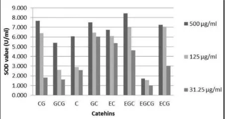
Table 2. SOD activity of catechins (U/ml)
Samples Concentrations
500 μg/ml 125 μg/ml 31.25 μg/ml CG 7.68±0.03de 6.39 ±0.39c 1.81±0.02b
GCG 5.4±0.16b 2.61±0.07b 1.62±0.11b
C 6.08±0.33bc 2.93±0.06b 2.59±0.03c
GC 7.52±0.22de 6.45±0.26c 6.03±0.02g
EC 6.74±0.47cd 6.12±0.07c 5.37±0.13f
EGC 8.44±1.01e 7±0.08d 4.64±0.2e
EGCG 1.71±0.06a 1.56±0.05a 1.02±0.03a
ECG 7.24±0.09cde 7.07±0.06d 3.02±0.18d
Data are presented as mean ± standard deviation. Different letters in the same column among catechins in high, moderate and low concentrations indicate significantly difference (Tukey’s HSD post hoc test).
Table 3. The IC50 value of catechins in T47D and MCF7
cells after 24 and 48 h incubation
Samples T47D (μM) MCF7 (μM)
24 h 48 h 24 h 48 h
C 70.33 72.97 563.01 128.86
ECG 2525.83 415.04 558.25 683.66
GCG 34.65 23.66 507.59 619.46
GC 51.54 35.68 1129.53 415.95
EC 109 946.6 7240.89 135.97
CG 162.35 130.86 2527.94 1400.25
EGCG 54.45 81.31 211.56 343.5
EGC 70.76 103.14 3421.03 1408.27
Doxorubicin 0.96 0.002 3.76 0.101
Each catechin compound was measured triplicate and rate inhibition was analyzed using probit.
SOD activity
The free radical O2•- is one of the most important
radicals formed in aerobic cells due to leakage of the electron transport chain. It is a precursor to form the highly reactive hydroxyl radical (•OH) through Fenton and Haber-Weiss reactions [19].
SOD is an endogenous antioxidant enzyme which catalyses the dismutation reaction of O2•- into hydrogen
peroxide (H2O2) and oxygen (O2) [20]. The activity of
catechins in scavenging O2•- can be seen in Table 2 and
Fig.2. Based on these the highest SOD activity at a
concentration of 500 µg/ml was recorded for EGC (8.44 U/ml), while the lowest was EGCG (1.71 U/ml). At 125 µg/ml, the highest SOD value was seen for ECG (7.07 U/ml) and EGC (7 U/ml), and the lowest was EGCG (1.56 U/ml). The highest SOD activity at 31.25 µg/ml was represented by GC (6.03 U/ml) and the lowest by EGCG (1.02 U/ml). Overall, at all three concentrations the lowest SOD activity was exhibited by EGCG.
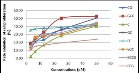
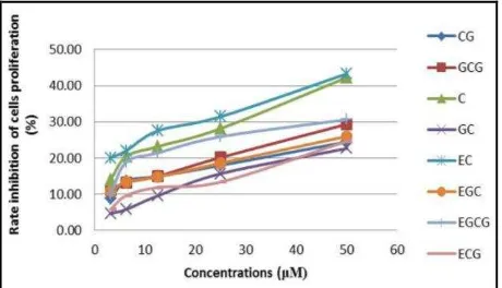
Cytotoxic activity
The MTS assay was used for measuring the number of viable cells in the cell culture; MTS tetrazolium compound was reduced into colored formazan product that is soluble in tissue culture medium, this reaction
was accomplished by nicotinamide adenine
dinucleotide (NADH) or NADH phosphate (NADPH) produced by dehydrogenase enzymes in viable cells [21]. The formazan product from MTS reduction giving color to violet can be read at 450-540 nm (max. absorbance 490 nm). IC50 value indicates the
concentration which is able to inhibit 50% of cell proliferation; the greater the value of IC50, the lower the
cytotoxic activity [22]. In the present study, cytotoxic activity was found to be concentration-dependent; higher concentrations inhibited proliferation strongly (Table 3, Figs.3&4).
Figure 4. Cytotoxic activity of catechins diluted in DMSO to 50, 25, 12.5, 6.25 and 3.125 μM and incubated for 48 h in T47D cell line. Inhibition of cell proliferation was interpreted as cytotoxic activity.
Figure 5. Cytotoxic activity of catechins diluted in DMSO to 50, 25, 12.5, 6.25 and 3.125 μM and incubated for 24 h in MCF7 cell line. Inhibition of cell proliferation was interpreted as cytotoxic activity.
Figure 6. Cytotoxic activity of catechins diluted in DMSO to 50, 25, 12.5, 6.25 and 3.125 μM and incubated for 48 h in MCF7 cell line. Inhibition of cell proliferation was interpreted as cytotoxic activity.
The results showed that longer contact of catechins with cancer cells will enhance the cytotoxic effect in both T47D and MCF7. The highest cytotoxic activity in T47D with short (24 h) and long (48 h) incubation was presented by GCG (34.65 µM and 23.66 µM, respectively). Catechins were not significantly active in MCF7 with both short and long incubation.
DISCUSSION
Based on Tables 1 and 2, and Figures 1 and 2 showing that catechins have high free radical scavenging activity and EGC is the most active free radical scavenger against both DPPH and O2•-, the present
result was consistent with previous studies reporting that: catechin epimers have good free radical scavenger activity against peroxyl radicals, singlet oxygen, peroxynitrite and hypochlorous acid [7-9, 12]; have high antioxidant efficacy based on Trolox equivalent antioxidant capacity (TEAC) [12], DPPH scavenging assay [23], O2•- scavenging activity (SRSA) [24], and
human LDL oxidation, ferric reducing-antioxidant power (FRAP) [11]. Catechins exhibited differential SOD value and DPPH scavenging activity; this result is consistent with a previous study reporting that lipid peroxidation activitiy of catechin’s epimers were different, suggesting that the electrochemical characteristic, steric structures of compounds may play role in their antioxidant actitvites [25].
Based on Table 3 and Figures 3-6 showing that catechins possess cytotoxic effect in T47D at both 24 h and 48 h incubation, only ECG was not active. This result is validated with previous research reporting that 100 μM of catechins including C, EC, GC, EGC, CG, ECG, GCG and EGCG inhibited colorectal cancer (HCT116) cell growth [6].
Tea polyphenols have antiproliferative effects on prostate, breast, lung and colorectal cancer. Green tea polyphenols have cytotoxic effects by histone deacetylase (HDAC) inhibition, due to increased proteasomal degradation [26]. EGCG inhibits breast cancer cells (Her2/neu) by activiting Forkhead box O (FoxO) proteins which protect cells from oxidative damage-induced apoptosis by inducing the antioxidant enzymes MnSOD and catalase (CAT) [27].
Catechins presented only insignificant cytotoxic effect in MCF7 cells at both 24 h and 48 h incubation. However, this results is not supported by a previous study in whicht catechin (C) effectively inhibited MCF7 cells proliferation at higher doses [34].
In summary, the most active antioxidant catechins of the present study are EGC and EGC. On the other hand, the most active cytotoxic activity in T47D cells at 24 h was recorded with GCG and at 48 h with GCG. Although insignificant, the highest cytotoxic activity in MCF7 cells was presented by EGCG at both 24 and 48 h.
ACKNOWLEDGEMENT
We gratefully acknowledge the financial support of Directorate General for Higher Education, National Ministry of Republic Indonesia for research grant of Hibah Bersaing 2012 (DIPA Kopertis Wilayah IV no 0561/023-04.2.01/12/2012).
REFERENCES
1. Li W, Ray RM, Lampe J W, Lin MG, Gao DL, Wu C, Nelson ZC, Fitzgibbons ED, Horner N, Hu YW, Shannon J, Satia JA, Patterson RE, Stalsberg H Thomas D. Dietary and other risk factors in women having fibrocystic breast conditions with and without concurrent breast cancer: A nested case-control study in Shanghai, China. Int J Cancer 2005; 115:981-93.
2. Singh A, Jain A, Sarma BK, Jha A, Singh HB. Natural antioxidants and their role in cancer prevention. In: Shankar S, Srivastava RK (eds) Nutrition, Diet and Cancer. Springer, Dordrecht, 2012.
3. Wilkes GM. Therapeutic options in the management of colon cancer: 2005 update. Clin J Oncol Nurs 2005; 9:31-44.
4. Kosmider S, Lipton L. Adjuvant therapies for colorectal cancer. World J Gastroenterol 2007; 13:3799-805.
5. Kan WLT, Yin C, Xu HX, Xu G, To KKW, Cho CH, Rudd JA, Lin G. Antitumor effects of novel compound, guttiferone K, on colon cancer by p21Waf1/Cip1-mediated G0/G1 cell cycle arrest and apoptosis. Int J Cancer 2013; 132:707-16.
6. Du GJ, Zhang Z, Wen XD, Yu C, Calway T, Yuan CS, Wang CZ. Epigallocatechin gallate (EGCG) is the most effective cancer chemopreventive polyphenol in green tea. Nutrients 2012; 4:1679-91.
7. Chun OK, Chung SJ, Song WO. Estimated dietary flavonoid intake and major food sources of U.S. adults. J Nutr 2007; 137:1244-52.
8. Cooper R, Morre J, Morre D. Medicinal benefits of green tea: part I. Review of noncancer health benefits. J Altern Complement Med 2005; 5:521-8.
9. Cooper R, Morre J, Morre D. Medicinal benefits of green tea: part II. Review of anticancer properties. J Altern Complement Med 2005; 11:639-52.
10. Demeule M, Michaud-Levesque J, Annabi B, Gingras D, Boivin D, Jodoin J, Lamy S, Bertrand Y, Beliveau R. Green tea catechins as novel antitumor and antiangiogenic compounds. Curr Med Chem Anticancer Agents 2002; 2:441-6.
11. Xu JZ, Yeung SYV, Chang Q, Huang Y, Chen ZY. Comparison of antioxidant activity and bioavailability of tea epicatechins with their epimers. Br J Nutr 2004; 91:873-81.
12. Muzolf-Panek M, Gliszczynska-Swiglo A, Szymusiak H, Tyrakowska B. The influence of stereochemistry on the antioxidant properties of catechin epimers. Eur Food Res Technol 2012; 235:1001-9.
13. Lee LS, Lee N, Kim YH, Lee CH, Jong SP, Jeon YW, Kim YE. Optimization of ultrasonic extraction of phenolic antioxidants from green tea using response surface methodology. Molecules 2013; 18:13530-45.
14. Hanani E, Mun’im A, Ryany S. Antioxidant activity and identification of antioxidative compounds of Callyspongia sponge from Seribu Island. Majalah Ilmu Kefarmasian 2005; 2:127-33.
15. Widowati W, Wijaya L, Wargasetia TL, Bachtiar I, Yellianty, Laksmitawati DR. Antioxidant, anticancer, and apoptosis-inducing effects of Piper extracts in HeLa cells. J Exp Integr Med 2013; 3:225-30.
16. Pokorny J, Yanishlieva N, Gordon M. Antioxidants in Food. CRC Press, Washington DC, 2001.
17. Prakash A, Rigelhof F, Miller E. Antioxidant Activity. Medallion Laboratories Analitical Progress.
19. Halliwell B, Gutteridge JMC. Free Radicals in Biology and Medicine. Oxford University Press, New York, 1999.
20. Sairam RK, Srivastava GC, Agarwal S, Meena RC. Differences in antioxidant activity in response to salinity stress in tolerant and susceptible wheat genotypes. Biologia Plantarium 2005; 49:85-91.
21. Berridge MV, Tan AS. Characterization of the cellular reduction of 3-(4,5-dimethylthiazol-2-yl)-2,5-diphenyltetrazolium bromide (MTT): subcellular localization, substrate dependence, and involvement of mitochondrial electron transport in MTT reduction. Arch Biochem Biophys1993; 303:474-82.
22. Kampa M, Alexaki VI, Notas G, Nifli AP, Nistikaki A, Hatzoglou A, Bakogeorgou E, Kouimtzoglou E, Blekas G, Boskou D, Gravanis A, Castanas E. Antiproliferative and apoptotic effects of selective phenolic acids on T47D human breast cancer cells: potential mechanisms of action. Breast Cancer Res 2004; 6:R63-74.
23. Rice-Evans CA, Miller NJ, Paganga G. Structure-antioxidant activity relationships of flavonoids and phenolic acids. Free Radic Biol Med 1996; 20:933-56.
24. Unno T, Yayabe F, Hayakawa T, Tsuge H. Electron spin resonance spectroscopic evaluation of scavenging activity of tea catechins on superoxide radicals generated by a phenazine methosulfate and NADH system. Food Chem 2002; 79:259-65.
25. Yang B, Kotani A, Aral K, Kusu F. Estimation of the antioxidant activities of flavonoids from their oxidation potentials. Anal Sci 2001; 17:599-604.
26. Thakur VS, Gupta K, Gupta S. Green tea polyphenols causes cell cycle arrest and apoptosis in prostate cancer cells by suppressing class I histone deacetylases. Carcinogenesis 2012; 33:377-384.
27. Belguise K, Guo S, Sonenshein GE. Activation of FOXO3a by the green tea polyphenol epigallocatechin-3-gallate induces estrogen receptor alpha expression reversing invasive phenotype of breast cancer cells. Cancer Res 2007; 67:5763-70.
28. Larsen CA, Bisson WH, Dashwood RH. Tea catechins inhibit hepatocyte growth factor receptor (MET kinase) activity in human colon cancer cells: Kinetic and molecular docking studies. J Med Chem 2009; 52:6543-45.
29. Manikandan R, Beulaja M, Arulvasu C, Sellamuthu S, Dinesh D, Prabhu D, Babu G, Vaseeharan B, Prabhu NM. Synergistic anticancer activity of curcumin and catechin: an in vitro study using human cancer cell lines. Microsc Res Tech 2012; 75:112-6
30. Wang YC, Bachrach U. The specific anti-cancer activity of green tea (-)-epigallocatechin-3-gallate (EGCG). Amino Acids 2002; 22:131-43.
31. Hayes CJ, Whittaker BP, Watson SA, Grabowska AM. Synthesis and preliminary anticancer activity studies of C4 and C8-modified derivatives of catechin gallate (CG) and epicatechin gallate (ECG). J Org Chem 2006; 71:9701-12
32. Azam S, Hadi N, Khan NU, Hadi SM. Prooxidant property of green tea polyphenols epicatechinand epigallocatechin-3-gallate: implications for anticancer properties. Toxicol In Vitro 2004; 18:555-61.
33. Kwak TW, Kim DH, Chung CW, Lee HM, Kim CH, Jeing YI, Kang DH. Synergistic anticancer effects of vorinostat and epigallocatechin-3-gallate against HuCC-T1 human cholangiocarcinoma cells. Evid Based Complement Alternat Med 2013; 2013:185158.
34. Alshatwi AA. Catechin hydrate suppresses MCF-7 proliferation through TP53/Caspase mediated apoptosis. J Exp Clin Cancer Res 2010, 29:167.