August 2013 113
Accredited by DGHE No: 66b/DIKTI/Kep/2011 DOI: 10.5398/medpet.2013.36.2.113
*Corresponding author: E-mail: [email protected]
Mucosal Mast Cells Response in the Jejunum of Ascaridia galli-Infected
Laying Hens
Darmawia*, U. Balqisa, M. Hambala, R. Tiuriab, Frengkia, & B. P. Priosoeryantob aFaculty of Veterinary Medicine, Syiah Kuala University
Jln. Tgk. H. Hasan Krueng Kale No. 8 Darussalam, Banda Aceh 23111, Indonesia bFaculty of Veterinary Medicine, Bogor Agricultural �ni�ersityBogor Agricultural �ni�ersity
Jln. Agatis, Kampus IPB Dramaga Bogor 16680, Indonesia (Received 26-06-2013; Reviewed 06-09-2013; Accepted 17-09-2013)
ABSTRACT
Intestinal defense mechanism against helminthes parasitic nematode to be associated with mu-cosal mast cells reaction. The aim of this research was to examine the effect of infection by Ascaridia galli parasite to trigger mucosal defense based on mucosal mast cells response in laying hens. Amount of ten head laying hens 12-wk old were divided into two groups containing five chickens of each. The first group, chickens were left as un-infected controls. The second group, chickens were infected orally with 1,000 embryonated eggs of A. galli. Mucosal mast cell responses were assayed by in situ jejunal mast cell counts in stained serial histological sections with Alcian blue �pH 0.��� andAlcian blue �pH 0.��� and Safranin-O �pH 0.1�� of the jejunum. Mucosal mast cells response were obser�ed and counted on daysof the jejunum. Mucosal mast cells response were obser�ed and counted on days 14 post infection. The result showed that A. galli infection was able to increase significantly (P<0.05) mast cells response. This research concluded that the A. galli infection can trigger the in�olment of mucosal mast cells response in jejunal defense of laying hens against parasitic diseases caused by A. galli.
Key words: Ascaridia galli, laying hen, mucosal mast cell
ABSTRAK
Mekanisme pertahanan intestinal terhadap cacing nematoda parasitik berkaitan dengan reaksintestinal terhadap cacing nematoda parasitik berkaitan dengan reaksi sel mast mukosa. Tujuan riset ini adalah menguji pengaruh infeksi parasit Ascaridia galli dalam merangsang pertahanan mukosa berdasarkan respons sel mast mukosa pada ayam petelur. Sebanyak sepuluh ekor ayam petelur yang berumur 12 minggu dibagi menjadi dua kelompok, masing-masing kelompok terdiri atas lima ekor ayam. Kelompok pertama, ayam tidak diinfeksi sebagai kontrol. Kelompok kedua, ayam diinfeksi melalui oral dengan 1.000 telur berembrio A. galli. Respons sel mast mukosa diuji in situ dengan menghitung sel mast pada jejunum melalui pewarnaan serial his-tologik dengan Alcian blue �pH 0,��� dan Safranin-O �pH 0,1�� dari sediaan preparat jejunum. ResponsAlcian blue �pH 0,��� dan Safranin-O �pH 0,1�� dari sediaan preparat jejunum. Respons jejunum. Respons sel mast mukosa diamati dan dihitung pada hari ke-14 pascainfeksi. Hasil menunjukkan bahwa infeksi A. galli dapat meningkatkan respons sel mast mukosa secara signifikan (P<0,05). Riset ini me -nyimpulkan bahwa infeksi cacing A. galli dapat merangsang keterlibatan respons sel mast mukosa pada pertahanan jejunum ayam petelur terhadap penyakit parasitik yang disebabkan oleh A. galli.
Kata kunci: Ascaridia galli, ayam petelur, sel mast mukosa
INTRODUCTION
Ascaridia galli is one of the major nematode para-sites causing substantial economic losses in domestical chickens farming worldwide. The normal habitat of the parasitic stages of A. galli is in the small intestine of
114 August 2013
infection. Thus, A. galli was capable of de�eloping in the lumen but could also enter the lining intestinal with migration to the tissue.
Characteristic immune responses occur during parasite infection in the small intestine. It has long been known that the mast cells are contributed in defense mechanism against parasite infection, particularly in locations that are in close contact with the external en�ironment such as intestines (�rb & Sheppard, 2012), with these responses peaking at the time of parasite expulsion from the host (McDermott et al., 2003), but the mucosal mast cells precise mechanisms in�ol�ed ha�e remained obscure. Infection induces mucosal mast cells degranulation in the intestinal that is considered to be a host defense mechanism against the parasite. In support of this hypothesis, �arious authors described that mast cells in�ol�ed in mucosal defense mechanism. Li et al. (2004) showed that mast cells are important for rapidly controlling murine infection with the protozoan parasite Giardia lamblia. Okayama & Kawakami (2006) described that the number of mast cells in inflamed tissue can be regulated by proliferation, migration, and sur�i�al (and apoptosis). Anthony et al. (2007) explained that many of effector cells are acti�ated in response to most helminth infections including mast cells. Mucosal mast cells contribute to expulsion of a number of gastrointestinal nematode parasites (Afferson et al., 2012).
Mast cells differentiate from multipotent hemato-poietic stem cells in the bone marrow that gi�e rise to committed mast cell progenitors in the blood and are re-cruited to tissues, where they mature. Franco et al. (2010) suggested that mast cell de�elopment is most closely associated with the megakaryocyte/erythrocyte lineage. The lambs infected with Haemonchus contortus had significantly greater numbers of mucosal mast cells in abomasal mucosa of lambs (Shakya et al., 2009). Ortolani et al. (2013) in�estigated the greater number of mucosal mast cells was able to decrease worm burden in the abomasums of sheep infected with H. contortus. Onah & Nawa (2004) obserbed that mastocytosis occurred in jejunal sections of mice challenged with Strongyloides venezuelensis.
Currently, little information is a�ailable describing the effects of A. galli infection on the mucosal defense regarding mast cells response of small jejunal of laying hens. Reference to mucosal mast cells mediate Trichinella spiralis, the nematode resides within enterocytes of the jejunum, expulsion from the intestine of mice has been reported (Ierna et al., 2005; Suzuki et al., 2008; Afferson et al., 2012); howe�er, De-yuan et al. (2003) reported that the mucosal mast cells in jejunum of chickens infected with A. galli were increased with no significantly differ-ence but a remarkable decrease of mast cells in the thy-mic medulla. Thus, the aim of the current study was to in�estigate the effects of A. galli infection on the mucosal defense based on mucosal mast cells response in jejunal of laying hens. Therefore, we in�estigated the distribu-tion of mucosal mast cells in laying hens wether these mucosal mast cells are strong associated for mucosal defence in the jejunal against A. galli infection.
MATERIALS AND METHODS
Chickens
Ten heads Isa Brown laying hens aged 12 wk were used in this study. All chickens maintained indi�idu-ally in laying hens battery cages at Faculty of Veterinary Medicine, Bogor Agricultural �ni�ersity. Feed and water were supplied ad libitum. Anthelminticum albendazole (PT. Mensana Aneka Satwa, Indonesia) dose 25 mg in 0.25 mL of each was gi�en to all chickens to ensure that they were free of helminth infection 2 wk before infec-tion carried out.
Ascaridia galli Parasite
A. galli adult worms were procured from the intes-tine of freshly slaughtered chickens. They were brought to the laboratory from local restaurant. Worms were washed with phosphate buffered saline (PBS). For col-lecting the egg worms, the selected A. galli female adult worms were wounded in half of body length using a needle with sharp tip under stereo microscope. The eggs obtained from uteri female adult worms were incubated in sterile aquadestilata at room temperature for 20-31 d till to de�elop embryonated eggs. The eggs were count-ed under stereomicroscope for preparing doses of 1,000 embryonated A. galli eggs (Darmawi et al., 2007; Balqis et al., 2009).
Ascaridia galli-Infected Laying Hens
The laying hens de�ided into two groups contained fi�e chickens of each. First groups, the chickens were introduced with 0.5 mL PBS. Second group, the chickens were orally infected with doses of 1,000 embryonated A. galli eggs contained in 0.5 mL PBS and introduced directly into the oesophagus using a needle with blunt o�al tip (Darmawi et al., 2007).
Tissue Preparation for Mast Cells Protocol
August 2013 115 (Sigma). After washing, sections were counterstained
with eosin and mounted. The number of mast cells per 10 �illus crypt units (VC�s) was counted on each section. Mast cell counts were performed under light microscopy using an eyepiece square graticule (eyepiece ×10, objecti�e ×40), and data expressed as mean number of mucosal mast cells (MMCs) per VC� as described byas described by pre�ious authors (McDermottMcDermott et al., 2003; Noviana et al., 2004; Li et al., 2004; Königová et al., 2008) with certain modifications.
Statistical Analysis
The MMC responses in different groups of laying hens were analyzed by the Student t test, where t test was used for comparisons of mast cell numbers. P value of <0.05 was taken to indicate a significant difference.
RESULTS AND DISCUSSION
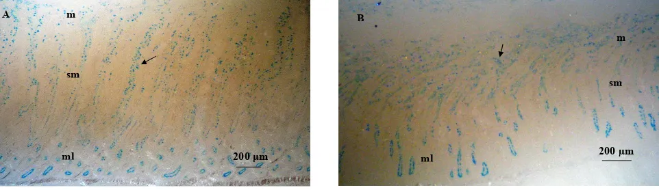
The simplest interpretation of the finding as seen in Figure 1, is that at least some mast cells go through mucosae in close contact with the external en�ironment, jejunum, mediating the expulsion of A. galli from the intestine. Staining with Alcian Blue–Safranin O re�ealed mast cells in all the organs examined. Mast cells were identified as blue granules against a pale brown back-ground. Here, we regarded them as mucosal mast cell. Two major subtypes of mast cells ha�e been identified in dogs: connecti�e tissue type, particularly localized in skin, around blood vessels, and in the peritoneal cavity; and mucosal type, which is associated with mucosal surfaces such as those in the gut or airways (No�iana et al., 2004). Regarding the distribution of mast cells within the �arious locations similar for and support those of Königová et al. (2008), who obserbed mucosal in the lamina propria mucosae, meanwhile connecti�e tissue mast cells were found in the tela submucosa in the stomach of Mongolian gerbils. In the present study, we recorded degranulated mucosal mast cells in jejunum of laying hens. The present study clearly demonstrate that the embryonated eggs of A. galli stimulated the immune mechanism particularly in mucosal defense by mean of
mucosal mast cells response in the jejunal of infected laying hens.
Embryonated eggs of A. galli were hatched in the small intestinal of chickens. The pre�ious study demon-strated that the A. galli lar�ae were succesfully isolated from intestinal of Isa brown laying hens infected with the ascending dose embryonated eggs of A. galli (Darmawi et al., 2007). In this study, we agree with and support those of Luna-Olivares et al. (2012) who found that the normal habitat of the parasitic stages of A. galli is in the profound crypt zone of the mucosa and in the tissue of the jejunum in layer pullets. Howe�er, the young lar�ae grown and sur�i�ed in the lumen to achie�ed aduld worm. The worm parasitic established in definiti�e host released antigenic materials in relationship between host-parasite interaction. Previously, investigators reported that the proteins were secreted by females and males adult worm of Syngamus trachea throughout amphidial glands, excretory/secretory gland cells, pha-ryngeal glands (Rica et al., 2005).
The common antigenic substances in many para-common antigenic substances in many para-sites were found in both somatic and excretory/secre-tory products. This hypothesis supported by many pre�iously reports exist about the role of somatic and or excretory/secretory released by nematode. Our pre�ious in�estigation showed that excretory/secretory protein
released by A. galli could be applicated for generating
the immune response by mean of immunoglobulin yolk (IgY) antibody formation in egg yolks (Darmawi et al., 2008; 2010) and serum (Darmawi et al., 2013) of immu-nized laying hens. Karimi et al. (2008) reported that in excretory/secretory and somatic of Ornithobilharzia turke-stanicum contained material antigenic substances, similar to the findings of Prasad et al. (2008) succesfully purified the fraction of excretory/secretory antigen of H. contortus in sheep. In the study of Smith et al. (2009) analyzed of excretory/secretory products released by Teladorsagia circumcincta. In another study using excretory/secretory antigen of Toxocara vitulorum infecti�e lar�ae, Hassan & Aziz (2010) noticed that the antigen was able to detect toxocariasis in buffalo cal�es. Pre�iously, Rokni & Kia (2005) have been reported the excretory/secretory and somatic antigen of Strongyloides stercoralis in human
Figure 1. Mast cells were identified as blue granules. The section was stained with Alcian Blue–Safranin O, m: mucosa, sm: submu -cosa, ml: muscularis, bar = 200 �m. A= Mast cells in jejunal of uninfected chicken (arrow); B= Mast cells in jejunal of infected with dose 1,000 embryonated of A. galli (arrow).
fi
μ
A
sm
ml m
200 μm
sm
ml B
m
fi
μ μ
sm
ml B
116 August 2013
intestinal nematode infection. More pre�iously, Choi et al. (2003) reported that the excretory/secretory antigen to be a better antigen for a serodiagnosis of clonorchiasis.
There are numerous studies regarding the secre -tory products of parasites in�ol�ed in the stimulating of immune response. Excretory/secretory product ofxcretory/secretory product of T. circumcincta were potentially in�ol�ed in immunity so targets of local immunoglobulin A (IgA) responses in mucus from sheep rendered immune to infection (Smith et al., 2009). Venom allergen-like (VAL) proteins from. Venom allergen-like (VAL) proteins from from gastrointestinal nematode Heligmosomoides polygyrus allow functional testing of the �arious potentially immu-nomodulatory (Hewitson et al., 2011). In confirmation of. In confirmation of our pre�ious study, we found that IgY antibody forma-tion in egg yolk of laying hens stimulated by excretory/ secretory was able to recognized the antigen in the tissue of A. galli (Darmawi et al., 2012).
In this study, the jejunal of both normal and in-fected chicken groups, mast cells were found in three tissue layers. Large numbers of mast cells were obser�ed in the mucosa. Fewer mast cells were apparent in the submucosa and tunica muscularis/serosa, respecti�ely.
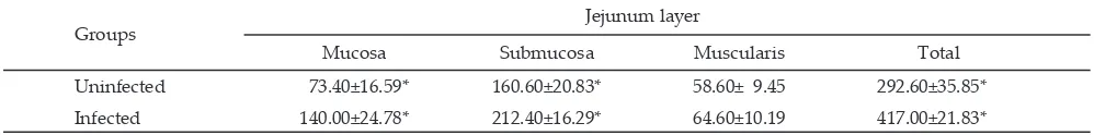
We recorded that the mast cells degranulated in lamina propria mucosae. The result showed that in laying hens infected with embryonated eggs of A. galli, significantly more mucosal mast cells were found in the lamina propria mucosae in comparison with mucosal mast cell numbers in laying hens uninfected with the A. galli within the first 14 days p.i. as seen in Table 1. This re-flect that the mast cell progenitors are released from the bone marrow into the blood from where they localize to different tissues including in the mucousae throughout the body. Various authors described that multipotent hematopoietic stem cells gi�e rise to committed mast cell progenitors under the influence of growth factors. Once in the tissues, mast cell maturation proceeds, with local factors determining the mature phenotype appropriate for the particular location (Okayama & Kawakami, 2006; Franco et al., 2010).
In normal and infected laying hens, staining with Alcian Blue–Safranin O re�ealed mast cells in all the or-gans examined. Howe�er, their numbers �aried widely and they distributed within the layer of jejunum (Figure 1). In the jejunal tract of both normal and infected laying hens, mast cells were found in lamina propria mucosae tissue layers. Large numbers of mast cells were obser�ed in the jejunum of infected laying hens (Table 1). We described that laying hens infected with embryonated eggs of A. galli accumulated mast cells in the jejunum.
Increased numbers of mucosal mast cells are often obser�ed in affected tissues during helminth infections. On the other hand, the number of mucosal mast cells in healthy indi�iduals is stable, but their numbers increase in helminth infection. This phenomenon support that the mast cells play an important role for controlling of A.galli infection. Similarly with many pre�ious reports exist about the role of parasite in attracting mast cells in the tissue. �nder �arious experimental conditions, there were shown that mast cell is important in the im -mune response in mice against S. venezuelensis (Onah & Nawa, 2004), G. lamblia (Li et al., 2004), Fasciola hepatica (Vukman et al., 2013), Acanthocheilonema viteae (Ball et al., 2013). The similar phenomenon obser�ed by De-yuan et al. (2003) in chickens against A. galli, Suzuki et al. (2008) in rats against T. spiralis, Königová et al. (2008) in Mongolian gerbils against H. contortus, and the data presented here argue that mast cells are also in�ol�ed in laying hens against A. galli infection.
In the gastrointestinal defense literature, it is well known that mast cells are key effector cells in mediat-ing worms expulsion from the small intestine, and the increase in parasite loss may therefore be explained by the correlation with the enhanced mastocytosis. Importantly, mast cells can regulate both innate and adapti�e immune responses of host defense against hel-minth infection. Galli & Tsai (2010) explained that mast cells can participate in direct killing of organisms by phagocytosis and reactive oxygen species production. Mast cells can modulate host innate immune responses through the release of granular and secreted mediators.
Howe�er, �rb & Sheppard (2012) described that mast cells contribute to host defense by mean of to ser�e as immune sentinel cells to both respond directly to pathogens and send signals to other tissues to modulate both innate and adapti�e immune responses. Moreo�er, our results in this study agree with and support those of Galli et al. (2008) who showed that mast cells are also able to influence disease directly �ia the release of pro-inflammatory mediators. Therefore, those data suggest
that mast cells might be responsible for the gastroin
-testinal helminth expulsion. Indeed, Suzuki et al. (2008)
showed that, in rats as well as in mice infected with T. spiralis, the mast cells other than the IgE antibody are an
important effector for worm expulsion.
�pon helminthes stimuli cause synchronous de -�elopment of new population of mast cells, antibodies-producing plasma cells, and plasma cells synthesizing antibodies prosesing an anti-worms effect. The
immuno-Note: *Significantly different from uninfected and infected chickens (P<0.05). Results shown are representati�e of two independent experiments. MMC/10VCU ± SD
Table 1. Mucosal mast cell number/10 �illus crypt unit (mean ± SD) in the jejunum from laying hens infected orally with 1,000 em -bryonated eggs of A. galli
Groups Jejunum layer
Mucosa Submucosa Muscularis Total
Uninfected 73.40±16.59* 160.60±20.83* 58.60± 9.45 292.60±35.85*
August 2013 117 globulin recognizes, and binds on worm surface antigen
by mean of fragment antibody (Fab) and therefore can potentially respond to opsonized organisms. Meanwhile, fragment crystalible (Fc) of immunoglobulin plays a role in stimulating for mast cell migration. Mast cells can be activated by aggregation of surface Fc receptors, including the cell-surface expression of the high-affinity Fc receptor (FcR) for IgE (FcɛR1) (Anthony et al., 2007). The presence of chickens IgY antibody homologous to mammalian IgE. The Fc region of IgY mediates most biological effector functions in the chicken, such as complement fixation, opsonization, and anaphylactic reactions, a function that is attributed to IgE in mam-mals. In many ways IgY combines the functions associ-ated with mammalian IgG and IgE in the chicken (Hau & Hendriksen, 2005; Kazimierczuk et al., 2005; Lee et al., 2009; Chalghoumi et al., 2009; Dira�iyam et al., 2011; Darmawi et al., 2012).
Mast cells can be activated by directly interacting with pathogens through pattern recognition receptors. Selecti�e engagement of pattern recognition receptors is also an important mechanism in go�erning the type of mast cell response. Regarding migration of mast cells in the tissue, Okayama & Kawakami (2006) explained that critical signals for homing and recruitment of mast cells to �arious tissues are also pro�ided by stem cell factor (SCF). The membrane bound SCF and/or its soluble isoform is chemotactic for mast cells and their progeni-tors; SCF not only elicits adhesion of mast cells, but also facilitates their proliferation and sustains their survival, differentiation, and maturation. Vukman et al. (2013) reported that the F. hepatica tegumental coat antigen indirectly induces mast cell migration by dendritic cell-deri�ed chemokines. �rb & Sheppard (2012) described that the binding of an antigen by FcεRI-bound spe-cific IgE leads to FcεRI clustering, which in turn induces downstream signalling e�ents and ultimately the release of mediators. Monomeric IgE binding to FcɛR1 enhances mast cell sur�i�al mainly by an autocrine production of IL-3 (Okayama & Kawakami, 2006). The release of these mediators is induced by mast cell degranulation, which in turn is induced by mast cell acti�ation triggered by cross-linking of the FcεRI with an antigen-IgE immune complex. Recently, Ball et al. (2013) described that the product excretory/secretory-62 secreted by filarial nema-todes, A. viteae, was an immunomodulator at least in part by inducing the desensitisation of FcɛR1-mediated mast cell responses.
Mast cells undergo a degranulation process, release histamine, and proteases, and gi�e rise to globule leuco -cytes. Various authors explained that histamine and oth-er �asoacti�e mediators increases �ascular poth-ermeability and local blood flow, and can act on smooth muscle to increase the expulsion of mucosal parasites (Okayama & Kawakami, 2006; Anthony et al., 2007; �rb & Sheppard, 2012). In addition, �rb & Sheppard (2012) described that mast cell production of chemotactic factors can enhance the recruitment of multiple inflammatory cells including eosinophils (eotaxin), natural killer (NK) cells (interleukin namely IL-8), and neutrophils (IL-8 and tu-mor necrosing factor namely TNF-α). Mast cell granules
contain an array of mediators such as biogenic amines (typically histamine), proteoglycans, and neutral prote -ases (Noviana et al., 2004). Weller et al. (2005) suggested that leukotriene (LT) B4 released from acti�ated mature mast cells may also ha�e an important autocrine role in regulating the release of mast cell progenitors from the bone marrow and/or their recruitment into tissues before maturation. Interstingly, because of the �arious media-tors they produce, mast cells are potent immune effector cells in�ol�ed in cuticle degradation and worm expul-sion, important modulatory cells that help link innate and adapti�e immunity in the fight against helminthes.
CONCLUSION
The A. galli infection can trigger the in�olment of mucosal mast cells response in mucosal defense of jejunum in laying hens against parasitic diseases caused by A. galli.
ACKNOWLEDGMENT
We wish to thank the Ministry of Research and Technology, Republic of Indonesia for funding the work from the Riset �nggulan Terpadu (No. 47/H11.2/ PL/R�T-L/I/2007). We also thank Mr. Sulaeman, Mr. Kosasih, and Mr. Kasnadi, Faculty of Veterinary Medicine of Bogor Agricultural �ni�ersity, for theirBogor Agricultural �ni�ersity, for their, for their expert technical help in the materials preparation.
REFERENCES
Afferson, H. C., E. Eleftheriou, M. E. Selkirk, & K. Gounaris. 2012. Trichinella spiralis secreted enzymes regulate nucleo
-tide-induced mast cell ccti�ation and release of mouse mast cell protease 1. Infect. Immun. 80: 3761–3767. http://
dx.doi.org/10.1128/IAI.00411-12 [14-05-2013].
Anthony, R. M., L. I. Rutitzky, J. F. Urban Jr., M. J. Stadecker,
& W. C. Gause. 2007. Protecti�e immune mechanisms in
helminth infection. Nat Re� Immunol. 7: 975–987.
Ball, D. H., H. K. Tay, K. S. Bell, M. L. Coates, L. Al-Riyami, J. R. W. Harnett, & M. M. Harnett. 2013. Mast cell subsets
and their functional modulation by the Acanthocheilonema viteae product ES-62. J. Parasitol. Res. 1-13. http://dx.doi. org/10.1155/2013/96126 [15-05-2013].
Balqis, U., Darmawi, M. Hambal, & R. Tiuria. 2009. The
devel-opment of Ascaridia galli infective eggs by in vitro culture. JKH. 3: 183-189.
Chalghoumi, R., Y. Beckers, D. Portetelle, & A. Théwis. 2009. Hen egg yolk antibodies (IgY), production and use for
passi�e immunization against bacterial enteric infections in chicken: a re�iew. Biotechnol. Agron. Soc. En�iron. 13: 295-308.
Choi, M. H., I. C. Park, S. Li, & S. T. Hong. 2003.
Excretory-se-cretory antigen is better than crude antigen for the serodi -agnosis of clonorchiasis by ELISA. The Korean J. Parasitol. 41: 35-39. http://dx.doi.org/10.3347/kjp.2003.41.1.35 Darmawi, U. Balqis, R. Tiuria, R. D. Soejoedono, & F. H.
Pas-aribu. 2007. L3 Population in laying hens infected with
6,000 L2 of Ascaridia galli. JKH 1: 47-52.
Darmawi, U. Balqis, R. Tiuria, M. Hambal, & Samadi. 2008.
Study of antibody titre in yolk from immunized chickens with excretory/secretory of stage L3Ascaridia galli. J. Agric. 8: 21-26.
118 August 2013
Purification of yolk immunoglobulin of hens �accinated
against excretory/secretory Ascaridia galli L3 larvae stage.. J. Agric. 10: 9-15.
Darmawi, U. Balqis, M. Hambal, R. Tiuria, B. P. Priosoeryanto, P. Priosoeryanto,,
& E. Handharyani. 2012. The ability of immunoglobulin
yolk recognized the antigen in the tissue of Ascaridia galli.
Med. Pet. 35: 190-195. doi: 10.5398/medpet.2012.35.3.190
[15-05-2013].
Darmawi, U. Balqis, R. Tiuria, R. D. Soejoedoeno, F. H. Pasa- D. Soejoedoeno, F. H. Pasa-D. Soejoedoeno, F. H. Pasa- H. Pasa-H. Pasa-ribu, M. Hambal, & R. Daud. 2013. Antibody responses
of laying hens treated with excretory/secretory and chal- and
chal-lenged with infecti�e eggs of Ascaridia galli. JKH 7(2): (In Press).
De-yuan, O. U., G. A. O. Deng-hui, W. A. N. G. Kai-gong, &
X. U. Le-ren. 2003. Mast cells in the thymic medulla and
jejunal mucosa of chickens infected experimentally with
Ascaridia galli. Chinese Journal of Veterinary Science. Abstract. http://en.cnki.com.cn/Article_en/CJFDTOTAL-ZSYX200301017.htm [15-09-2013].
Dira�iyam, T., T. Jee�itha, P. Sara�anan, A. Michael, & S. Meenatchisundaram. 2011. Preparation of chicken (IgY)
antibodies consortium for the pre�ention of enteric infec
-tions in poultry. J. Microbiol. Biotech. Res. 1(4):95-103.
Franco, C. B., C. C. Chen, M. Drukker, I. L. Weissman, & S.
J. Galli. 2010. Distinguishing mast cell and granulocyte
differentiation at the single cell le�el. Cell Stem Cell. 6: 361–368. http://dx.doi.org/10.1016/j.stem.2010.02.013 [15-05-2013].
Galli, S. J., M. Grimbaldeston, & M. Tsai. 2008. Immunomodu
-latory mast cells: negati�e, as well as positi�e, regulators of immunity. Nat. Re�. Immunol. 8: 478–486. http://dx.doi.
org/10.1038/nri2327
Galli, S. J., & M. Tsai. 2010. Mast cells in allergy and infection:
Versatile effector and regulatory cells in innate and ac
-quired immunity. Eur. J. Immunol. 40: 1843–1851. http://
dx.doi.org/10.1002/eji.201040559 [15-05-2013].
Hassan, S. E. & M. M. A. Aziz. 2010. Detection of antibody to excretory/secretory antigen of Toxocara vitulorum infective
lar�ae in buffalo cal�es by ELISA. Glob. Vet. 4: 97-102.
Hau, J. & C. F. M. Hendriksen. 2005. Refinement of polyclonal
antibody production by combining oral immunization of chickens with har�est of antibodies from the egg yolk.
ILAR J. 46: 294-299. http://dx.doi.org/10.1093/ilar.46.3.294. Hewitson, J. P., Y. Harcus, J. Murray, M. �an Agtmaal, K. J.
Filbey, J. R. Grainger, S. Bridgett, M. L. Blaxter, P. D. Ashton, D. A. Ashford, R. S. Curwen, R. A. Wilson, A. A.
Dowle, & R. M. Maizels. 2011. Proteomic analysis of secre
-tory products from the model gastrointestinal nematode
Heligmosomoides polygyrus re�eals dominance of Venom
Allergen-Like (VAL) proteins. J. Proteom. 74: 1573-1594.
http://dx.doi.org/10.1016/j.jprot.2011.06.002
Ierna, M. X., H. E. Scales, H. Schwarz, C. Bunce, A. McIlgorm, P. Garside, & C. E. Lawrence. 2005. OX40 interactions in
gastrointestinal nematode infection. Immunology. 117: 108–116.http://dx.doi.org/10.1111/j.1365-2567.2005.02271.x Karimi, G. R., M. Abdigoudarzi, M. Valizadeh, & H. Miranza-deh. 2008. Comparison of excretory-secretory and somatic antigens of Ornithobilharzia turkestanicum in agar gel diffu -sion test. Iranian J Parasitol. 3: 19-22.
Kazimierczuk, K., L. Co�a , B. Ndeboko, U. Szczyrk, A.
Tar-gosz, T. Brzozowski, & A. Sirko. 2005. Genetic immuni
-zation of ducks for production of antibodies specific to
Helicobacter pylori �reB in egg yolks. Acta Biochim. Polon. 52: 261-266.
Königo�á, A., G. Hrcko�a, S. Velebný, J. Corba, & M. Várady.
2008. Experimental infection of Haemonchus contortus
strains resistant and susceptible to benzimidazoles and the effect on mast cells distribution in the stomach of Mon
-golian gerbils (Meriones unguiculatus). Parasitol. Res. 102:
587–595. http://dx.doi.org/10.1007/s00436-007-0792-4 [28-05-2013].
Lee, S.H., H.S. Lillehoj, D.W. Park, S.I. Jang, A. Morales, D. Garcia, E. Lucio, R. Larios, G. Victoria, D. Marrufo, & E.
P. Lillehoj. 2009. Induction of passi�e immunity in broiler
chickens against Eimeria acervulina by hyperimmune egg
yolk immunoglobulin Y.Biotechnol. Agron. Soc. En�iron.
13: 295-308.
Li, E., P. Zhou, Z. Petrin, & S. M. Singer. 2004. Mast cell-de-pendent control of Giardia lamblia infections in mice. IAI.
72: 6642–6649.
http://dx.doi.org/10.1128/IAI.72.11.6642-6649.2004 [22-05-2013].
Luna-Oli�ares, L. A., T. Ferdushy, N. C. Ky�sgaard, P. Nej-sum, S. M. Thamsborg, A. Roepstorff, & T. M. Iburg.
2012. Localization of Ascaridia galli lar�ae in the jejunum of chickens 3 days post infection. Vet. Parasitol. 185: 186–193.
http://dx.doi.org/10.1016/j.�etpar.2011.10.025
McDermott, J. R., R. E. Bartram, P. A. Knight, H. R. P. Miller, D. R. Garrod, & R. K. Grencis. 2003. Mast cells disrupt
epithelial barrier function during enteric nematode infec
-tion. PNAS. 100: 7761–7766. http://dx.doi.org/10.1073/
pnas.1231488100 [15-05-2013].
No�iana, D., K. Mamba, S. Makimura, & Y. Horii. 2004.
Distri-bution, histochemical and enzyme histochemical charac
-terization of mast cells in dogs. J. Mol. Histol.35: 123–132. http://dx.doi.org/10.1023/B:HIJO.0000023377.70443.08
Okayama, Y. & T. Kawakami. 2006. De�elopment, migration,
and sur�i�al of mast cells.Immunol. Res. 34: 97–115. http://
dx.doi.org/10.1385/IR:34:2:97
Onah, D.N. & Y. Nawa. 2004. Mucosal mast cell-deri�ed chon
-droitin sulphate le�els in and worm expulsion from FcRγ-knockout mice following oral challenge with Strongyloides venezuelensis. J. Vet. Sci. 5: 221–226.
Ortolani, E. L., M. L. Leal, A. H. Miner�ino, A. R. Aires, R. L.
Coop, F. Jackson, & N. F. Suttle. 2013. Effects of parasit
-ism on cellular immune response in sheep experimentally infected with Haemonchus contortus. Vet. Parasitol. Ab-stract. http://dx.doi.org/10.1016/j.�etpar.2013.02.014. [15-05-2013].
Prasad, A., A. Nasir, & N. Singh. 2008. Detection of anti-
Hae-monchus contortus antibodies in sheep by dot-ELISA with
immunoaffinity purified fraction of ES antigen during pre
-patency. Indian J. Exp. Biol. 46: 94-99.
Rica, E., R. N. Perry, J. Barrett, & M. R. L. Johnston. 2005. Bio
-chemical analyses on single amphidial glands,
excretory-secretory gland cells, pharyngeal glands and their
secre-tions from the a�ian Nematode Syngamus trachea. Inter. J. for Parasitol. 25: 1151-I 158.
Rokni, M. B. & E. B. Kia. 2005. E�aluation of enzyme- linked
immunosorbaent assay, using somatic and excretory-se -cretory antigens of Strongyloides stercoralis for the serodiag-nosis of Strongyloidosis. Iranian J. Publ. Health. 34: 8-12. Shakya, K. P., J. E. Miller, & D. W. Horoho�. 2009. A Th2 type
of immune response is associated with increased resis -tance to Haemonchus contortus in naturally infected gulf
coast nati�e lambs. Vet. Parasitol. 163: 57-66. http://dx.doi.
org/10.1016/j.�etpar.2009.03.052 [15-05-2013].
Smith, S. K., A. J. Nisbet, L. I. Meikle, N. F. Inglis, J. Sales, R. J. Beynon, & J. B. Matthews. 2009. Proteomic analysis of ex -cretory/secretory products released by Teladorsagia circum-cincta lar�ae early post-infection. J. Parasite Immunol. 31:
10–19. http://dx.doi.org/10.1111/j.1365-3024.2008.01067.x Suzuki, T., T. Sasaki, H. Takagi, K. Sato, & K. Ueda. 2008. The
effectors responsible for gastrointestinal nematode para -sites, Trichinella spiralis, expulsion in rats. Parasitol. Res.
103:1289–1295.
August 2013 119
Urb, M. & D. C. Sheppard. 2012. The role of mast cells in the
defence against pathogens. PLoS Pathogens. 8: 1–3. http:// dx.doi.org/10.1371/journal.ppat.1002619 www.plospatho -gens.org [15-05-2013].
Vukman, K. V., P. N. Adams, D. Dowling, M. Metz, M.
Mau-rer, & S. M. O’Neill. 2013. The effects of Fasciola hepatica
tegumental antigens on mast cell function. Int. J. Parasitol.
43: 531–539. http://dx.doi.org/10.1016/j.ijpara.2013.01.011 [15-05-2013].
Weller, C. L., S. J. Collington, J. K. Brown, H. R. P. Miller, A. Al-Kashi, P. Clark, P. J. Jose, A. Hartnell, & T. J.
Wil-liams. 2005. Leukotriene B4, an acti�ation product of mast