Corresponding author: [email protected]
Assessing downgrading of esophageal
adenocarcinoma after neoadjuvant
chemotherapy: a case report
Indrasto Histopaedianto1, Adeodatus Yuda Handaya2*,Hendro Wartatmo2
1Trainee in Digestive Surgery, 2Digestive Surgery Division, Department of Surgery, Faculty of Medicine/Dr. Sardjito General Hospital, Universitas Gadjah Mada Yogyakarta, Indonesia
DOI: http://dx.doi.org/10.19106/JMedSci004901201706
ABSTRACT
Esophageal carcinoma, squamous cell carcinoma, and adenocarcinoma, are a deadly
serious malignancy. The delay in diagnosis due to the lack of speciic symptoms in the
early stages of the disease and the nature of esophageal cancer is very aggressive lead to a poor prognosis with a survival rate of small. Current management of esophageal cancer is recommended multimodal approach in the form of neoadjuvant chemotherapy or combined radiochemotherapy (CRT) and with surgery. In this article, multimodality treatment using chemotherapy and surgery on esophageal adenocarcinoma was reported. A 52-year-old male presented with a total obstruction of the esophagus and was planned to perform temporary gastrostomy for chemotherapy preparation. Gastrostomy found a
solid and ixed tumor located in the gastroesophageal junction, with the size of 7 x 6 x
5 cm3. He underwent a chemotherapy using the regimen of 8 cycles paclitaxel without radiotherapy due to the long queue of radiotherapy schedule. After chemotherapy, we re-evaluated and planned to perform surgical removal of the tumor. During surgery, we found
a total obstruction along with ibrosis of the esophagus but found no tumor/mass. We
performed partial esophagectomy and end to side anastomosis using anastomosis end to side using the CDH25 circular stapler. Surgery was carried out by the thoracoabdominal approach. The patient was discharged on day 12 in a stable condition and was undergoing
soft diet. We planned to evaluate the patient using Carcinoembryonic Antigen (CEA) test
and Positron Emission Tomography (PET) scan. In conclusion, multimodality treatment using chemotherapy and surgery on a case of one-third distal esophageal adenocarcinoma provides good results.
ABSTRAK
Karsinoma esofagus, baik karsinoma sel skuamosa, atau adenokarsinoma merupakan keganasan yang serius dan mematikan. Lambatnya diagnosis akibat kurangnya gejala
spesiik pada stadium awal penyakit dan sifat kanker esofagus yang sangat agresif
INTRODUCTION
Esophageal carcinoma and squamous cell carcinoma as well as adenocarcinoma are a serious malignancy with a poor prognosis and possibility of lethal in most cases. Esophageal carcinoma affects more than 450,000 people worldwide with a rapid incidence.1 Although
the incidence is relatively rare when compared to other cancers, according to the National Cancer Institute, in 2012, in the United States about 35,781 patients with esophageal cancer were estimated and in 2015 about 16.980 new cases with 15,590 deaths were reported.2
Currently, esophageal cancer is the number 6 leading cause of cancer death worldwide because its aggressivity and poor survival rate. Although it has been many developments in the diagnosis and management, the 5-year survival rate for patients diagnosed with esophageal cancer only range between 15% - 20%.1,3
Esophageal cancer generally consists of two forms, squamous cell carcinoma, and adenocarcinoma.1 Squamous cell carcinoma
is the most common histologic type (95%). The incidence of esophageal squamous cell carcinoma increases with age with a peak
kemoterapi. Saat operasi gastrostomi teraba tumor keras di gastroesophageal junction, berukuran 7 x 6 x 5 cm3, padat teriksir. Kemudian dilakukan kemoterapi menggunakan paslitaksel sebanyak 8 siklus tanpa radioterapi karena antrian jadwal terlalu panjang. Setelah kemoterapi pasien di re-evaluasi dan direncanakan operasi pengangkatan tumor. Pada
operasi ditemukan obstruksi total esofagus dengan jaringan ibrotik sepanjang esofagus,
namun tidak ditemukan masa tumor. Selanjutnya dilakukan partial esophagectomy
dan anastomosis end to side menggunakan stapler CDH25. Operasi dilakukan dengan pendekatan thoracoabdominal. Pasien dipulangkan hari ke 12, kondisi baik dan sudah diet lunak. Direncanakan untuk evaluasi dengan pemeriksaan Carcinoembryonic Antigen
(CEA) dan Positron Emission Tomography (PET) scan. Dapat disimpulkan, penatalaksanaan multimodalitas dengan kemoterapi dan pembedahan pada kasus adenokarsinoma esofagus 1/3 distal memberikan hasil baik.
Keywords: esophageal adenocarcinoma – chemotherapy – paclitaxel – esophagectomy - gastrectomy
squamous cell carcinoma increased 3-fold in dark-skinned individuals, whereas the type of adenocarcinoma is more common in fair-skinned individuals.2,4
Increased incidence of esophageal cancer is associated with risk factors for these cancers. Squamous cell carcinoma of the esophagus is associated with smoking and alcohol consumption, genetic factors such as tylosis, Plummer-Vinson syndrome, and environmental exposure such as ingestion of alkali and radiotherapy. Esophageal adenocarcinoma is often grown in the distal esophagus and is associated with esophagitis, Barrett’s esophagus, a metaplasia occurring in the distal gastroesophageal junction because of chronic irritation from gastroesophageal
relux disease (GERD). Obesity, whether related to their GERD or due to consumption
of western-style diet is poor in vitamins,
vegetables, fruit, ish, and poultry; is also a
risk factor for esophageal adenocarcinoma.1,3,5
Esophageal cancer is often called the “silent cancer”. Most patients are diagnosed later because of the lack of symptoms in the early stages of the disease.6 Dysphagia and
weight loss occurs in about 90% cases of
odynophagia, hoarseness, cough, chest pain, melena, and residual symptoms (sequelae) of the spread of disease both locally advanced and distant spread. Approximately 75% of patients have lymph node involvement at the time of diagnosis.5,7
Esophagectomy is a primary management of early-stage esophageal cancer although
its speciic role in supericial cancer is still
being debated ever since the development of endoscopic mucosal treatment. Nowadays, the
scientiic evidence is enough to recommend
the multimodal treatment of locally advanced cancers using the combination of neoadjuvant
chemotherapy or chemoradiotherapy (CRT)
followed by surgery. Some clinicians just
use deinitive CRT to avoid mortality due to
surgery.8
We report a case of adenocarcinoma of the esophagus treated with multimodal approach of chemotherapy followed by surgery.
CASE REPORT
A-52-years-old male was consulted
to the Department of Digestive Surgery of Dr. Sardjito General Hospital as an already
diagnosed case of esophageal tumor. He
was consulted to the department at 9 March 2015. The patient came with dysphagia as a chief complaint accompanied with nausea, vomiting, and weight loss. The onset of the
symptoms started from December 2014,
and gradually became more severe. At the beginning of the symptoms, he was capable of ingesting solid diet, but at the time he was consulted, he was only capable of ingesting
liquid diet. During the illness, he lost his
weight as much as 20 kg, which was from 72 to 52 kg. Physical examination did not
ind any abnormality. Routine laboratory test
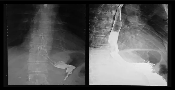
found hemoglobin level was 9.7 mg/dL. The patient had been undergoing barium meal and endoscopic examination at prior hospital. Barium meal test revealed a suspected distal esophageal tumor with a malignant
tendency (FIGURE 1). Endoscopy revealed
an esophageal tumor at 35 cm depth. Biopsy on the mass was conducted during endoscopy
procedure. Histopathology analysis result was
moderately-differentiated adenocarcinoma of the esophagus.
The patient was diagnosed as distal esophageal tumor and elected to undergo gastrostomy and exploration laparotomy on 13
March 2015. During exploration laparotomy, ixed, 7 x 6 x 5 cm3 esophagogastric junction mass was found. Nasogastric tube (NGT)
could not pass through the tumor. Temporary gastrostomy was done and it was concluded that the esophageal tumor was unresectable and planned to undergo post-operative
chemotherapy. He was consulted to
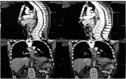
Hemato-oncology department to receive chemotherapy. Eight cycles of paclitaxel were administered and completed at 2 November 2015. Serial CT scan evaluation was done 3 times. First
CT scan (29 May 2015) revealed narrowing of esophageal lumen, total obstruction on the level of 7th thoracic vertebrae, and no periesophageal iniltrating mass (FIGURE 2).
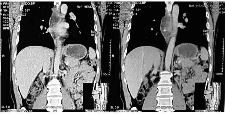
Second CT scan (17 September 2015) showed intraluminal mass causing total obstruction below the level of 9th thoracic vertebrae, and no periseophageal iniltrating mass (FIGURE
3). Third CT scan (9 November 2015) revealed distal esophageal mass with 1.4 x 1.2 x 3.4 cm3 dimension, smaller than prior dimension (FIGURE 4). Esophagography (19 November
2015) revealed stenosis of third distal of esophagus.
FIGURE 3. Second CT scan (17 September 2015), CT coronal view showed intraluminal tumor of esophagus
causing total obstruction on the level of 9th thoracic vertebra.
FIGURE 4. Third CT scan (9 November 2015), CT coronal view showed decreasing of the intraluminal tumor
dimension.
On 17 December 2015, he underwent
laparotomy, thoracotomy, intraoperative endoscopy, resection and anastomosis of esophagus. Tumor mass was not found during
the procedure, there was ibrotic tissue around
esophagus and esophagogastric junction. Intraoperative endoscopy found stenosis of esophagus. Partial esophagectomy and gastrectomy was done, followed by
Patient clinical appearance following the procedure was better than before.
Post-operative anatomic pathology evaluation revealed adenocarcinoma with clean border. PET scan and tumor marker assay were planned as follow-up plan. Multimodality approach (chemotherapy and surgery) to management esophageal cancer gave a good outcome in this case.
DISCUSSION
Tumor or esophageal cancer has a high mortality rate and also known as “silent
cancer” due to unspeciic symptoms of an
early stage of cancer and results in delayed diagnosis. Esophageal cancer is often to be
found in 50-60 years old patient. Dysphagia
and weight loss are the most reported symptoms in as much as 90% of the total case. The dysphagia is reported to be progressive,
from dificult to ingest solid diet to liquid diet due to obstruction caused by cancer. Other
symptoms are odynophagia, hoarseness, coughing, chest pain, and melena. Lymph node involvement is found in more than 75% cases. Physical examination in esophageal cancer shows no abnormality, except cervical and supraclavicular lymph nodes enlargement.5,7
Radiologic examination may support the
esophageal cancer diagnosis. Barium contrast
may show illing defect to differ obstruction
related dysphagia or neurogenic related dysphagia. Esophagogastroduodenoscopy
(EGD) is the easiest way to observe dimension,
circumferential and linear spread of the tumor, and may allow biopsy of the tumor.
Endoscopic ultrasound (EUS) is a deinitive
method to establish tumor stage. According to TNM tumor staging of the American Joint Commission on Cancer (AJCC), tumor stage is determined based on invasion depth (T) and lymph node involvement (N). Computed tomography (CT) scan with contrast of chest
metastasis compared to luorodeoxyglucose positron emission tomography (FDG-PET)
scan.
According to Guidelines for Clinical
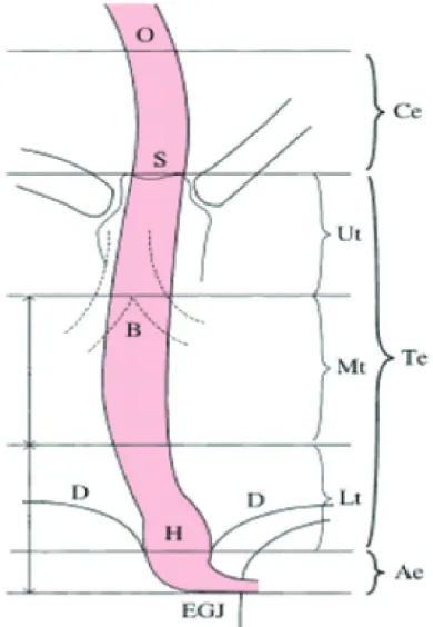
and Pathologic Studies on carcinoma of the Esophagus on 2007, the esophagus is divided into 3: proximal, medial, and distal. Proximal part is also known as cervical esophagus (Ce), a short segment located on the median line goes along to posterior of the membranous trachea and anterior of the vertebral column. It ends on the same level as manubrium sternum. Thoracic part (Te) goes down on the anterior of the vertebral column and thoracic aorta through esophageal hiatus of the diaphragm. Thoracic part is divided into: the upper thoracic esophagus (Ut), the middle thoracic esophagus (Mt) and lower thoracic esophagus (Lt). The distal part of the esophagus is also called abdominal esophagus (Ae) which is the shortest part. It goes down from the level of esophageal hiatus to gastric cardia on the level of 11th or 12th thoracic vertebra. Overall,
the esophagus is divided into 5 parts: Ce< Ut, Mt, Lt, and Ae which is related to esophageal
cancer predilection (FIGURE 5).10
Histologically, the esophagus consists
of 4 main layers: mucosal, submucosal, muscularis propia, and adventitia. There is no serous membrane in esophagus, which makes
cancer cell is easier to spread. Stratiied
epithelia of esophageal mucosal layer consist of 3 compartments: basal layer, supra basal layer (intermedium or prickle layer), and
supericial layer. Cells proliferate on basal
layer and differentiate when migrating from
suprabasal to supericial layer. Submucosal
FIGURE 5. Five parts of the esophagus. O: esophageal oriice; S: upper margin of the sternum; B: tracheal bifurcation; D: diaphragm; EGJ: esophagogastric junction; H: esophageal hi
-atus. (Guidelines for Clinical and Pathologic
Studies on Carcinoma of the Esophagus (9th edition), the Japanese Society for Esophageal
Diseases)
Histologically, esophageal cancer mainly divided into 2, squamous cell carcinoma and adenocarcinoma. Less than 1-2% is sarcoma or small cell carcinoma. Lymphoma, carcinoids, and melanoma is very rare.3
Natural history of squamous cell carcinoma and adenocarcinoma of esophagus is different. Squamous cell carcinoma is started
from inlammation of squamous epithelial
and progressively change to dysplasia and in situ cancer. Adenocarcinoma tends to grow at distal esophagus from columnar-lined metaplastic epithelium which is also known as
Barret’s esophagus. Gastro esophageal relux
disease (GERD) can damage esophageal
surface. Approximately 75% of all esophageal adenocarcinoma are found at distal esophagus and squamous cell carcinoma is commonly found between middle part and third distal esophagus.1,3
Otherwise, tumor which involves other
structures such as aorta, trachea, vertebra, and etc., is unresectable.11
Locally advanced tumor is treated using resection surgery since esophagectomy with radical lymphadenectomy is the best choice to achieve local control.8 Surgical resection must involve radical, complete, RO, en bloc
esophagectomy associated to an extended
two-ield lymphadenectomy.12
However, resection is associated with poor
survival rate, metastasis risk, or locoregional
recurrence post resection. On locoregional
tumor, post resection 5-year survival rate is 10-40%, with a risk of therapeutic failure due to far metastasis. Therefore, it is recommended to use multimodality management on locally advanced cancer which is neoadjuvant therapy
or combined chemoradiotherapy (CRT)
followed by resection. Some experts prefer
deinitive CRT to avoid surgical mortality risk. Radiotherapy only has 6% of 3-year
survival rate. Prior study shows chemotherapy for locally advanced esophageal cancer has
45%-75% of response rate. However, it has
high relapse rate with poor long-term survival rate.8,13
Chemotherapy without radiotherapy before surgery has some advantages such as improvement in dysphagia symptom, better survival rate, disease-free survival before surgery, better control of local tumor or downstaging tumor, better resectability, and chance to eradicate micrometastasis by lowering cancer cell dissemination.8,13
Most commonly used chemotherapy
regimen is cisplatin and 5-lorouracil infusion. Other regimens are bleomycin, vinblastine,
carboplatin, etoposide, and paclitaxel.11
Although chemotherapy has high advantage, its use is limited by toxicity risk, especially in elderly. The more chemotherapy regimen combination we use, the more toxicity risk increase.14,15
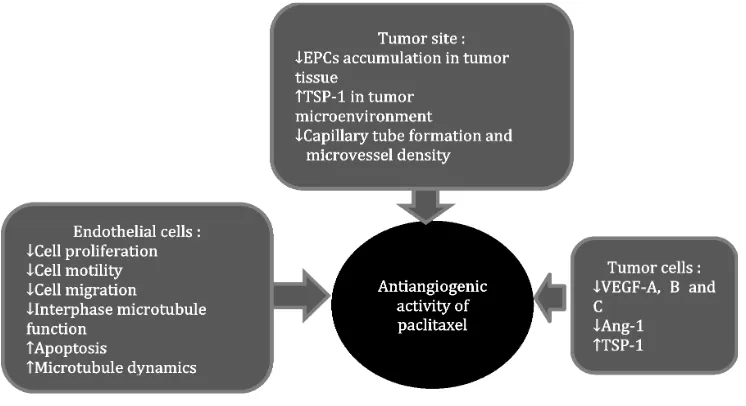
Paclitaxel is a taxane anticancer drug. It is commonly used as chemotherapeutic agent in malignancy. It is a microtubule stabilizer that selectively inhibit cell on mitosis phase (M) without interfering synthesis phase (S). it may cause cytotoxicity, depends on administration time and concentration which is radiation sensitizer.16 It also has antiangiogenic activity,
so that, it is a rational therapy to control cancer
progressivity (FIGURE 6).17
CONCLUSION
Multimodality management using chemotherapy and surgery gives a good outcome. Chemotherapy given prior to the surgery may decrease the dimension of the tumor, so that tumor resectability will increase. Post-operative anatomic pathology reveals adenocarcinoma with clean border. Post chemotherapy total stenosis is treated by partial esophagectomy and gastrectomy procedures. Esophago-gastric anastomosis
using stapler CDH25 shows a good outcome.
ACKNOWLEDGEMENT
We would like to thank the patient who has participated in this study.
REFERENCES
1. Zhang Y. Epidemiology of esophageal cancer. World J Gastroenterol 2013;19 (34): 5597-606. https://doi.org/10.3748/wjg.v19.i34.5598 2. Surveillance, Epidemiology, and End Result
Program Turning. SEER Stat Fact Sheet: Esophageal Cancer. Retrieved December 26, 2015. Available from URL: http://seer.cancer. gov/statfacts/html/esoph.html
3. Napier KJ, Scheerer M, Mison S. Esophageal cancer: a review of epidemiology, pathogenesis, staging workup and treatment modalities. World J Gastrointest Oncol. 2014; 6(5): 112-20. https://doi.org/10.4251/wjgo.v6.i5.112 4. Pennathur A, Gibson MK, Jobe BA, Luketich
JD. Oesophageal carcinoma. Lancet 2013; 381: 400-12. https://doi.org/10.1016/S0140-6736(12)60643-6
5. Lin SH, Chang JY. Esophageal cancer: diagnosis and management. Chin J Cancer 2010; 29(10):843-54. https://doi.org/10.5732/ cjc.010.10151
6. Tétreault M. Esophageal cancer: insights from mouse models. Cancer Growth and Metastasis.
2015; 8(S1): 37-46. https://doi.org/10.4137/ CGM.S21218
7. Cho JW, Choi SC, Jang JY, Shin SK, Choi KD, Lee JH, et al. Lymph node metastases in esophageal carcinoma: an endoscopist’s view. Clin Endosc 2014; 47:523-9. https://doi. org/10.5946/ce.2014.47.6.523
8. D’Journo XB, Thomas PA. Current management of esophageal cancer. J Thoracic Dis 2014; 6 (S2): S253-64.
9. Moore KL, Dalley AF, Agur AMR. Clinically Oriented Anatomy, 6th ed. Baltimore:
Lippincott William & Wilkins, 2010.
10. Takubo K. Pathology of the esophagus: an atlas and textbook, 2nd ed. Hongkong: Springer
Publishing, 2007.
11. Keditsu KK, Jiwnani S, Karimundackal G, Pramesh CS. Multimodality management of esophageal cancer. Indian J Surg Oncol 2013;4(2):96-104. https://doi.org/10.1007/ s13193-013-0216-0
12. Low DE. Evolution in surgical management of esophageal cancer. Dig Dis 2013; 31:21-9. https://doi.org/10.1159/000343650
13. Shah RD, Cassano AD, Neifeld JP. Neoadjuvant therapy for esophageal cancer. World J Gastrointest Oncol 2014;6(10):403-6. 14. Kothari N, Almhanna K. Current status of
novel agents in advanced gastroesophageal adenocarcinoma. J Gastrointest Oncol 2015;6(1):60-74.
15. Balducci L. Systemic treatment of gastric snd esophageal adenocarcinoma in elderly patients. J Gastrointest Oncol 2015;6(1):75-8.
16. Barbuti AN, Chen Z. Paclitaxel through the ages of anticancer therapy: exploring its role in chemoresistance and radiation therapy. Cancers 2015;7:2360-71. https://doi. org/10.3390/cancers7040897