Plant Science 157 (2000) 33 – 41
Lytic activity in pearl millet: its role in downy mildew disease
resistance
S. Umesha, M. Shylaja Dharmesh, H. Shekar Shetty *
Downy Mildew Research Laboratory,Department of Studies in Applied Botany,Seed Pathology and Biotechnology,Uni6ersity of Mysore, Manasagangotri,Mysore 570 006, India
Received 10 January 2000; received in revised form 14 March 2000; accepted 27 March 2000
Abstract
Sclerospora graminicola causes downy mildew disease in susceptible pearl millet. Molecular basis of downy mildew disease resistance has been studied. Coleoptile region has been shown earlier to be the most susceptible site for attack by the pathogen. Lytic activity is differentially expressed in the coleoptile region of 3-day-old pearl millet seedlings of resistant and susceptible cultivars. Significantly higher levels of lytic factors were measured in the coleoptile region of resistant cultivars (100%) than in that of susceptible cultivars (20%). Both constitutive and inducible lytic factors were observed in different resistant cultivars, and they were able to lyse the pathogen. The level of lytic activity correlated well with the degree of resistance as evaluated by field screening studies. The present study, therefore, proposes that lytic factors found in the coleoptile region of the pearl millet seedling, are responsible for the lysis of the pathogen in the resistant plant, and may therefore provide resistance to downy mildew disease. This study also provides a simple method to evaluate downy mildew resistance in pearl millet cultivars. © 2000 Elsevier Science Ireland Ltd. All rights reserved.
Keywords:Pearl millet; Downy mildew; Lytic factors; Resistance; Susceptibility;Sclerospora graminicola;Pennisetum glaucum
www.elsevier.com/locate/plantsci
1. Introduction
Pearl millet (Pennisetum glaucum (L.) R. Br.) is
grown for grain and forage over 26 million hectares in the tropical and subtropical areas of the world. Downy mildew disease of pearl millet
caused by an oomycetous biotrophic fungus Scle
-rospora graminicola (Sacc.) Shro¨ter, is the most
widespread and destructive disease of pearl millet in India and West Africa [1 – 4]. The disease is economically important, since it causes more than 60 – 70% loss of yield in susceptible hybrids [5,6]. Although downy mildew has been recognized as a potentially important disease of pearl millet since a long time, the disease continues to be a major threat, as a basic understanding of host – pathogen interactions, and of resistance and
sus-ceptible mechanisms is lacking. Biochemical
studies carried out by various investigators have revealed that certain enzymes have the ability to lyse the cell [7,8], and such enzymes may be in-volved in lysing the pathogen, thereby enabling the plant to become resistant.
Lytic factors are either proteinaceous or non-proteinaceous [8 – 10] small or large molecular weight biochemical components that has the abil-ity to act upon the chemical components of the cell wall, and thereby lyse the cell [11,12]. Such components are produced both by micro-organ-isms and plants [7,12,13]. Autolysis of fungal mycelia by the enzymes produced by themselves
such as proteases and/or chitinases and hydrolysis
of fungal cell walls by enzymes obtained from microorganisms has been well documented [14,15]. Further, lytic factors have also been reported to be responsible for exolysis followed by breaking down the walls of fungal pathogens [16,17].
Vari-* Corresponding author. Tel.: +91-821-515126; fax: + 91-821-411467.
E-mail address:[email protected] (H. Shekar Shetty).
ous examples of biological control have been re-lated to the activity of hydrolytic enzymes [8]. Lytic activity is also responsible for the cleavage of red blood cells, and hence the lysis is easy to monitor [18,19].
The present study attempts to demonstrate (i) the differential expression of lytic factors in resistant and susceptible cultivars of pearl millet;
(ii) their involvement in downy mildew
disease resistance and (iii) the potential use of a simple lytic assay for the evaluation of resistant cultivars.
2. Materials and methods
2.1. Collection of seeds
Pearl millet cultivars showing varying degrees of resistance and susceptibility to downy mildew dis-ease were obtained from the Project Co-ordinator, All India Co-ordinated Pearl Millet Improvement Project (AICPMIP), Pune, and the International Crops Research Institute for Semi-Arid Tropics (ICRISAT), Hyderabad, India. The seeds were sown in the downy mildew sick plot, which is
being maintained at the department, and
their reaction to the disease was tested and confi-rmed by adopting the procedure of Williams et al. [20].
2.2. Collection of downy mildew pathogen — S.
graminicola
Pearl millet leaves infected with S. graminicola
pathotype 1 were collected from the greenhouse of the Applied Botany Department, University of Mysore, India. Leaves of infected pearl millet plants, showing symptoms of downy mildew dis-ease, were collected in the evening, washed in
running tap water to remove pre-existing
sporulation, blot-dried and placed in a moist chamber for sporulation [21]. Fresh sporangia were collected in distilled water, and washed with 20 mM phosphate buffer saline (PBS), pH 7.4. Washings were discarded after centrifugation at 3000 rpm for 15 min. Fresh sporangia, free from other possible contaminants, were collected in PBS and were allowed to release zoospores. The
pathogen suspension, containing
sporangio-phores, sporangia and zoospores, was used for the studies.
2.3. Collection of infected and non-infected
seedlings
Seeds of both resistant and susceptible cultivars were plated on sterile wet blotters in Petri dishes, and germinated according to the standard blotter method [22].
Seeds were germinated at 2591°C, under near
ultraviolet light, with alternating periods of 12/12
h light and darkness. In order to collect infected seedlings, 2-day-old seedlings were dipped in a fresh suspension of pathogen zoospores (50 000
ml−1). After 24 h of infection, the seedlings were
used for further study. Coleoptile, root, and shoot portions were separated from infected and non-in-fected seedlings, and used for the study.
2.4. Preparation of host extracts for enzyme assay
Resistant and susceptible pearl millet seeds, root, shoot and coleoptile portions of infected and non-infected seedlings were homogenized in 1:2
(w/v) 20 mM sodium phosphate buffer, pH 7.4
containing 0.1% triton X-100. The homogenate was centrifuged at 1500 rpm for 5 min to remove the debris, and the supernatant was dialysed against 20 mM sodium phosphate buffer, pH 7.4 using 6000 – 12 000 cut-off dialysis tubing (Sigma Chemical Co., St. Louis, MO; USA), over night at 4°C, the dilution was noted down and used for enzyme assay.
2.5. Assay of lytic acti6ity
RBC lysis and pathogen lysis assays were car-ried out by spectrophotometric method [23,24]. Studies were conducted in quadruplicates and ex-periments were repeated in five different batch seeds, which were obtained during different har-vest time.
2.5.1. By using red blood cells
Fresh sheep blood was collected from a local slaughter house, in a bottle containing 4% sodium citrate solution. 50 ml of blood was centrifuged at 10 000 rpm for 15 min at 20°C. Cell pellet was washed four or five times in PBS, and suspended
in 10 ml of PBS. 50 ml of RBC suspension was
incubated with 25 and 50 ml of various resistant
seed extracts and with susceptible seed extracts at
S.Umesha et al./Plant Science157 (2000) 33 – 41 35
final reaction volume of 250 ml PBS. The samples
were incubated at 37°C for 30 min. Reaction mixtures were then centrifuged at 3000 rpm for 5
min. The supernatants were separated and 100 ml
of each were diluted to 2.5 ml in PBS; the quantity of red haemoglobin pigment released by RBC due to lysis was read at 415 nm, in a spectrophotome-ter (U-2000, Hitachi, Japan). The release of haemoglobin pigment by distilled water was con-sidered as positive control with 100% lytic activity. The relative absorbence was measured for all sam-ples, the relative percent lytic activity is calculated and presented.
2.5.2. Using S. graminicola zoospore suspension
Washed zoospores ofS. graminicola were tested
as potential substrates for lytic activity of resistant pearl millet, since lysis of the pathogen observed
previously [24]. S. graminicola zoospore
suspen-sions were incubated with host extracts at 37°C for 30 min. At the end of the incubation period, the reaction mixture was centrifuged at 1500 rpm for 10 min, to separate sedimented zoospore from the supernatant, which contained amino acids and sugars released from zoospores. The quantities of
amino acids and sugars released from the
zoospores were estimated, by means of spec-trophotometric method, and the phenol – sulphuric acid method respectively, and the results were compared. The quantitative release of amino acids and sugars were calculated using the calibration curves obtained from aromatic aminoacid at 280
nm (5 – 300mg/ml) andD-glucose (0 – 100mg/ml) by
phenol – sulphuric acid method at 490 nm respec-tively. The release of amino acids and sugars from zoospores by susceptible and resistant host ex-tracts indicate the comparative levels of lytic activ-ity in them.
2.6. Dose dependent lysis of RBC by resistant
seed extract
To establish the dose dependent nature of the host extract, equal amounts of RBCs were
incu-bated with 0 – 50 ml of resistant (SDN 503R) seed
extract for 30 min at 37°C, and the linearity in the release of haemoglobin pigment of RBC was mon-itored at 415 nm. The thermostability of lytic activity was examined by measuring lytic activities, after boiling at 100°C for 3 min.
2.7. Correlation between lytic acti6ity and degree
of resistance in the field
Infected and non-infected seedlings of various resistant and susceptible pearl millet seeds (Table 1) were assayed for the amount of lytic activity, as described above. Same cultivar seeds were sown in the downy mildew sick plot and the degree of resistance and susceptibility were determined fol-lowing the procedure of Williams et al. [20]. Seeds showing maximum lytic activity in the laboratory and maximum resistance in the sick plot were taken as 100, and the relative per cent lytic activity and resistance were calculated and compared.
2.8. Statistical analysis
Data on percentages were transformed to arc-sine and standard analysis of variance was carried out with transformed values. The means were compared for significance using Duncan’s new
Multiple Range Test (DMRT; P=0.005).
3. Results
3.1. Demonstration of lytic acti6ity in resistant
pearl millet
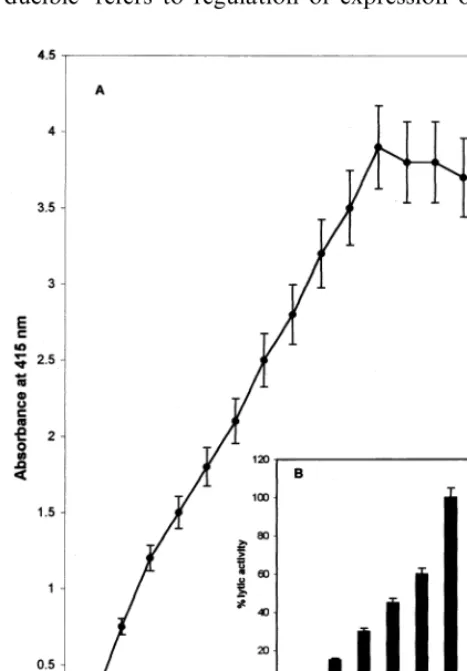
Table 1, Figs. 1 and 2 indicate the expression of lytic activity in resistant and susceptible pearl mil-let. Lytic activity was demonstrated by using its ability to lyse RBC (Fig. 1) and pathogen (Fig. 2) respectively. Dose dependent increase in the lysis of RBC with the increase in concentration of resistant extract suggested that the RBC-lysis as-say can be used to quantitate the level of activity and its ability to lyse RBC. RBC-lysis assay corre-lated well with the results of pathogen lysis assay. Further RBC-lysis assay is simpler, rapid and less time consuming. Hence it has been used in the present study.
S
.
Umesha
et
al
.
/
Plant
Science
157
(2000)
33
–
41
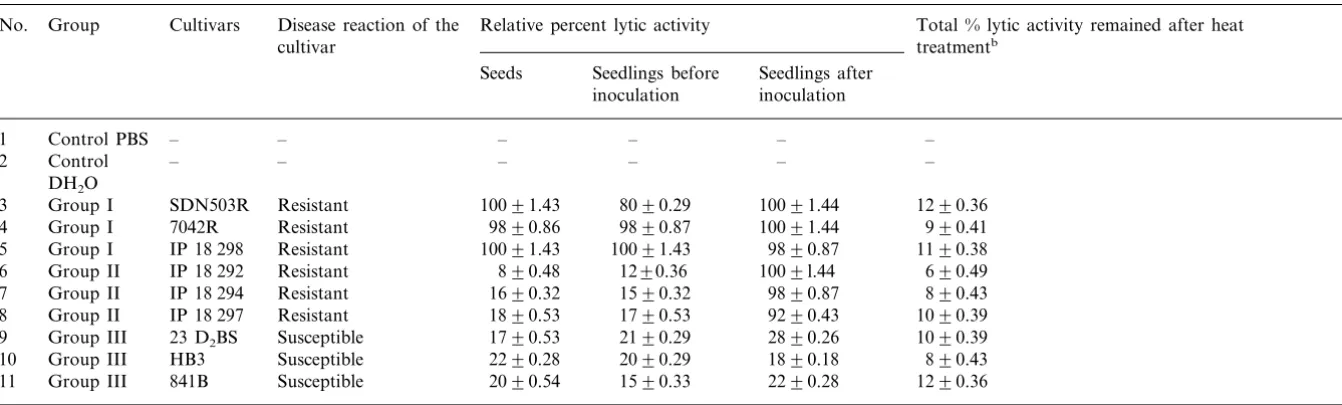
Table 1
Differential expression of lytic enzyme activity in various resistant and susceptible pearl millet seeds and seedlingsa
No. Group Cultivars Disease reaction of the Relative percent lytic activity Total % lytic activity remained after heat treatmentb
cultivar
Seedlings after Seeds Seedlings before
inoculation inoculation
– –
– –
– 1 Control PBS –
– – –
2 Control – – –
DH2O
10091.44 1290.36 10091.43
Resistant 8090.29
SDN503R Group I
3
10091.44 990.41
4 Group I 7042R Resistant 9890.86 9890.87
9890.87 1190.38 10091.43
5 Group I IP 18 298 Resistant 10091.43
1290.36
Group II IP 18 292 Resistant 890.48 1009l.44 690.49
6
1590.32
Group II IP 18 294 Resistant 1690.32 9890.87 890.43
7
9290.43 1090.39 1790.53
1890.53 8 Group II IP 18 297 Resistant
Group III 23 D2BS Susceptible 2190.29 2890.26 1090.39
9 1790.53
1890.18 890.43 2090.29
2290.28 Susceptible
10 Group III HB3
1590.33
Group III 841B Susceptible 2090.54 2290.28 1290.36
11
aVarious inoculated and uninoculated resistant and susceptible seedlings were analyzed for lytic enzyme activity. Appropriate controls were set up. The lytic activity was
tested at equal protein concentration level (10mg/ml) and the lysed product of RBC was determined at 415 nm. Distilled water lysed all RBCs taken in the assay. The maximum absorbance at 415 nm obtained by distilled water was taken as 100 and the relative per cent lysis was calculated for all other samples. The effect of thermal denaturation was also examined. Based on the level of expression of lytic activity before and after inoculated samples, the nature of expression is depicted as constitutive and inducible enzyme activity respectively. Values of quadruplicate samples in each group are expressed as S.E. of the mean according to the analysis of variance test.
S.Umesha et al./Plant Science157 (2000) 33 – 41 37
activity before and after inoculation. Such culti-vars expressing lytic activity constitutively are called constitutive resistant cultivars. On the other hand, the group II resistant (IP 18 292, IP 18 297) cultivars expressed basal levels of lytic activity, similar to group III susceptible cultivars. How-ever, there was a remarkable increase in lytic activity after inoculation, suggesting the induction of lytic activity by the pathogen. Such resistant cultivars, showing significant expression of lytic activity only after inoculation, are called inducible resistant cultivars. The term ‘constitutive’ and ‘in-ducible’ refers to regulation of expression of lytic
Fig. 2. Differential distribution of lytic activity in 3-day-old pearl millet seedlings. Three-day-old inoculated seedlings of resistant SDN 503R () and susceptible 23 D2BS () were
collected and coleoptile, shoot and root portions were sepa-rated and homogenised in 1.0 ml of 20 mM PBS, pH 7.4. The extracts were examined for the lytic activity. The total activity in a seedling (coleoptile+shoot+root) was taken as 100% and relative percent distribution of activity was calculated and compared between resistant and susceptible pearl millet seedlings. The lines on each bar represent S.E. when subjected to analysis of variance (DMRT; P=0.005)
Fig. 1. Resistant extract exhibited dose dependent lysis of red blood cells. (A) Various doses of resistant extracts (10 mg/ml protein concentration) were incubated with red blood cells (RBCs) under assay conditions. The release of the haemoglobin pigment from the lysed RBC was monitored at 415 nm. Dose dependent lysis was observed. The curve was saturated at \50 ml concentration of extract. (B). To under-stand the time dependent reactivity 25ml of resistant extract was added to RBC and incubated up to 60 min. Aliquots were drawn at various time intervals and extent of lysis was measured. Relative percent was calculated and compared to 100% lysis of RBC by water.
activity. Lytic enzyme activity of 80% reduction was noticed when extracts were boiled for 3 min in a boiling water bath. Results suggest that the lysing components are heat labile.
3.2. Lysis of the pathogen by resistant extract
The lysis of the pathogen was confirmed by measuring and quantitating the release of amino acids and carbohydrates by the pathogen. The protease and glycosidase enzymes, which are in-volved in degradation of proteins and
polysaccha-rides/glycoconjugates of the pathogen, released
Lytic activity is localized in the coleoptile region of 3-day-old resistant seedlings. In order to under-stand the physiological relevance of expression of lytic activity, root, shoot and coleoptile regions were separated from inoculated 3-day-old seedling of the resistant (SDN 503R) and susceptible
(23D2BS) cultivars.
Fig. 2 indicates that lytic activity in the coleop-tile tissue of resistant seedlings was 8 and 2.5 times as high as in the roots and shoots of resistant seedlings respectively. The lytic activity in the coleoptile tissue of the resistant seedlings (SDN 503R) was eight times greater than in the
coleop-tile tissue of the susceptible seedlings (23D2BS).
The data suggest that overwhelming increase of lytic activity in resistant cultivar to that of suscep-tible is significant, since coleoptile portion is the susceptible tissue for the entry of the pathogen. Although, two-fold increase in the shoot portion
Fig. 4. Correlation between the level of expression of lytic activity and degree of resistance in pearl millet. Different degree resistant and susceptible pearl millet genotypes: (1) 841 B, (2) 7042 (S)-1, (3) 23 D2BS, (4) HB3, (5) 7042(S), (6) 843
B, (7) 81B, (8) 7042R and (9) SDN 503R were assayed for lytic activity using RBC lysis assay. The same genotypes were tested for their resistance and susceptibility in the sick plot under field conditions. Percent resistance () and percent lytic activity () were plotted in the graph. The maximum activity and resistance shown in SDN 503R was taken as 100% lytic activity and 100% resistance respectively. Various test samples T1, T2, T3 and T4 obtained from ICRISAT were tested for their lytic activity and percent lytic activity as well as its resistance in the field is indicated in the graph. They were compared with the degree of resistance. A good correla-tion exists between the expression of lytic activity and the degree of resistance. The lines on each bar represent S.E. when subjected to analysis of variance (DMRT; P=0.005)
Fig. 3. Lytic factors in resistant pearl millet lysed the patho-gen. Resistant-SDN 503R, IP 18 292 and susceptible-23 D2BS and HB3 seedlings were extracted with PBS. Extracts were dialysed and incubated with the washed pathogen. Equal protein concentration :10 mg/ml of each of these extracts were incubated with zoospore for 90 min at 37°C. Pathogens were removed by centrifugation at 3000 rpm for 10 min. Supernatants were examined for the release of amino acid () and carbohydrate () from the pathogen. Controls with only zoospore, resistant extracts and susceptible extracts were set up. The quantitative release of amino acids and carbohy-drates were considered as lysed products of the pathogen. Results are expressed as mmol of amino acid and glucose released per mg protein and compared between resistant and susceptible pearl millet. The lines on each bar represent S.E. when subjected to analysis of variance (DMRT; P=0.005).
of resistant seedling was observed over that of susceptible variety. No change was seen in the root portion of resistant and susceptible varieties.
3.3. Validity of lytic acti6ity in screening resistant
and susceptible seedlings
S.Umesha et al./Plant Science157 (2000) 33 – 41 39
samples 1 to 9, which were numbered on the basis of their increase in resistance to downy mildew disease. A good correlation between the degree of resistance under field condition and levels of lytic activity was established. Under similar conditions test samples (T1 – T3), which were previously known to be highly resistant, showed a higher level of lytic activity than the highly susceptible sample (T4). T1 – T3 were later identified as resis-tant cultivars such as IP 18 298, IP 18 297 and IP 18 292 respectively and T4 was identified as 852B, which is a susceptible cultivar. These results show the validity of the lytic assay in screening for resistance or susceptibility in pearl millet.
4. Discussion
The results of the present investigation proposes the probable involvement of lytic factors in the downy mildew disease resistance in pearl millet, and also provides a simple and reliable assay for the evaluation of pearl millet cultivars for downy
mildew disease resistance/susceptibility. Lytic
fac-tors have been shown to be present at the infection sites of pearl millet seedlings based on the correla-tion analysis (Fig. 4).
RBC-lysis assay and pathogen-lysis assays have been used to measure the lytic activity quantita-tively; these measurements have clearly demon-strated differential expression of resistant and susceptible cultivars. Based on these differences, pearl millet cultivars could be categorised into three groups (Table 1): group I, constitutive resis-tant variety where the lytic activity is expressed even before the cultivar is infected with the patho-gen; group II, inducible resistant variety, where lytic enzyme activity is induced, after the cultivar has been inoculated with the pathogen; and group III, susceptible variety where only basal levels
(920%) lytic activity is manifested even after
inoculation.
The RBC and pathogen-lysing factors are be-lieved to be high molecular weight components, rather than cell-lysing factors of low molecular weight. The possibility of low molecular weight lytic factors being responsible for the lysis of the pathogen is ruled out by following observations: (1) dose-dependent (Fig. 1A) and time-dependent (Fig. 1B) lysis of RBC as well as pathogen (Fig. 3), (2) the retention of lytic activity even after
thor-ough dialysis, using 12 000 cut-off dialysis tubing; (3) further, the data on the loss of 90% lytic activity on boiling for 3 min (Table 1) suggested that lysing factors could be heat labile compo-nents. Since, degradation of protein and carbohy-drate component of the pathogen was evident (Fig. 3), proteases and glucosidases may be in-volved in the lysis. The possible involvement of high molecular weight thermolabile non-proteina-ceous components may not be ruled out. However, purification and charecterization of these compo-nents may provide more detailed information re-garding the molecular understanding of downy mildew disease resistance in pearl millet.
Further, the physiological relevance of the lytic activity has been explored. The present study re-veals that lytic activity can be observed within 30 min, while pathogen requires 6 – 24 h to establish itself in the host. Moreover, the activity is local-ized at the site of action, i.e. in the coleoptile region of 3-day-old seedlings where pathogen en-ters first, and then gets established systemically in the host tissues. The present study also reveals that lytic factors acting in resistant coleoptile re-gion are responsible for the lysis of the pathogen. Susceptible cultivars that already harbour the pathogen in their coleoptile region revealed estab-lishment of the pathogen. The findings may indeed suggest that the pathogen enters into a resistant cultivar in the same way as in a susceptible variety, but then takes different routes in the two types of cultivars to follow susceptibility and resistance. These studies are supported by histological obser-vations that were made in the course of experi-ments carried out earlier in our laboratory [25,26]. In the coleoptile of resistant pearl millet after 24 h of inoculation, the lysed fungal hyphae was ob-served, while in the coleoptile of susceptible vari-ety establishment of fungal hyphae and mycelia were observed. We predicted that the lysis of fungal cell wall is due to lytic enzymes.
shape, zoospore-cleavage, etc. [25,26]; (b) proteases and glycosidases may help the lysis of pathogen’s surface [25 – 28].
The RBC-lysis assay developed for lytic enzyme is simple, rapid and inexpensive. Therefore, it is proposed to use it in routine screening of a large number of samples of pearl millet seeds for downy mildew disease resistance. The assay may eliminate the laborious and time-consuming field trials now required for the determination of resistant and susceptible nature of the host cultivars. Similar attempts were made earlier to assay chitinases [29],
b-1,3-glucanases [30], and other glycosidases [31].
Selection of such specific enzymes for the assess-ment of lysis of the pathogen is often difficult, when the pathogen composition is not known. The assay proposed by us has advantages in that the total lysing ability can be evaluated using intact RBC as well as intact pathogen. The studies estab-lish a good correlation between degree of resis-tance and lytic factors, rather than adopting individual enzyme assays.
The validity of the assay in determining degree of resistance has also been proved. The results of lytic assays were correlated with the degree of resistance observed in the field. Fig. 4 clearly shows a very good correlation between the degree of resistance and the level of lytic activity. In addition, the randomly selected test samples, when examined by the proposed assay, showed perfect correlation between lytic activity levels and resis-tance in the field. The susceptible or resistant nature of seeds can therefore be evaluated.
Based on the findings, it is proposed that lytic factors expressed in coleoptile region of resistant cultivar may lyse the pathogen once it enters. This inturn may provide resistance to downy mildew disease. On the contrary in susceptible cultivar, lack of lytic factors may not hinder the growth of the pathogen, and therefore disease establishment occurs.
References
[1] S.G. Bangar, W.D. More, Pathogenic variability ofScle
-rospora graminicola, in: Abstract of Papers. Proceedings of the 43rd and 44th Annual Meetings, Indian Phyto-pathological Society, Indian Phytopathology (Supple-mentary issue), 1993, p. 19.
[2] H.S. Shetty, Basic research on downy mildews of cereals and disease management, in: P. Vidhyasekaran (Ed.),
Basic Research for Crop Disease Management, Daya Publishing House, New Delhi, 1990, pp. 315 – 342. [3] S.D. Singh, S.B. King, J. Werder, Downy mildew disease
of pearl millet, in: English Summaries in French and Espanol, Patancheru, A.P., India, 1993.
[4] K.R. Kini, N.S. Vasanthi, S. Umesh-kumar, H.S. Shetty, Purification and properties of a major isoform ofb -1,3-glucanases from pearl millet seedlings, Plant Sci. 150 (2000) 139 – 145.
[5] S.A. Shetty, H.S. Shetty, S.B. Mathur, Downy mildew of pearl millet, in: Technical Bulletin. Downy Mildew Re-search Laboratory, Department of Studies in Applied Botany, University of Mysore, Manasagangotri, Mysore, 1995.
[6] S.D. Singh, Downy mildew of pearl millet, Plant Dis. 79 (1995) 545 – 550.
[7] G.F. Pegg, J.C. Vessey, Chitinase activity inLycopersi
-con esculentumand its relationship to the in vivo lysis of
Verticillium albo-atrummycelium, Physiol. Plant Pathol. 3 (1973) 207.
[8] I. Larena, P. Melgarejo, Biological control ofMonilinia luxaandFusarium oxysporumf.sp. lycopersiciby a lytic enzyme producingPenicillium purpurogenum, Biol. Con-trol 6 (1996) 361 – 367.
[9] G.F. Pegg, H.D. Young, Changes in glycosidase activity and their relationship to fungal colonization during in-fection of tomato by Verticillium albo-atrum, Physiol. Plant Pathol. 19 (1981) 371.
[10] T. Larena, P. Melgarejo, The lytic enzymatic complex of
Penicillium purpurogenum and its effects on Monilinia laxa, Mycol. Res. 97 (1993) 105 – 110.
[11] P. Melgarejo, E.M. Sagasta, Destructive morphological changes induced inMonilinia laxain peach twigs, Crop Protection 5 (1986) 422 – 426.
[12] P. Vidyasekaran, Host enzymes and disease resistance, in: Physiology of Disease Resistance in Plants, CRC Press, Boca Raton, FL, 1988, pp. 5 – 17.
[13] V.V. Lozovaya, A. Waranyuwat, M. Widholmj, b -1,3-glucanases and resistance to Aspergillus fla6us infection
in maize, Crop Sci. 38 (1998) 936 – 943.
[14] S.J. Taj-Aldeen, W.N. Jaffar, Cellulose activity of the thermotolerant Aspergillus ni6eus isolated from desert
soil, Mycol. Res. 96 (1992) 14 – 18.
[15] S.J. Taj-Aldeen, I.K. Aklenany, Properties of the cellu-lolytic system from Aspergillus ni6eus, Mycol. Res. 97
(1993) 15 – 22.
[16] R. Campbell, J.M. Ephgrave, Effect of bentonite clay on the growth ofGaeumannomyces graminisvar. triticiand its interaction with antagonistic bacteria, J. Gen. Micro-biol. 129 (1993) 771 – 777.
[17] R. Campbell, Biological Control of Microbial Plant Pathogens, Cambridge University Press, Cambridge, UK, 1989.
[18] C. Howe, K.O. Llyod, L.T. Kee, Isolation of glyco-proteins from red cell membranes using phenol, Methods Enzymol. 28 (1972) 236 – 244.
S.Umesha et al./Plant Science157 (2000) 33 – 41 41 [21] K.M. Safeeulla, Biology and Control of the Downy
Mildews of Pearl Millet, Sorghum and Finger Millet, Wesley Press, Mysore, 1976, pp. 2 – 89.
[22] ISTA, Proceedings of International Seed Testing Associ-ation, International Rules for Seed Testing, Seed Sci. Technol. 21 (1993) 25 – 30.
[23] R.C. McKellar, Novel mechanisms for the cAMP reac-tion between Listeria monocytogenes and Corynebac
-terium, Int. J. Food Microbiol. 18 (1993) 77 – 82. [24] S. Umesha, Molecular mechanisms of downy mildew
disease resistance in pearl millet, University of Mysore, Mysore, 1993 M.Phil. Dissertation.
[25] K.C. Nagarathna, S.A. Shetty, H.S. Shetty, Hypersensi-tive reaction in Pennisetum glaucum and associated changes in defense-related enzymes in response to infec-tion by Sclerospora graminicola, in: 6th International Congress of Plant Pathology, Montreal, Canada, July 28 – Aug 6, 1993 Abstr. No. 12.3.37.
[26] M.S. Sharada, S.A. Shetty, H.S. Shetty, Infection pro-cesses ofSclerospora graminicolaonPennisetum glaucum
lines resistant and susceptible to downy mildew, Mycol. Res. 99 (1995) 317 – 322.
[27] P. Nigam, D. Singh, Enzyme and microbial systems involved in starch processing, Enzyme Microb. Technol. 17 (1995) 770 – 778.
[28] M. Shylaja, H.S. Sheshadri, Studies on some enzymes of the toad (Bufo melanostictus) testis and their probable role at the time of fertilization, Experientia 41 (1985) 1113 – 1118.
[29] A. Ordentlich, Y. Elad, I. Chet, The role of chitinase of
Serratia marcesens in biocontrol of Sclerotium rolfsii, Phytopathology 78 (1988) 84 – 88.
[30] M.J. Cardero, D. Raventos, B.S. Segundo, Differential expression and induction of chitinase and b -1,3-glu-canase in response to fungal infection during germination of maize seeds, Mol. Plant Microbe Interactions 7 (1994) 23 – 31.
[31] P. Ahl Goy, G. Felix, J.P. Metraux, F.J. Meins, Resis-tance to disease in the hybrid Nicotiana glutinosa X Nicotina dibneyiis associated with high constitutive levels of b-1,3-glucanase, chitinase, peroxidase and polyphe-noloxidase, Physiol. Mol. Plant Pathol. 41 (1992) 11 – 21.