1 CHAPTER 1
STRUCTURE AND FUNCTION OF CELLS
T
he cell (from Latin cella, meaning "small room") is the basic structural, functional and biological unit of all known living organisms. Cells are the smallest unit of life that can replicate independently, and are often called the "building blocks of life" (http://en.wikipedia.org/wiki/Cell_biology). The cell is the structural integrity, functional. and hereditary smallest of living creatures in the form of a small space bounded by membranes and contains a concentrated liquid. In Becker. et al (2000:2) mentioned that the cell is the basic unit of biology.Cells consist of a protoplasm enclosed within a membrane, which contains many biomolecules such as proteins and nucleic acids. Organisms can be classified as unicellular (consisting of a single cell; including most bacteria) or multicellular (including plants and animals). While the number of cells in plants and animals varies from species to species, humans contain about 100 trillion (1014) cells.The cells come from preexisting cells and have a life of their own in addition to their joint role in the multicellular organism. Most living things are composed of single cells. or so-called unicellular organisms. such as bacteria and amoeba. Other living things. including plants. animals. and humans, are multicellular organisms composed of many specialized cell types with their respective functions Most plant and animal cells are visible only under the microscope, with dimensions between 1 and 100 micrometres. The human body is composed of more than 1013 cells. Nevertheless. the whole body of all organisms derived from a single cell division results. For example, the body of bacteria derived from the parent bacterial cell division, while the bodies of mice derived from cell division of the fertilized egg parent.
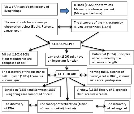
The cell was discovered by Robert Hooke in 1665. The cell theory, first developed in 1839 by Matthias Jakob Schleiden and Theodor Schwann, states that all organisms are composed of one or more cells, that all cells come from preexisting cells, that vital functions of an organism occur within cells, and that all cells contain the hereditary information necessary for regulating cell functions and for transmitting information to the next generation of cells.[5] Cells emerged on Earth at least 3.5 billion years ago.
Approximately 200 years later, Dutrochet. von Scheleiden. and Schwaan Hook confirms discovery. In 1824. R.J.H. Dutrochet cells expressed the principle which states that all animals and plants are composed of cells that stick together by the force of the adhesive. Then in 1838. M.J. Scheleiden published a book that includes an understanding of the genesis of plant tissue. Scheleiden finding suggests the presence of nucleoli and cell theory in plants. Meanwhile next year. T. Schwaan put forward the theory in animal cells. Cell theory states that living things are composed of cells. The discovery of the cell theory above Durjadin line with findings in 1835 that found that in the cell there is a viscous substance. which is now known as protoplasm.
2 R. Virchow put forward the theory that corrects the theory of abiogenesis biogenesis. Biogenesis theory states that all living cells come from cells that already exist. The concept was popular with Omnis cellula cellulae. Later in the 20th century that many experts find various types of structures and formations contained within the cell. For example,in 1867, L. ST. George found the cell organelles are now called - Golgi complex. In 1869. F. Meischer find nuclein. and in 1887. van Beneden find centrioles.
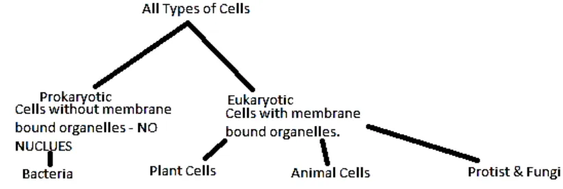
The cell is the smallest unit of life. All organisms alive today. derived from a stem cell existing in millions of years ago. These cells undergo a gradual evolution going to adjust to its environment. Based on these changes. it is now the cell can be grouped into two major groups. namely prokaryotic cells (prokaryotic) and eukaryotic cells (eukaryotic). Prokaryotic and eukaryotic term first used by Hans Ris in 1960.
Eukaryotic cells are distinguished from the more primitive prokaryotic cells by the presence of 1) cytoplasmic membranous organelles, 2) a nuclear membrane (i.e. a true nucleus), and 3) chromosomal proteins. In this lab we will focus primarily on organelles, their functions within the cell and how they differ between plant and animal cells.
Figure 1.1. All types of cells( http://mrsgiegler.weebly.com/2/archives/10-2011/1.html)
3 Figure 1.2. Schemes cell development and cell theory
A. Prokaryotic Cells
Cells that lack a membrane-bound nucleus are called prokaryotes (from the Greek meaning before nuclei). These cells have few internal structures that are distinguishable under a microscope. Cells in the monera kingdom such as bacteria and cyanobacteria (also known as blue-green algae) are prokaryotes. Prokaryotes are single-celled organisms that are the earliest and most primitive forms of life on earth. As organized in the Three Domain System, prokaryotes include bacteria and archaeans. Prokaryotes are able to live and thrive in various types of environments including extreme habitats such as hydrothermal vents, hot springs, swamps, wetlands, and the guts of animals. Most are unicellular, but some prokaryotes are multicellular.
Prokaryotic cells are the simplest systems that exhibit all of the signs of life. They are the smallest types of cell, averaging 2-5 µm in length, which makes them just visible under the light microscope. Prokaryotic cells differ significantly from eukaryotic cells. They don't have a membrane-bound nucleus and instead of having chromosomal DNA, their genetic information is in a circular loop called a plasmid. Bacterial cells are very small, roughly the size of an animal mitochondrion (about 1-2µm in diameter and 10 µm long). Prokaryotic cells feature three major shapes: rod shaped, spherical, and spiral.
4 Instead of going through elaborate replication processes like eukaryotes, bacterial cells divide by binary fission.
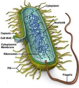
((http//www.cellsalive.com/cells) Figure 1.3. The structure of the bacterial cell.
Bacteria perform many important functions on earth. They serve as decomposers, agents of fermentation, and play an important role in our own digestive system. Also, bacteria are involved in many nutrient cycles such as the nitrogen cycle, which restores nitrate into the soil for plants. Unlike eukaryotic cells that depend on oxygen for their metabolism, prokaryotic cells enjoy a diverse array of metabolic functions. For example, some bacteria use sulfur instead of oxygen in their metabolism (http://library.thinkquest.org/C004535/prokaryotic_cells.html).
Despite their small size, inside each cell there is the complete chemical and biochemical machinery necessary for growth, reproduction and the acquisition and utilization of energy. Prokaryotes have a large array of abilities. Some of them live in the absence of oxygen, some live in extreme conditions of heat or cold, others at the bottom of oceans where the only source of energy is hot hydrogen sulfide bubbling up from the core of the earth.
5 1. Capsule – A gelatinous capsule is present in some bacteria outside the cell membrane and cell wall. The capsule may be polysaccharide as in pneumococci, meningococci or polypeptide as Bacillus anthracis or hyaluronic acid as in streptococci. Capsules are not marked by normal staining protocols and can be detected by India ink or methyl blue; which allows for higher contrast between the cells for observation. Found in some bacterial cells, this additional outer covering protects the cell when it is engulfed by other organisms, assists in retaining moisture, and helps the cell adhere to surfaces and nutrients.
2. Cell Wall - Outer covering of most cells that protects the bacterial cell and gives it shape. The cell wall acts to protect the cell mechanically and chemically from its environment, and is an additional layer of protection to the cell membrane. Different types of cell have cell walls made up of different materials; plant cell walls are primarily made up of pectin, fungi cell walls are made up of chitin and bacteria cell walls are made up of peptidoglycan.
3. Cytoplasm - A gel-like substance composed mainly of water that also contains enzymes, salts, cell components, and various organic molecules. The cytoplasm in prokaryotic cells is a gel-like, yet fluid, substance in which all of the other cellular components are suspended. Jello for cells. It is very similar to the eukaryotic cytoplasm, except that it does not contain organelles. Recently, biologists have discovered that prokaryotic cells have a complex and functional cytoskeleton similar to that seen in eukaryotic cells.2 The cytoskeleton helps prokaryotic cells divide and helps the cell maintain its plump, round shape. As is the case in eukaryotic cells, the cytoskeleton is the framework along which particles in the cell, including proteins, ribosomes, and small rings of DNA called plasmids, move around.
4. Cell Membrane or Plasma Membrane - Surrounds the cell's cytoplasm and regulates the flow of substances in and out of the cell. Prokaryotic cells can have multiple plasma membranes. Prokaryotes known as "gram-negative bacteria," for example, often have two plasma membranes with a space between them known as the periplasm. Just inside the cell wall, the plasma membrane is a selective barrier which regulates the passage of materials to from the cell. It is through this membrane that a cell must exchange food molecules, gases and other vital ingredients. Composed of phospholipid and protein membranes form thin, flexible, self-sealing, highly selective barriers between the inside of the cell and the outside world. As in all cells, the plasma membrane in prokaryotic cells is responsible for controlling what gets into and out of the cell. A series of proteins stuck in the membrane (poor fellas) also aid prokaryotic cells in communicating with the surrounding environment. Among other things, this communication can include sending and receiving chemical signals from other bacteria and interacting with the cells of eukaryotic organisms during the process of infection. Infection is the kind of thing that you don't want prokaryotes doing to you. Keep in mind that the plasma membrane is universal to all cells, prokaryotic and eukaryotic. Because this cellular component is so important and so common, it is addressed in great detail in its own In Depth subsection.
6 that prokaryotic plasmids can be genetically engineered. Today, they are isolated, changed to carry other interesting information and then reintroduced into new cells. In this way unique and usefull little bacterial factories can be designed, created and put to work.
5. Fimbriae (pili) - Hair-like structures on the surface of the cell that attach to other bacterial cells. Shorter pili called fimbriae help bacteria attach to surfaces. Fimbriae are responsible for attachment of bacteria to specific receptors of human cell (adherence). There are special types of pili called (sex pili) involved in conjunction. 6. Flagella - Long, whip-like protrusion that aids in cellular locomotion. Flagella are
organelles for cellular mobility. The bacterial flagellum stretches from cytoplasm through the cell membrane(s) and extrudes through the cell wall. They are long and thick thread-like appendages, protein in nature. Are most commonly found in bacteria cells but are found in animal cells as well. These are strands of protein that pass though the outer surface of the cell body either either singly or in tufts. Energy provided by the plasma membrane rotates the flagellum by means of a unique rotating 'joint' and this in turn moves the bacterium through its liquid world. Prokaryotic flagella are very different from similar looking structures used by eukaryotic cells. 7. Ribosomes - Cell structures responsible for protein production. Prokaryotic
ribosomes are smaller and have a slightly different shape and composition than those found in eukaryotic cells. Bacterial ribosomes, for instance, have about half of the amount of ribosomal RNA (rRNA) and one third fewer ribosomal proteins (53 vs. ~83) than eukaryotic ribosomes have.3 Despite these differences, the function of the prokaryotic ribosome is virtually identical to the eukaryotic version. Just like in eukaryotic cells, prokaryotic ribosomes build proteins by translating messages sent from DNA.
8. Plasmids - Gene carrying, circular DNA structures that are not involved in reproduction.
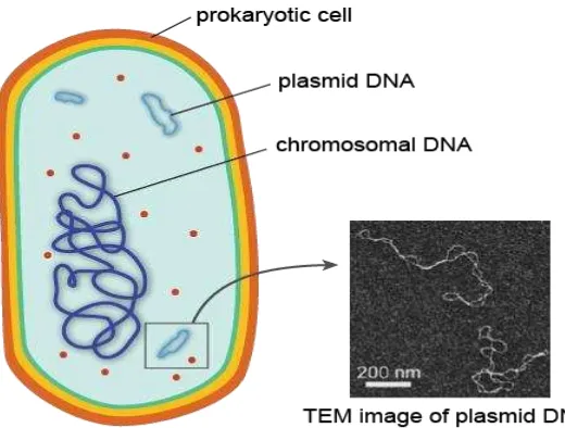
7 Figure 1.4. Transmission electron micrograph image (www.ncbi.nlm.nih.gov)
Usually, the DNA is spread throughout the entire cell, where it is readily accessible to be transcribed into messenger RNA (mRNA) that is immediately translated by ribosomes into protein. Sometimes, when biologists prepare prokaryotic cells for viewing under a microscope, the DNA will condense in one part of the cell producing a darkened area called a nucleoid (http://www.shmoop.com/biology-cells/prokaryotic-cells.html).
As in eukaryotic cells, the prokaryotic chromosome is intimately associated with special proteins involved in maintaining the chromosomal structure and regulating gene expression. In addition to a single large piece of chromosomal DNA, many prokaryotic cells also contain small pieces of DNA called plasmids. These circular rings of DNA are replicated independently of the chromosome and can be transferred from one prokaryotic cell to another through pili, which are small projections of the cell membrane that can form physical channels with the pili of adjacent cells.
The transfer of plasmids between one cell and another is often referred to as "bacterial sex." Sounds dirty. The genes for antibiotic resistance, or the gradual ineffectiveness of antibiotics in populations, are often carried on plasmids. If these plasmids get transferred from resistant cells to nonresistant cells, bacterial infection in populations can become much harder to control. For example, it was recently learned that the superbug MRSA, or multidrug-resistant Staphylococcus aureus, received some of its drug-resistance genes on plasmids.
Prokaryotic cells are often viewed as "simpler" or "less complex" than eukaryotic cells. In some ways, this is true: prokaryotic cells usually have fewer visible structures, and the structures they do have are smaller than those seen in eukaryotic cells. Don’t be fooled, however, into thinking that just because prokaryotic cells seem "simple" that they are somehow inferior to or lower than eukaryotic cells and organisms. Making this assumption can get you into some serious trouble.
8 cells are able to thrive in environments so hostile that no eukaryotic cell or organism would survive for more than a few seconds.
Prokaryotic cells are also able to pull off stuff that eukaryotic cells could only dream of, in part because of their increased simplicity. Being bigger and more complex is not always better. These cells and organisms are just as adapted to their local conditions as any eukaryote, and in that sense, are just as “evolved” as any other living organism on Earth.
Reproduction in prokaryotic cells is by binary fission; a process of growth, enlargment and division. The DNA molecule of the cell is accurately duplicated and the two copies separated form each other by movement of the cell membrane to which they are attached. The cell then divides into two smaller but identical cells and each begins its own independent existence.
Figure 1.5. Reproduction in prokaryotic cells is by binary fission (www.brooklyn.cuny.edu/bc/ahp/LAD/C5/C5_Prokary.html)
B. Eukaryotic Cells
Eukaryotes are organisms whose cells are organized into complex structures by internal membranes and a cytoskeleton. The most characteristic membrane bound structure is the nucleus. This feature gives them their name, (also spelled "eucaryote,") which comes from the Greek ευ, meaning good/true, and κάρυον, meaning nut, referring to the nucleus. Animals, plants, fungi, and protists are eukaryotes. Eukaryotes is wrapped by a nuclear membrane so that it does not mix with the cytoplasm. The most striking difference from prokaryotic cells is the true nucleus that encloses most of the cell's DNA so that the DNA is stored in a different compartment of the cytoplasm.
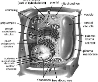
9 Figure 1.6.
Eukaryotic Cells models in three dimensions (animal cells).
10 Table 1.1. Comparison of features of prokaryotic and eukaryotic cells
(http://en.wikipedia.org/wiki/Cell_biology)
Prokaryotes Eukaryotes
Typical
organisms bacteria, archaea protists, fungi, plants, animals Typical size ~ 1–5 µm[9] ~ 10–100 µm[9]
Type of nucleus nucleoid region; no
true nucleus true nucleus with double membrane
DNA circular (usually) linear molecules (chromosomes) with histone proteins Ribosomes 50S and 30S 60S and 40S
Cytoplasmic
Mitochondria None one to several thousand (though some lack mitochondria)
Chloroplasts None in algae and plants
Organization usually single cells single cells, colonies, higher multicellular organisms with specialized cells
In addition we can also see the difference between plant and animal cells by observing the models listed below.
Figure 1.8. Plant cells (http.www.cellsalive.com/cells.)
11 A cell structure is illustrated in Figure 1.9. Plant cells and animal cells Figure 1.6. which describes the shape of various types of cells.
If we look at the plants and animals have a very big difference. where plants can not move with such active animals. This is because the shape of plant cells are rigid so it is not flexible. in contrast to animal cells that are flexible and can change shape. Aside from the shape. the differences plant cells and animal cells can also be differentiated from the following:
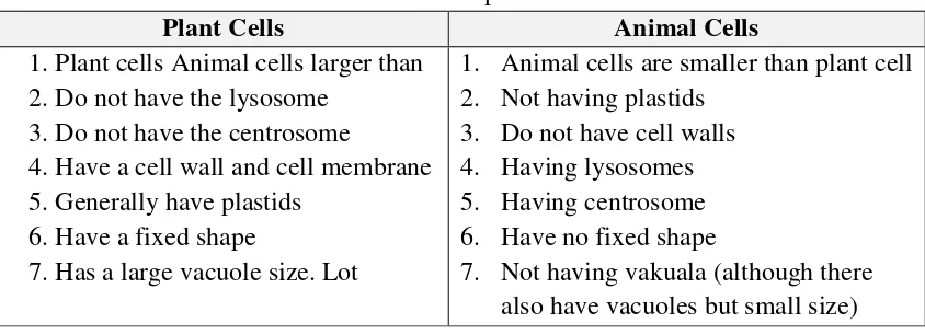
Table 1.2. Differences as plant and animal cells
Plant Cells Animal Cells
1. Plant cells Animal cells larger than 2. Do not have the lysosome
3. Do not have the centrosome
4. Have a cell wall and cell membrane 5. Generally have plastids
6. Have a fixed shape
7. Has a large vacuole size. Lot
1. Animal cells are smaller than plant cell 2. Not having plastids Broadly speaking, the structure and function of each cell component is as follows
1. Nuclei
12 Figure 1.10. The structure of the nucleus (micro.magnet.fsu.edu).
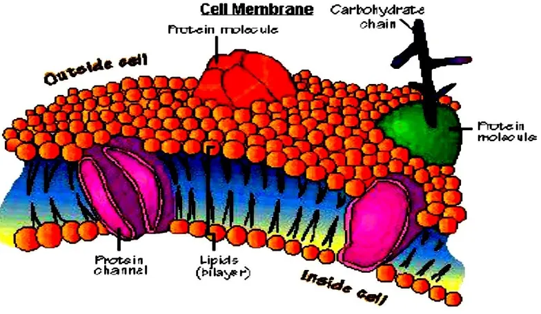
2. Plasma membrane
Cell membranes are found in animal cells whereas cell walls are found in plant cells. Cell walls and membranes have similar functions. Like a city perimeter, cell membranes surround the cell and have the ability to regulate entrance and exit of substances, thereby maintaining internal balance. These membranes also protect the inner cell from outside forces. Cell walls, as the city analogy implies, are much stronger than cell membranes and protect cells from lysing (exploding) in extremely hypotonic (diluted) solutionsMembrane is very thin and is selectively permeable to the size of 7.5-10 nm. The plasma membrane is a lipid double layer (bilayer) is the molecular structure of two layers. Lipids are important are glycolipids and phospholipids and little chance of containing cholesterol. The structure of the plasma membrane of cells such support to be able to take advantage of changes in ion permeability control at the plasma membrane of cells for communication purposes. In addition, it also serves as a protective organelles within the cell.
13 Figure 1.11. Plasma Membrane
3. The cell organelles
Organelles are parts of the cell which are adapted and/or specialized for carrying out one or more vital functions, analogous to the organs of the human body (such as the heart, lung, and kidney, with each organ performing a different function). Both eukaryotic and prokaryotic cells have organelles, but prokaryotic organelles are generally simpler and are not membrane-bound.
There are several types of organelles in a cell. Some (such as the nucleus and golgi apparatus) are typically solitary, while others (such as mitochondria, chloroplasts, peroxisomes and lysosomes) can be numerous (hundreds to thousands). The cytosol is the gelatinous fluid that fills the cell and surrounds the organelles.
14 Figure 1.12. Organelles Cell.
a. Endoplasmic Reticulum (Endoplasmic reticulum)
There are two types of endoplasmic reticulum (ER) – Smooth ER and Rough ER. This extensive network makes up approximately one half of all membranous tissue of the cell and is the site of membrane and protein synthesis. The ER system is much like a road system along which industry can be found. Goods are manufactured and shipped to needed areas via the road system. Rough ER is named for the presence of ribosomes along its membrane and is the source of proteins. Smooth ER lacks ribosomes and is responsible for lipid synthesis and processes a variety of metabolic processes such as drug detoxification.
Endoplasmic reticulum (ER) membrane is a maze so much so that it covers more than half the total membrane in eukaryotic cells. The word ` endoplasmic means within the cytoplasm ' and reticulum derived from the Latin word which means the network. RE consists of a network of tubules and membrane bubbles called sisternal or lumen. ER membrane separates the internal space. namely the space sisternal from the cytosol. And because the ER membrane continuous with the nuclear envelope. the room between the two membrane sheath was continuous with the RE sisternal room.
In general, the RE has the following functions;
1) Performers synthetic metabolic activity. because it contains a variety of enzymes.
2) denaturation and elongation of fatty acids.
3) Providing a wide surface for enzymatic reactions.
4) An ultra- structural skeleton that provides the mechanical strength of the cell. the cytoplasm koloidalnya matrix.
5) As a place of exchange of molecules through a process of osmosis. diffusion and active transport to the ER membrane and eksosistosis.
6) Establish a new core wrap on cell division.
15 The Science of Biology. 4th Edition. by Sinauer Associates. www.sinauer.com)
and WH Freeman (www.whfreeman.com).
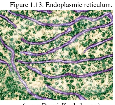
Figure 1.13. Endoplasmic reticulum.
(www.DennisKunkel.com.)
Figure 1.14. Photos using Scanning Electron Microscopy of reticulum endoplasmic and the ribosomes. (TEM x 61.560).
b. Golgi apparatus (Golgi Apparatus/Golgi Complexes)
16 Based on morphological observations and in situ cytochemistry and biochemical studies indicate that the Golgi apparatus is involved in a large number of cell activities include assembly of proteins and lipids high carbohydrate or better known as glycosylation process. recovery of the cell membrane. and secretion.
In general, the function of the Golgi apparatus. among others: 1) forming cell walls in plants
2) produces lysosomes
3) forming acrosome in spermatozoa containing enzymes to break down the cell wall of the egg.
4) Places such as mucus synthesis of polysaccharides. cellulose. hemicellulose. and pectin (constituent of plant cell walls).
5) Forming the plasma membrane.
6) Forming bag to wrap secretion of substances to be issued a cell. such as proteins. glycoproteins. carbohydrates. and fats.
Figure 1.15a. EM picture of a golgi Figure 1.15.b. Artist rendition of the Apparatus Golgi Complex
17 c. Mitochondria
Mitochondria are found in both plant and animal cells and is the site of cellular respiration. Through this process that will be covered in the Photosynthesis and Respiration lab ATP is created which is used for energy by the cell. The size and shape of mitochondria. as well as the numbers in the cells. tissues and varies according to the physiological state of the cell by. By using visible light microscopy oval mitochondria. but mitochondria can also dumbbell -shaped. spherical. or racket. with a diameter of 0.5-1.0 and length up to 7 lm. Due to the very small size of new structures can be viewed using an electron microscope. Mitochondria contain small amounts of DNA. RNA and ribosomes. Mitochondrial DNA provide the password for the synthesis of certain specific proteins on the inner membrane. Most mitochondrial proteins are encoded by nuclear DNA and synthesized by ribosomes are present in the cytosol or in the endoplasmic reticulum. This shows that there is a connection / transfer of information from DNA to the nucleus of mitochondrial later emerged from DNA found in the mitochondria themselves.
Figure 1.17. Electron microscope picture of a mitochondria
d. Chloroplasts
18 Figure 1.18. Cloroplast
4. In the membrane
Eukaryotic cells typically have a much larger volume than prokaryotic cells. typically a thousand times or more. Material or any material of cells it contains many times more. For example that the human body cells contain DNA that is a thousand times more than that of bacteria.
Membranes in various organelles such as the mitochondria membrane. vacuole membrane (in plant cells). the Golgi apparatus and the other is the venue for important reactions. Due to the addition of the cell volume must be balanced with the addition of the cell surface area by maintaining the ratio of surface area to volume ratio. This explains why all eukaryotic cells have many characteristics of the basic form and complexity of the membrane in the form of:
a. In the endoplasmic reticulum membrane that forms a maze -like compartments. b. Golgi membranes in the body that make up the pile of bags deflated play a role in
the conversion of product molecules from the endoplasmic reticulum. c. Lysosomal membrane of cells that contains digestive enzymes.
d. Peroxisome membrane wrapping where the formation and decomposition of H2O2 are reactive and dangerous during the oxidation of a variety of molecules by O2.
e. Vacuole membrane (tonoplas) in plant cells that form small bubbles and large cavity filled with fluid.
19 mechanisms (transport) is relentless. The mechanism of endocytosis and exocytosis is. which only occurs in eukaryotic cells.
In endocytosis. the parts forming the outer membrane curvature toward the later rounded and separated into bubbles of membrane-bound cytoplasm and contain substances that come from outside of cells and molecules that have been previously absorbed on the surface of cells.
Exocytosis is the reverse process of endocytosis. In this case. bubbles encased in a cell membrane. referring to the plasma membrane and release their contents into the outer environment. That way. the membranes around the compartment which is located deep within the cell to function effectively increase the cell surface area for the exchange of materials from outside.
5. Cytoskeleton
The cytoskeleton acts to organize and maintain the cell's shape; anchors organelles in place; helps during endocytosis, the uptake of external materials by a cell, and cytokinesis, the separation of daughter cells after cell division; and moves parts of the cell in processes of growth and mobility. The eukaryotic cytoskeleton is composed of microfilaments, intermediate filaments and microtubules. There are a great number of proteins associated with them, each controlling a cell's structure by directing, bundling, and aligning filaments. The prokaryotic cytoskeleton is less well-studied but is involved in the maintenance of cell shape, polarity and cytokinesis.
The cyotoskeleton represents the cell's skeleton. Like the bony skeletons that give us stability, the cytoskeleton gives our cells shape, strength, and the ability to move, but it does much more than that. The cytoskeleton is made up of three types of fibers that constantly shrink and grow to meet the needs of the cell: microtubules, microfilaments, and actin filaments. Each type of fiber looks, feels, and functions differently. Microtubules consists of a strong protein called tubulin and they are the 'heavy lifters' of the cytoskeleton. They do the tough physical labor of separating duplicate chromosomes when cells copy themselves and serve as sturdy railway tracks on which countless molecules and materials shuttle to and fro. They also hold the ER and Golgi neatly in stacks and form the main component of flagella and cilia.
Figure 1.19. Cytoskeleton (www.sinauer.com).
20 move from one part of the cell to another. This is caused by the growing size of a cell. the more complicated and more specialized structures in it.Thus the greater the necessity to keep these structures remain as they are and adjust their movements.
Cell skeleton is composed of a network of protein filaments. Three of the most important of which are actin filaments (also called microfilaments). intermediate filaments. and Microtubules.
Microfilaments are long thin fibers with a diameter of 5-6 nm. Composed of a protein called actin. Many microfilaments form a collection or network at various places in the cell. That coupled with the presence of cell motion. When an animal cell divides into two. for example. a beam forming microfilaments and separates the two daughter cells.
In many cells. the cytoplasm moved and this phenomenon called cytoplasmic flow. Motion depends on the presence of microfilaments. Microfilaments is also a feature that is important in the cell move and change shape. This does not just apply to the free movement of independent cells as well as amoeba. but also in most animal cells during embryo formation.
Figure 1.20. Scheme of cytoskeleton.
21 Cylindrical microtubule protein is found in most animal and plant cells. There are two kinds of - tubulin and - tubulin. Each with a molecular weight of about 55,000 daltons. Microtubules also play a very important role in cell division. Successful cell
division requires proper distribution of chromosomes into each daughter cell. Each chromosome moves to the goal ended in a bundle of microtubules. Microtubules are
also used in the formation of centrioles. basal bodies and flagella.
There are two groups of microtubules: 1) microtubules are stabilized microtubules can be preserved with a fixative solution of any sort. for example: OsO4. MnO4. or aldehydes at any temperature. 2) microtubules are labile microtubules can be preserved only by the solution of Figure 1.16. Aldehyde fixative microtubules and at about 4 ° C.
Figure 1.21a. Microtubules.
22 Table 1.3. Comparison between the properties of microtubules. microfilaments and
intermediate filaments.
To improve your understanding of the material above, do the exercises below!
1) Explain the four main parts of a prokaryotic cell structure and function with each one!
2) Explain briefly the difference between animal cells and plant cells ? 3) Explain how the transport mechanism in eukaryotic cells ?
4) Explain briefly how the function of the Golgi apparatus (Golgi apparatus) in a cell ! 5) Briefly describe the difference between stable microtubules and microtubule
instability!
If you have difficulty in answering the questions above consider the answers below as a reference.
1) In general, prokaryotic cells have four main parts to the structure and function of each part is as follows: (1) the cell wall. which consists of a variety of organic materials. such as cellulose. hemicellulose. and chitin. its function is to give a particular shape to the cells. as a powerful protector. also to regulate the entry and exit of chemicals into the cell. (2) the plasma membrane is wrapping the protoplasm and is often referred to plasmalema or hyaline layer. composed of proteins and lipids. In certain places the plasma membrane folds and form a building called the mesosoma. Mesosoma often
EXERCISE
23 called kondrioid which acts as a regulator for the division and photosynthesis photosynthetic bacteria. (3) is often called the protoplasm or cytoplasm of plasma cells. is a colloid that contains a lot of carbohydrates. proteins. enzymes. sulfur. calcium carbonate and volutin which contains ribonucleic acid (ARN) and easy to suck the color is alkaline. (4) the flagella. the structure in the form of a rope coming out of the surface of the cell. the cell is able to move to move. this tool is derived from the basal granules found in the cytoplasm. in the middle there is a filament consisting of compounds protein called flagelin.
2) Plants and animals have a very big difference. where plants can not move with such active animals. This is because the shape of plant cells are rigid so it is not flexible. in contrast to animal cells that are flexible and can change shape. Aside from the shape. the differences plant cells and animal cells can also be differentiated from the following:
Plant Cells Animal Cells
1. Plant cells Animal cells larger than 2. Do not have the lysosome
3. Do not have the centrosome
4. Have a cell wall and cell membrane 5. Generally have plastids
6. Have a fixed shape
7. Has a large vacuole size. Lot
8. Animal cells are smaller than plant cell 9. Not having plastids endocytosis. the parts forming the outer membrane curvature toward the later rounded and separated into bubbles of membrane-bound cytoplasm and contain substances that come from outside of cells and molecules that have been previously absorbed on the surface of cells. While exocytosis is the reverse process of endocytosis. In this case. bubbles encased in a cell membrane. fused with the plasma membrane and release their contents into the outer environment. That way. the membranes around the compartment which is located deep within the cell to function effectively increase the cell surface area for the exchange of materials from outside.
4) Golgi apparatus is involved in a large number of cell activities include assembly of proteins and lipids high carbohydrate or better known as glycosylation process. recovery of the cell membrane. and secretion.
In general, the function of the Golgi apparatus. among others: a) forming cell walls in plants ;
b) produces lysosomes ;
c) forming the sperm acrosome contains enzymes to break down the cell wall of the egg.
d) The synthesis of polysaccharides such as mucus. cellulose. hemicellulose. and pectin (constituent of plant cell walls).
24 f) Establish bag to wrap the secretion of substances to be issued a cell. such as
proteins. glycoproteins. carbohydrates. and fats.
5) Stable microtubules. the microtubules can be preserved with a fixative solution of any sort. for example: OsO4. MnO4. or aldehydes at any temperature. Microtubules are labile microtubules can be preserved only by the aldehyde fixative solution and at about 4° C.
The cell is the structural integrity. functional. and hereditary smallest of living creatures in the form of a small space bounded by membranes and contains a dense fluid. the cell is the basic unit of biology. Biogenesis theory states that all living cells come from cells that already exist. The concept was popular with Omnis cellula e cellula. Cells can be grouped into two major groups. namely prokaryotic cells (prokaryotic) and eukaryotic cells (eukaryotic).
Prokaryotic cells are cells that do not have nuclear membrane. this causes the nucleus mixed or hold a direct relationship with the cytoplasm. The size of prokaryotic cells is very small. ie 1-10 lm. Examples of prokaryotic cells is the mycoplasma. bacteria and algae blue. In general, prokaryotic cells have four main parts to that: the cell wall. plasma membrane. cytoplasm. and flagella.
Is a eukaryotic cell with a true nucleus. This cell is wrapped by a nuclear membrane so that it does not mix with the cytoplasm. There are two main parts of the cell. namely: core and its contents are often called nucleoplasm. and the remaining part is called the cytoplasm. Nucleus and cytoplasm were surrounded by a membrane. as well as smaller parts such as mitochondria and Golgi bodies.