L
Journal of Experimental Marine Biology and Ecology 248 (2000) 191–205
www.elsevier.nl / locate / jembe
Growth increments and biomineralization process in
cephalopod statoliths
a,b ,* a
Vera Bettencourt , Angel Guerra a
Instituto de Investigaciones Marinas(CSIC), Eduardo Cabello 6, 36208 Vigo, Spain b
Universidade do Algarve, UCTRA, Campus das Gambelas, 8000 Faro, Portugal Received 15 September 1999; received in revised form 7 December 1999; accepted 18 January 2000
Abstract
A study on morphological, structural and biochemical composition of Sepia officinalis and
Loligo vulgaris statoliths and statocyst endolymph was undertaken with the aim of determining the
major factors affecting the deposition process of statolith formation and to clarify the cause for the poor definition of the growth increments in S. officinalis statoliths. It is suggested that the different biochemical composition of the statocyst endolymph found in the two species accounts for distinct statolith crystallisation processes, which results in a different microstructure. This explains the better definition of growth increments in L. vulgaris statoliths comparing with those of S.
21 21
officinalis. The protein content as well as Ca and Mg concentrations in the endolymph are 21
more implicated in growth increments formation than Sr ion concentration. Moreover, the daily
variations of the three factors mentioned, allowed us to formulate a working hypothesis to explain the daily deposition of growth increments: a dark ring (rich in organic matter) is deposited during
daylight whereas a light ring (rich in CaCO ) during darkness. These results are discussed in the3
light of alternative hypotheses explaining the deposition mechanisms in statoliths. 2000
Elsevier Science B.V. All rights reserved.
Keywords: Biomineralization; Cephalopods; Growth increments; Statoliths
1. Introduction
Cephalopod statolith formation results from the deposition of a mineral structure in a living creature. This mineralization process in a biological environment is called biomineralization. Gautron (1994) defined biomineralization as a sequence of events, in
*Corresponding author. Tel.: 134-986-231-930; fax: 134-986-292-762.
E-mail address: [email protected] (V. Bettencourt)
among others) little is known about the processes involved in increment deposition in the statoliths. However, the clarification of this point is essential, since it could provide answers to several questions about the probable daily origin of the increments, allowing us to use the statoliths as an extremely useful tool for bioecological studies in the majority of cephalopod species.
Cephalopod statoliths are paired calcareous concretions essentially composed of calcium carbonate crystallised as aragonite with only a small percentage of organic material that has been ascertained to be protein (Radtke, 1983).
Presently, two alternative hypotheses to explain the deposition mechanisms in statoliths have been developed. Morris (1988, 1991a,b) hypothesised that the low
21
concentration of Mg ions in the statocyst fluid allows the CaCO precipitation in the3
form of aragonite, the process being controlled by the pH of the statocyst endolymph and the organic matrix. Lipinski (1993) proposed that strontium is directly or indirectly responsible for the definitions of growth layers and increments in the statoliths.
Considering both hypotheses, a study on morphological, structural and biochemical composition of the statoliths and statocyst endolymph of cuttlefish Sepia officinalis and the long-finned squid Loligo vulgaris was undertaken. Our goals in this paper are to determine major factors affecting the deposition process in the statolith and to determine the causes for the poor definition of the growth increments in S. officinalis statoliths compared to those of L. vulgaris.
2. Material and methods
2.1. Specimen collection and terminology used
Sepia officinalis (Linnaeus, 1758) and Loligo vulgaris (Lamarck, 1797) specimens
were collected from commercial fisheries in the Galician Rias (NW Iberian Peninsula) in summer of 1996.
V. Bettencourt, A. Guerra / J. Exp. Mar. Biol. Ecol. 248 (2000) 191 –205 193
linear structures visible inside the statolith (Lipinski, 1993), the ‘light rings’ are
essentially composed of CaCO and the ‘dark rings’ are composed of organic matter.3
Finally, an ‘increment’ or a ‘growth increment’ is an area of the statolith composed of a light ring and a dark ring (Dawe et al., 1985, Rodhouse et al., 1994).
2.2. Scanning electronic microscopy (SEM)
Ten statoliths of Sepia officinalis and ten of Loligo vulgaris were prepared for SEM (Philips SC-30). The statoliths were extracted from the cephalic cartilage (Lipinski, 1980) and stored in 96% ethanol. After removal, statoliths were cleaned in distilled water and then broken along the frontal plane (terminology after Clarke, 1978) using a scalpel. The statoliths were then etched in HCl (0.27 M) for 2–3 min and again cleaned in distilled water. Time differences in etching were random. The statoliths were then mounted on an aluminium stub and vacuum-coated with gold before viewing in the
SEM. Magnifications did not usually exceed 10003.
2.3. Energy dispersive X-ray analyses
Twenty statoliths of S. officinalis and ten statoliths of L. vulgaris covering the widest length range possible were used. Statoliths were prepared as for SEM. Energy dispersive X-ray analyses were performed on an EDAX analyser attached to the SEM. The analyses were taken in duplicate from two distinct points of each statolith. One point was located
in the lateral dome at 50 mm from the exterior margin and the other one chosen as a
central point in the concave side of the wing (Fig. 1). A Student’s t-test was used to compare data between wing and lateral dome of both species.
2.4. Atomic absorption spectrophotometry
Calcium, magnesium and strontium concentrations in the endolymph of both species were determined using an atomic absorption spectrophotometer (Varian 250 Plus). Six samples of each species were taken. Three samples of 40 individuals each were collected at 07:00–10:00 h (morning specimens) on distinct days, and another three samples of 40 individuals each were taken at 19:00–21:00 h (evening specimens). After capture,
individuals were immediately processed and 10–20 ml of endolymph were extracted
from the statocyst with the help of a microsyringe. The statoliths were removed and then
frozen at 2208C until required for protein analysis.
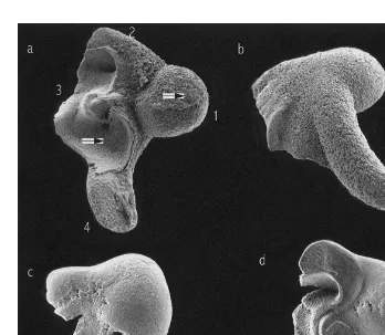
Fig. 1. SEM pictures of (a) anterior view, (b) posterior view of Sepia officinalis statoliths (ML5105 mm), and (c) anterior view, (d) posterior view of Loligo vulgaris statolith (ML5152 mm). 1, Lateral dome; 2, dorsal dome; 3, wing; 4, rostrum, and arrows indicate the points where the EDAX analysis were done.
2.5. Organic matter determination
Statoliths were ashed in order to determine the percentage of organic matter present.
Water cleaned statoliths were dried at 608C for 24 h and then weighed individually.
Dried statoliths were place in a muffle furnace at a temperature of 5008C for 3 h and
again weighed. The organic content was expressed as the percentage of weight lost due to ashing compared to the original dry weight. The percentage of organic matter and size of the individuals relationship was determined and the differences between species were tested using an ANCOVA.
2.6. Protein content
V. Bettencourt, A. Guerra / J. Exp. Mar. Biol. Ecol. 248 (2000) 191 –205 195
determined together, in the morning and in the evening specimens, since the protein in the statolith accumulated throughout growth. The total protein content was determined by quantification of the total nitrogen using the Havilah et al. (1977) method. The soluble protein content was determined by the Bradford and Smith (Smith et al., 1985) method. However, first it was necessary to solublise and purify the sample, etching the statoliths in 1 M EGTA for 32 h. The solution was dialysed (MWCO: 3.500d) against
ultra-pure water for 24 h and finally lyophilised (Christ Beta 2-16) at 2808C for 18 h.
ANOVAs test were used to check differences between species and between morning and evening specimens it was used an ANOVAs
2.7. Biomineralization in vitro
A comparative study of the crystallisation rate of dissolved calcium in the endolymph was carried out in both species. The crystallisation rate was determined by measuring pH (Sentron 1001pH) variation in time (Wheeler et al. (1981) in Wright, 1991; Garcia-Ruiz, pers. commun.). The in vitro crystallisation method consists of diffusing ammonia, coming from a deposit-solution of ammonium bicarbonate, through a solution of calcium
chloride and endolymph, in a closed system. The gradual dissolution of NH raised the3
pH of CaCl solution until it reached basic values, allowing the conversion of CO in to2 2
carbonate. All the assays were completed in triplicate.
3. Results
3.1. Scanning electronic microscopy (SEM)
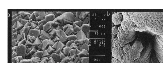
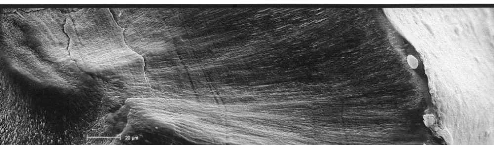
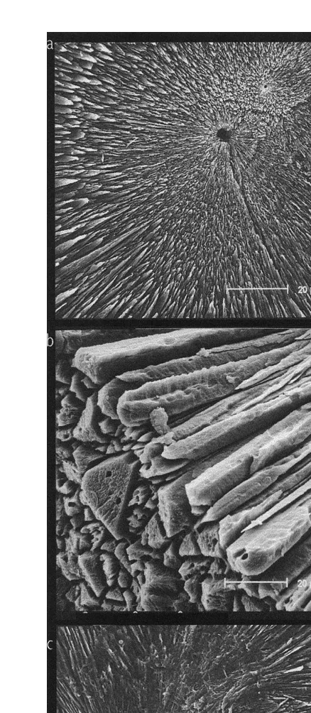
Specific morphological differences between the statoliths of the two species were observed. Thus, S. officinalis statoliths have the four main parts,: lateral dome, dorsal dome, rostrum and wing, very well defined and separated by pronounced angles. However, these parts are not so well defined in L. vulgaris statoliths and they are not separated by pronounced angles, but by smooth outlines (Fig. 1). The crystalline microstructure of the external surfaces of S. officinalis statoliths is very heterogeneous with crystals showing prominent angles and being deposited irregularly. In contrast, the crystalline microstructure of the external surfaces of L. vulgaris statoliths is quite homogeneous with crystals regularly deposited and forming slight curvatures (Fig. 2). These crystal curvatures provide a relatively broad surface, continual and uniform, that allows organic matter deposition, developing a more conspicuous and homogeneous dark ring than in crystal growth of S. officinalis statoliths where their pronounced angles prevent the formation of any broad and uniform surface. This seems to be the explanation for the better visualisation of increments in L. vulgaris statoliths than in those of S. officinalis (Fig. 3). The focus is clearly visible in the statoliths of S.
officinalis. Nevertheless, growth increments are very difficult to visualise, being obvious
Fig. 2. SEM pictures of external surface of (a) Sepia officinalis and (b) Loligo vulgaris statoliths.
deposition does not exist (Fig. 4c). Consequently, it is practically impossible to visualise growth increments in this part of the statolith.
3.2. Energy dispersive X-ray analyses
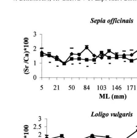
Fig. 5 shows the relationship between Sr / Ca ratios in the wing and in the lateral dome of S. officinalis and L. vulgaris. There was significantly more strontium in the wing than
in lateral dome of statoliths of both species (t53.55, P,0.05 in S. officinalis and
t52.64, P,0.05 in L. vulgaris). Although, there was more calcium in the lateral dome
than in the wing of both species, these differences were non-significant (P.0.05).
21 21
There were no significant differences in absolute concentration of Sr and Mg ion
between the two species, but the Sr / Ca ratio was significantly higher in the wing than in
the lateral dome of both species (t53.66, P,0.05 in S. officinalis and t52.71,
P,0.05 in L. vulgaris). The Sr / Ca ratio was also higher in the wing (t5 22.09,
P,0.05) and in the lateral dome (t5 21.81, P,0.05) of L. vulgaris than in S.
officinalis. This ratio did not depend on the length of the individuals, in either species.
3.3. Atomic absorption spectrophotometry
Calcium, magnesium and strontium concentrations in the endolymph of S. officinalis and L. vulgaris are shown in Table 1. There were significantly higher values of calcium in the morning specimens than in the evening ones in both species, and the opposite trend was seen for magnesium ions. Comparing these ion concentrations between species and considering specimens captured at the same period of the day it can be seen that both, calcium and magnesium showed significantly higher concentrations in S. officinalis than in L. vulgaris. Furthermore, strontium was more concentrated in the morning specimens of S. officinalis, but more concentrated in the evening individuals of L.
V
.
Bettencourt
,
A
.
Guerra
/
J.
Exp
.
Mar
.
Biol
.
Ecol
.
248
(2000
)
191
–
205
197
V. Bettencourt, A. Guerra / J. Exp. Mar. Biol. Ecol. 248 (2000) 191 –205 199
Fig. 5. Relationship between Sr / Ca ratio, in the wing and lateral dome of Sepia officinalis and Loligo vulgaris statoliths and the size ML (mm) of the individuals.
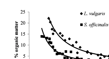
3.4. Organic matter determination
The percentage of organic matter in the statoliths of both species decreased as a power of the size of the individual (Fig. 6). However, the percentage of organic matter was
significantly higher in the statoliths of L. vulgaris than in S. officinalis ones (F5224.7,
P,0.0001).
Table 1
21 21 21 21
Ca , Mg , Sr and protein concentration (mg ml , means6S.D.) in Sepia officinalis and Loligo vulgaris 21
endolymph and total and soluble protein content (mg mg ) in statoliths of both species Concentration Sampling period (h)
Sepia officinalis Loligo vulgaris
07:00–10:00 19:00–21:00 07:00–10:00 19:00–21:00
21 Endolymph (mg ml )
21
Ca 502.6636.1* 835.8624.7* 372.669.9* 602.563.5*
21
Mg 89.062.6* 71.962.4* 42.863.1* 8.460.4*
21
Sr 4.360.64* 1.560.14* 1.660.19* 3.860.30*
Protein 1820.06468.8* 661.76204.6* 5566.06552.2* 2003.16553.0* 21
Statolith (mg mg )
n.s. n.s.
Total protein 43.7614.5 53.3610.4
Protein soluble 15.260.3* 18.660.5*
Fig. 6. Relationship between the percentage of organic matter, in Sepia officinalis and Loligo vulgaris statoliths, and the size (ML, mm) of the individuals.
3.5. Protein content
The total protein content in L. vulgaris endolymph was significantly higher than in S.
officinalis, independent of the hour of the day (Table 1). Protein content was
significantly higher in the morning than in the evening, in both species. Although the total protein content was not significantly higher in the statoliths of L. vulgaris than in those of S. officinalis, the soluble protein in L. vulgaris statoliths was significantly higher than that of S. officinalis.
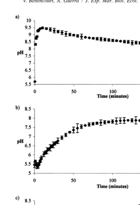
3.6. Biomineralization in vitro
The rate of crystallisation was measured by pH variation in time (Fig. 7). The increase of pH was very fast in the blank assay, reaching a maximum of 9.48 after 10 min. Subsequently, pH decreased slow. This revealed a very short induction time for nucleation, followed by a rapid crystallisation rate. The pH variation followed a different pattern in the S. officinalis endolymph assay. First, the pH decreased a little and then started to increase gradually, reaching maximum of 7.89 after 120 min. The induction time was longer and the crystallisation rate slower than in blank assay. The overall pH variation in L. vulgaris endolymph assay was similar to S. officinalis. However, the increase of pH was slower and not so constant. Induction of nucleation required 150 min and after that, the crystallisation process occurred very slowly as indicated by the very low rate of reduction of pH.
4. Discussion
V. Bettencourt, A. Guerra / J. Exp. Mar. Biol. Ecol. 248 (2000) 191 –205 201
Fig. 7. Rate of crystallisation (mean and standard deviation) measured by the pH variation in time in a closed system. (a) Blank assay, (b) Sepia officinalis endolymph assay, and (c) Loligo vulgaris endolymph assay. The decrease in pH trend was significant (P,0.0001) in all assays.
more calcified as individuals grow since the proportion of organic matter declines with age. Consequently, ageing results in a lost of increments definition.
and feeding) and of endogenous factors, such as physiological responses associated with growth, stress and reproductive cycles (Gallahar and Kingsford, 1996). Since all the individuals used in our analysis were collected at the same time and place, it seems less likely that environmental factors could be the potential source of variation of strontium. Our results showed that the Sr / Ca ratio is not related to growth in either species. Therefore the hypothesis that strontium is responsible for growth increments definition is, by it self, inadequate to explain the differences.
Most of the authors that have studied the biomineralization process (Crenshaw, 1982; Weiner and Traub, 1984; Williams, 1984 and Simkiss and Wilbur, 1989; among others) indicated that the organic matrix could form the substrate to, firstly, promote and secondly, regulate the direction of growth of the crystals and also could act as inhibitor of crystallisation. It has been demonstrated (Addadi and Weiner, 1985) that inhibition of crystal growth occurs when proteins interact with some axes of the crystal, converting its form. Moreover, Wright (1991) showed the existence of a glycoprotein in the soluble matrix of the otoliths that could inhibit the crystal nucleation, even after crystallisation has started. Our findings are in agreement with these results. Thus, addition of endolymph to the crystallisation system increased the required time to begin the crystallisation process and decreased the precipitation rate. The delay in the increase of pH and subsequently slow decrease in the assay for L. vulgaris endolymph as compared with the assay using S. officinalis endolymph suggested a longer induction time and a subsequent reduction in the crystallisation rate. This delay could be explained because the total protein content is higher in L. vulgaris endolymph than in S. officinalis, either in the morning or in the evening specimens. The fact that the L. vulgaris curve is not unimodal could suggest the presence of multiple proteins or activities sites. Furthermore, growth increments could be more defined because the aragonite crystal edges are smoothened in presence of more proteins, allowing for a uniform and continuous deposition of organic matter.
The relationship between calcium and protein concentrations in the endolymph inverted between the morning and the evening individuals of both species. The evening specimens showed more calcium and less protein than the morning ones. This seems to support the existence of a rapid precipitation of the aragonite crystals during the night, which agrees with the increase of feeding activities of S. officinalis species during the darkness in Ria de Vigo (Castro and Guerra, 1989). Sauer et al. (1997) also showed that
V. Bettencourt, A. Guerra / J. Exp. Mar. Biol. Ecol. 248 (2000) 191 –205 203
presumably to feed. Calcium concentration in the endolymph decreased towards dawn while protein contents increased. These proteins probably inhibit the precipitation of
CaCO crystals and modify the form of the crystals allowing a uniform deposition of the3
organic matter during daylight. This modification accounted for the formation of growth
increments constituted by alternate light rings (rich in CaCO ) and dark rings (rich in3
organic matter). These results agree with the findings of Mugiya et al. (1981) who indicated that the concentration of total calcium in the endolymph of gold fish, Carassius
auratus, showed a clear daily variation and calcium precipitation decreased or stopped at
sunrise and resumed in 3 h.
21 21
The role of Mg and Sr ions in the deposition process of the statolith is also
controversial. Magnesium is considered by Morris (1988, 1991a) to be the key factor in calcification, whereas statolith biomineralization is controlled by strontium concentration in the endolymph, according to Lipinski (1993). With the results of the present study we can not reject any of the hypotheses, since the concentrations of both ions in the endolymph were within the range of values that affect calcification in a crystallisation
21
system. However, Sr showed a different pattern of concentration in the endolymph of
21
the two species. Although Sr ions are vital for the development of cephalopods’
21
statoliths (Hanlon et al., 1989), Mg ions seem to be more closely related with growth
increment definition, since their concentration changes inversely in relation to calcium
21
concentration, in the endolymph of both species. In this way, the Mg ions were more
21
concentrated, and associated with low Ca concentration during daylight allowing
organic matter deposition while delaying calcification. The inverse process would occur
21 21
at sunset when Ca ion concentration was high, protein content was low and Mg ion
concentration was low. Conjunction of these three factors gives the possibility of a rapid
CaCO precipitation in aragonite form.3
Acknowledgements
´
The authors would like to express their gratitude to Dr J.M Garcia Ruız for his hospitality and support during our research visit to the Instituto Andaluz de Ciencias de la Tierra, University of Granada, Spain in June 1997 for biomineralization studies, and to CACTI, University of Vigo, Spain for the analysis of elements. Drs G. Pierce and
¨
H.O. Portner are thanked for its helpful criticism and corrections on a first draft of the manuscript. We also thanks two anonymous referees whose comments helped to improve the manuscript. This work is a part of a PhD thesis by V. Bettencourt, funded by FCT-Portugal (Praxis / BD/ 4561 / 94) and supported by CICYT (MAR 95-1919-C05). [SS]
References
Addadi, L., Weiner, S., 1985. Interactions between acidic proteins and crystals: Stereochemical requirements in biomineralization. Proc. Natl. Acad. Sci. USA 82, 4110–4114.
Arkhipkin, A.I., 1993. Age, growth stock structure and migratory rate of pre-spawning short-finned squid Illex
´ ´ ´ ˆ ´ Gautron J., 1994. Mineralisation de la coquille de l’oeuf dans l’uterus: Mise en evidence du role de la matiere
`
organique. Diplome D’Ingenieur CNAM en Biochimie Industrielle et Agro-Alimentaire, Conservatoire National des Arts et Matiers, 146 pp.
Hanlon, R.T., Bidwell, J.P., Tait, R., 1989. Strontium is required for statolith development and thus normal swimming behaviour of hatchling cephalopods. J. Exp. Biol. 141, 187–195.
Havilah, E.E.J., Wallis, D.M., Morris, R., Woolnough, J.A., 1977. A micro-colorimetric method for determination of ammonia in Kjeldahl digests with a manual spectrophotometer. Lab. Pract. 1, 545–547. Jackson, G.D., 1998. Age, growth and maturation of the deepwater squid Moroteuthis ingens (Cephalopoda:
Onychoteuthidae) in New Zealand waters. Polar Biol. 17, 268–274.
Kristensen, T.K., 1980. Periodical growth rings in cephalopod statoliths. Dana 1, 39–51.
Lipinski, M.R., 1980. Statolith as a possible tool for squid age determination. Bull. Acad. Pol. Sci. (ClassII) 28, 569–582.
Lipinski, M.R., Dawe, E., Natsukari, Y., 1991. Introduction to Section 2. In: Proc. Int. Workshop held in the Instituto di Tecnoligia della Pesca e del Pescatto (ITPP–CNR), Mazara del Vallo, Italy, 9–14 October 1989, Squid Age Determination Using Statoliths, pp. 77–81, NTR–ITPP Spec. Publ. No. 1.
Lipinski, M.R., 1993. The deposition of statoliths: a working hypothesis. In: Okutani, T., O’Dor, R.K., Kubodera, T. (Eds.), Recent Advances in Cephalopods Fisheries Biology, Tokai University Press, Tokyo, pp. 241–262.
Lipinski, M.R., Durholtz, M.D., Underhill, L.G., 1998. Field validation of age readings from the statoliths of chokka squid (Loligo vulgaris reynaudii d’Orbigny, 1845) and an assessment of associated errors. ICES J. Mar. Sci. 55, 240–257.
Morris, C.C., 1988. Statolith growth lines and statocyst function in the cephalopoda. PhD thesis, University of Cambridge, UK 140 pp.
Morris, C.C., 1991a. Statocyst fluid composition and its effects on calcium carbonate precipitation in the squid
Alloteuthis subulata (Lamarck, 1798): towards a model for biomineralization. Bull. Mar. Sci. 49 (1-2),
379–388.
Morris, C.C., 1991b. Methods for in situ experiments on statoliths increment formation, with results for embryos of Alloteuthis subulata. In: Proc. Int. Workshop held in the Instituto di Tecnologia della Pesca e del Pescatto (ITPP–CNR), Mazara del Vallo, Italy, 9–14 October 1989, Squid Age Determination Using Statoliths, pp. 67–72, NTR–ITPP Spec. Publ. No. 1.
Morris, C.C., Aldrich, F.A., 1985. Statolith length and increment number for age determination of Illex
illecebrosus (LeSeur, 1821) (Cephalopoda, Ommastresphidae). NAFO Sci. Counc. Stud. 9, 101–106.
Mugiya, Y., Watabe, N., Yamada, J., Dean, J.M., Dunkelberger, D.G., Shimizu, M., 1981. Diurnal rhythm in otolith formation in the goldfish, Carassius auratus. Comp. Biochem. Physiol. 68A, 659–662.
Natsukari, Y., Nakanose, T., Kazunari, O., 1988. Age and growth of loliginid squid Photololigo edulis (Hoyle, 1885). J. Exp. Mar. Biol. Ecol. 116, 177–190.
Radtke, R.L., 1983. Chemical and structural characteristics of statoliths from the short-finned squid Illex
illecebrosus. Mar. Biol. 76, 47–54.
V. Bettencourt, A. Guerra / J. Exp. Mar. Biol. Ecol. 248 (2000) 191 –205 205 Sauer, W.H.H., Roberts, M.J., Lipinski, M.R., Smale, M.J., Hanlon, R.T., Webber, D.M., O’Dor, R.K., 1997.
Choreography of the squid’s ‘nuptial dance’. Biol. Bull. 192, 203–207.
Smith, P.K., Krohn, R.I., Hermanson, T.I., Mallia, A.K., Gartner, F.H., Provenzano, M.D., Fujimoto, E.K., Goeke, N.M., Olson, B.J., Klenck, D.C., 1985. Measurement of protein using bicinchoninic acid. Anal. Biochem. 150, 76–85.
Simkiss, K., Wilbur, K.M., 1989. Biomineralization, Academic Press, San Diego.
Weiner, S., Traub, W., 1984. Macromolecules in mollusc shells and their functions in biomineralization. Philos. Trans. R. Soc. Lond., Ser. B. 304, 425–434.
Williams, R.J.P., 1984. An introduction to biominerals and the role of organic molecules in their formation. Philos. Trans. R. Soc. Lond., Ser. B 304, 411–424.