Research Report
Elastin Expression and Elastic Fiber Thickness in
The Uterosacral Ligament of Women With Uterine Prolapse
Kandungan Elastin dan Tebal Serat Elastis Ligamentum Sakrouterina pada Perempuan dengan Prolaps Uteri
Erwin Supriadi, Supriadi Gandamihardja, Edwin Armawan
Department of Obstetrics and Gynecology Medical Faculty of Padjadjaran Uneversity/
Dr. Hasan Sadikin Hospital Bandung
INTRODUCTION
Pelvic-organ prolapse (POP) is a common condition, often treated with surgery. Ageing of the population has resulted in an increasing incidence of surgical re-pairs for POP. By the age of 80 years, women have an 11% risk of surgery for prolapse or urinary incon-tinence, and a third of these women will have another repair after failed initial POP surgery. In addition, women with POP have a worse quality of life. Thus, there is a need for a better understanding of the sup-portive structures of the pelvic organ are involved in the pathophysiology of POP.1,2
Uterine prolapse is a condition of decline in the uterine into the vagina or out of the vagina due to the weakening of the connective tissue of the uterine backers. Uterine prolapse is a disease that is widely available in society and the incidence increases with age and parity. Incidence is not known with certainty. Study in the United States in 2004 on 16,000 patients found 14.2% suffered from uterine proplapse. In the United States, age is associated with the incidence of uterine prolapse surgery are women aged above 50 years is 2.7 to 3.3 per 1000 women. In a British study in 2001, the annual incidence of uterine prolapse sur-gery is 2 per 1000 people. This shows a high
opera-Abstract
Objective: To measure and correlate bet-ween elastin expres-sion and elastic fibre thickness in the uterosacral ligaments of women with uterine prolapse.
Method: This analytic with cross sectional study was done in January - March 2011. Specimens were obtained from the parafin block of uterosacral ligaments of women having uterine prolapse (prolapse group, n = 16) and the same location in patients with no prolapse having simple hysterectomy, radical hysterectomy or sta-ging laparotomy (control group, n = 16) who came to Dr. Hasan Sadikin hospital and its network. The percentage of elastin-positive tissue and elastic fibre thickness were measured by immunohisto-chemistry from random field readings per sample. The examiner was unaware of sample identity and the patients’ clinical history.
Result: The risk factors (age, parity and vaginal delivery) were compared between the control and prolapse groups, and there were no difference of body mass index between the groups. Immunohis-tochemical staining and morphometric analysis indicated there were significant different beween elastin expression in the prolapse group compare with the control group (p < 0.001). The mean thickness of elastic fibres was 0.938 μm in the group with uterine prolapse and 2.244 μm in the control groups (p = 0.02). Elastin expression was associated with elastic fiber thickness (r = 0.589 and p = 0.016).
Conclusion: In this cross-sectional study investigating elastin changes in women with prolapse, there was correlation between elastin expression and elastic fiber thickness in uterosacral ligament of women with uterine prolapse.
[Indones J Obstet Gynecol 2011; 35-2: 79-83]
Keywords: elastin, uterosacral ligament, uterine prolapse
Abstrak
Tujuan: Untuk mengukur dan menghubungkan kandungan elastin dengan tebal serat elastis ligamentum sakrouterina pada perempuan dengan prolaps uteri.
Metode: Jenis penelitian ini adalah studi analitik secara potong silang yang dilakukan selama bulan Januari - Maret 2011. Sampel diambil dari blok parafin ligamentum sakrouterina perempuan de-ngan prolaps uteri (kelompok penelitian dede-ngan prolaps n = 16) dan sampel yang sama juga dari ligamentum sakrouterina perem-puan tanpa prolaps uteri yang dilakukan histerektomi totalis, his-terektomi radikal atau staging laparotomy (grup kontrol n = 16) yang datang ke Rumah Sakit Dr. Hasan Sadikin Bandung dan ru-mah sakit jejaring. Persentase kandungan elastin dan tebal serat elastis dari pewarnaan imunohistokimia diukur secara random dan kemudian dihitung reratanya perlapang pandang. Pemeriksa tidak diberitahu identitas dan riwayat klinis pasien yang menjadi sampel penelitian.
Hasil: Terdapat perbedaan rata-rata faktor risiko (umur, pari-tas dan jumlah melahirkan pervaginam) antara kelompok penelitian perempuan dengan dan tanpa prolaps uteri, tetapi faktor indeks massa tubuh tidak terdapat perbedaan pada kedua kelompok peneli-tian. Berdasarkan pewarnaan imunohistokimia dan uji statistik me-nunjukkan perbedaan yang bermakna kandungan elastin antara kelompok penelitian perempuan dengan dan tanpa prolaps uteri (p < 0,001). Rata-rata tebal serat elastis kelompok prolaps uteri ada-lah 0,938 μm dan kelompok kontrol 2,244 μm (p = 0,02). Terdapat hubungan antara kandungan elastin dengan tebal serat elastis (r = 0,589 dan p = 0,016)
Kesimpulan: Pada studi potong silang memperlihatkan peru-bahan elastin pada perempuan dengan prolaps uteri, terdapat kore-lasi antara kandungan elastin dengan tebal serat elastis ligamentum sakrouterina pada perempuan dengan prolaps uteri.
[Maj Obstet Ginekol Indones 2011; 35-2: 79-83]
Kata kunci: elastin, ligamentum sakrouterina, prolaps uteri
tion rate compared to other surgical procedure per-formed on women. This is a phenomenon of the ice-berg because these figures only show a woman who had surgery alone. This figure does not include wo-men with uterine prolapse who did not undergo sur-gery, the woman who cared not for prolapsed and women who never seek treatment. Around 200,000 uterine prolapse surgery performed in the United Sta-tes each year.3,4
Gregory et al. in 2005 said that the incidence of uterine prolapse vary in certain races. Caucasians and Ras Spain have a higher risk than the races of Asia, Africa and India.5 There is only a few data of uterine
prolapsed incidence in Indonesia. According to the annual report of Obstetrics and Gynecology RSHS 2006, from 1455 cases treated gynecological there were 30 cases of uterine prolapse and 13 cases of which performed vaginal hysterectomy.6
Some important factors in the incidence of uterine prolapse is the age factor, the hormone estrogen, ex-tracellular matrix, childbirth, obesity, chronic cough and chronic constipation. In Women’s Health Initia-tive study, women with obesity have a higher risk of getting uterine prolapse than women with normal weight. According to some researchers say that the injury factors associated with labor and gave birth to three children or more will increase the occurrence of uterine prolapse.3,7
Pelvic organ prolapse occurs because the system backers disappeared, usually this damage due to trau-ma or due to atrophy of the pelvic tissues backers. Especially when the process is contained in one of the most important part of the pelvic support system such as the cardinal or uterosacral ligament or because of damage to the pelvic and urogenital diaphragm.8
The extracellular matrix determines tissue strength and is predominantly made of collagen, but glycopro-teins, e.g. elastin, are also important for cell scaffold-ing and might have a role in the biomechanical be-haviour of the uterosacral ligament. In addition, changes in elastic fibres with ageing might be respon-sible for some of the connective tissue changes seen in the development of uterine prolapse, similar to that reported in sun damaged skin. Basic research on elas-tic fibres has established that mature elaselas-tic fibres have a very intricate ultrastructure which follows tight developmental regulation, and that they are highly tis-sue-specific, as they seem to adapt to tissue function. Elastic fibres are complex structures consisting of amorphous elastin and microfibrils. Elastic fibres are difficult to repair, as damaged fibres require highly coordinated expression of the molecules that consti-tute the microfibrils. A recent report described mice lacking the protein lysyl oxidase-like 1 (LOXL1), which normally interacts with fibulin-5 at sites of elastogenesis, resulting in lack of normal elastic fibre deposition into pelvic organs postpartum, and POP in some of the mice. This report provided an additional insight into the importance of elastin in POP.8-11
There is only a few data of relationship between the expression of elastin and elastic fibers thickness with the incidence of uterine prolapsed. Some re-searchers assume that if there is decreased expression of elastin and elastic fibers thickness, it can happen connective tissue weakness of pelvic organ and
uter-ine prolapse can then occur. Some research on the comparative elastin expression and elastic fibers thickness showed that patients with uterine prolapse had elastin expression and elastic fibers thickness which decreased compared to patients without uterine prolapse uteri.12
SUBJECTS AND METHODS
Subjects of study were paraffin block of uterine pro-lapse patient with and without uterine propro-lapse who came to Dr. Hasan Sadikin Hospital Bandung and its networking. Samples were recent paraffin block which qualified of include and exclude criteria until achieved number of minimal samples. This analytic with cross sectional study was done in January -March 2011.
RESULT
Table 1. Subject characteristic
Characteristic
• Mean (SD) 61.69 (7.255) 46.94 (7.434)
• Median 62.00 46.50
• Range 44 - 73 32 - 59
Parity
• Mean (SD) 6.06 (2.489) 3.56 (1.209)
• Median 6.00 3.50
• Range 3 - 12 1 - 6
Number vaginal delivery
• Mean (SD) 5.13 (1.708) 3.13 (1.088)
• Median 5.00 3.00
• Range 3 - 9 1 - 5
BMI
• Mean (SD) 21.96 (1.793) 23.32 (2.172)
• Median 21.35 23.30
• Range 20 - 26.84 20 - 29.28
Table 1 shows the characteristics of both study groups. In the study group with uterine prolapse ob-tained mean maternal age was 61.69 years, age range 44 - 73 years, whereas the study group without uterine prolapse that mean maternal age was 46.94 years, age range 32 - 59 years. Mean parity for uterine prolapse research group was 6 times, in the range from 3 to 12 times give birth, while the parity for the study group without uterine prolapse was 3.5 times, in the range of 1 - 6 times the birth. Mean number of vaginal delivery with uterine prolapse research group was 5.13 times, in the range of 3 - 9 times. While in the study group without uterine prolapse was 3.13 times, in the range of 1 - 5 times. Mean body mass index with uterine prolapse uteri research group average was 21.96 in the range of 20 to 26.84. While in the study group without uterine prolapse was 23.3 in the range of 20 to 29.28.
Table 2. Comparison of elastic fiber thickness (EFT) of women with and without uterine prolapse.
EFT (μm)
Mean (SD) 0.938 (0.9811) 2.244 (0.9919) 0.02
Median 0.5 2.5
Range 0 C 2 - 3 0 - 1 C 4 - 5
Table 2 shows the differences in elastic fiber thick-ness data both research groups. There are significant differences in the elastic fiber thickness between the two research groups (p = 0.02). The mean elastic fi-bers thickness for uterine prolapse group was 0.938 m and the control group was 2.244 m.
Table 3. Comparison elastin expression score of women with or without uterine prolapse.
Elastin Score
Mean (SD) 2.50 (1.966) 6.56 (3.010) < 0.001
Median 2 7
Range 1 - 8 3 - 12
Table 3 showed a difference score of the elastin expression of the research group.There were signifi-cant differences between the two study groups (p < 0.001). In the study group with uterine prolapse ob-tained the median score of elastin expression was 2 (range 1 - 8), whereas the study group without uterine prolapse, the median score was 7 (range 3 - 12). It can be concluded that, there was a decrease of elastin expression uterosacral ligament in the study group with uterine prolapse.
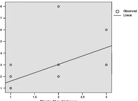
Figure 1 showed that from linier regression analy-sis, elastin expression was associated with elastic fi-ber thickness in uterosacral ligament of women with uterine prolapse.(r = 0.589 and p = 0.016).
DISCUSSION
Subject Characteristic
In this study, characteristic of subjects analyzed were age, parity, number of vaginal delivery and body mass index. This was stated by the idea that age, parity, number of vaginal delivery and body mass index are risk factors that will influence the incidence of uterine prolapse.
Karam et al13 in their research focused on a
ho-mogeneous group of postmenopausal women; selec-tion bias was minimized by assessing a consecutive group of patients. The prolapse and control groups had similar baseline characteristics, e.g. age, parity, number of vaginal deliveries, BMI, use of hormonal replacement therapy and smoking status. This allowed us to isolate the association of elastin with POP, with a minimal confounding effect from other important causative variables.
Table 1 shows the characteristics of both study groups. After comparing the characteristics of re-search subjects, it was found that the mean of the uterine prolapsed study group was older. More num-ber of parity and numnum-ber of vaginal delivery com-pared with the study group without uterine prolapse. But the characteristics of body mass index in both groups the study found relatively similar results. The age range (44-73 years) obtained in this showed al-though the mean incidence of uterine prolapse in older age, but not impossible uterine prolapse may occur at a young age. Karam et al stated that there was no significant correlation between the expression of elastin and thick elastic fibers with age women.
Elastin Expression
Although much work has been devoted to the study of collagen in prolapse tissues, little is known about elastin in the human vaginal wall and the changes oc-curring with tissue ageing. However, tissue flexibility and extensibility depends on this important extracel-lular matrix protein. Collagen and elastin functions are complementary, the former being involved with tensile strength and the latter with deformation and recoil. The formation of elastic fibres is extremely complex and has been partly elucidated in animal mo-dels. Elastic fibre degradation has been linked to se-veral human conditions, such as emphysema, aortic aneurysms, sun-damaged skin, and in some inherited connective tissue diseases such as Marfan syndrome, which is caused by a fibrillin-1 mutation. Elastin is formed from tropoelastin, its soluble precursor, which is deposited on microfibril bundles. LOX, a mono-amine oxidase secreted by fibrogenic cells such as fibroblasts or smooth muscle cells, produces an oxi-dative deamination of the lysine residues, resulting in cross-linking of the tropoelastin and the formation of an elastin core. Of the four LOXLs, LOXL-1 was shown to co-locate with fibulin-5 at sites of elasto-genesis. Once elastin is formed, degradation and re-newal is controlled by elastolytic enzymes.9,14
The study of elastin in human tissues remains chal-lenging. Elastin can be measured at the mRNA level, precursor protein level, or mature elastin level. Mea-8
suring elastin mRNA expression might not reflect the true status of elastin in the tissue itself, as mRNA still needs to be translated and will undergo further post-translational modifications and turnover that will not be assessed by studying mRNA. The tropoelastin can be measured, but this is only a protein precursor of elastin, and therefore mature elastin is not being assessed. A common way to quantify mature elastin is by indirectly measuring its cross-links with desmo-sine. However, this measurement could be inaccurate, as the tissue deterioration associated with POP can affect these cross-links.15,16
Yamamoto et al. reported a decrease in elastin mRNA expression and elastin synthesis in cultured fibroblasts of women with POP. In their study, fibro-blasts were harvested from the cardinal ligaments of patients with uterine prolapse, and not from the va-gina, as used in the present study. Ewies et al. as-sessed elastin expression in the cardinal ligament of postmenopausal women with and without prolapse, and found that it was lower in prolapsed cardinal liga-ments. Lin et al.17 used immunohistochemistry to
compare elastin expression in 23 women with anterior vaginal wall prolapse, vs 15 control women. They found that elastin expression in the vaginal wall was actually higher in patients with prolapse.
In the study by Karam et al.13, patients with POP
had lower elastin expression and lower EFW than controls of comparable age. They used in situ elastin expression and EFW as estimates of the quantity of elastin in the tissue, and elastin organization/architec-ture, respectively. They also found that elastin expres-sion and EFW were not significantly associated with age in postmenopausal women, possibly because the damage to elastin occurs earlier in life and effective elastogenesis is then hindered by incomplete degra-dation or elimination of native elastin.
Table 3 shows the difference score data on the elastin expression of the research group. There are significant differences between the two study groups (p < 0.001). In this study looks significantly decreased elastin expression of patients with uterine prolapse than without uterine prolapse.
Elastic Fiber Thickness
The results of Goepel et al. study, indicate that the ligaments of the prolapsed uterus are characterized by higher immunoreactivity for tenascin, and lower abun-dance of elastin. This is the first immunohistochemi-cal report that evaluates the difference in immunola-beling of tenascin in the uterosacral ligament of wo-men with POP versus those with no prolapse. The data suggest that elastin might play an important role in the pathophysiology of POP and that lacking ap-propriate expression of elastin may predispose woman to develop POP. Of note, striking differences of elas-tin immunolabeling was seen. In non-POP postmeno-pausal women, a normal elastin-labeling pattern was seen. In women with POP, however, elastin was either completely absent or showed clear signs of fragmen-tation.13
Table 2 shows the differences in elastic fiber thick-ness data both research groups. There are significant differences between the elastic fiber thickness two
re-search groups (p = 0.02). Obtained mean elastic fibers thickness for uterine prolapse group (M = 0.938 m) and the control group (M = 2.244 m). Even from the entire sample group contained six samples uterine prolapse that there were no elastic fibers at all. Based on these data, it can be concluded that the more thicker the elastic fibers of patients without uterine prolapse than patient with prolapse.
Elastin plays a major functional role in the main-tenance of the integrity of the ligaments, but the fac-tors involved in elastic fiber formation are generally unknown. The lack of regeneration of functional elas-tic fibers in adults is a major problem, and once this ability to regenerate is lost, the restoration of normal function is not possible. Adult tissues synthesize elas-tin in response to cyclic stretching, injury, UV radia-tion and in many diseases, including emphysema. Adults, however, cannot rebuild the elastic fiber as-sembly mechanisms and, consequently, function is not restored.12,17
SUGGESTION
There is a need of similar studies to assess the cor-relation between elastin with other components of the extra-cellular matrix of connective tissue in relation to protein synthesis of elastin. Research of nutritional status with impaired synthesis and extra-cellular ma-trix protein damage particularly elastin can be con-sidered.
CONCLUSIONS
In this cross-section study investigating elastin chang-es in women with prolapse, there was correlation bet-ween elastin expression and elastic fiber thickness in ute-rosacral ligament of women with uterine prolapse.
REFERENCES
1. Bump RC, Norton PA. Epidemiology and natural history of pelvic floor dysfunction. Obstet Gynecol Clin North Am 1998; 25: 723-46
2. Dietz HP, Haylen BT, Vancaillie TG. Female pelvic organ prolapse and voiding function. Int Urogynecol J Pelvic Floor Dysfunct 2002; 13: 284-8
3. Anne MW. An overview of Pelvic Organ Prolapse. In: Anne MW, Linda B, Joseph S, Mare RT, Editor. Office Urogynecology. New York: MacGraw-Hill; 2004: 189-96 4. Walter MD, Weber AM. Anterior Vaginal Prolapse with
and without genuine stress incontinence. In: Linda C, David S, Editor. Textbook of female urology and urogynecology. London: Martin Dunitz Ltd; 2001: 587-97
5. Gregory SJ, Ganka N, Eric V, Shlomo R, Lanssa VR. Fa-milial transmission of genitovaginal prolapse. Int Urogyne-col J. 2005; 16(17): 489-501
6. Laporan Tahunan OBGIN RSHS. Bagian OBGIN Univer-sitas Padjadjaran/RS Dr. Hasan Sadikin Bandung. 2006 7. Sandra RV. Anterior compartement prolapse, Urinary
in-continence and the Effect of Anterior colporraphy and paravaginal repair. In: Gretehen ML, Editor. Urogynecol-ogy. USA: Oxford University Press Inc; 2000: 118-31 8. Weber AM, Buchsbaum GM, Chen B. Basic science and
translational research in female pelvic floor disorders: pro-ceedings of an NIH-sponsored meeting. Neurourol Urodyn 2004; 23: 288-301
9. Kielty CM, Sherratt MJ, Shuttleworth CA. Elastic fibres. J Cell Sci 2002; 115: 2817-28
10. Liu X, Zhao Y, Gao J. Elastic fiber homeostasis requires lysyl oxidaselike 1 protein. Nat Genet 2004; 36: 178-82 11. The anatomy and Dynamics of Pelvic Floor and
dysfunc-tion. In: Peter P, editor. The Female Pelvic Floor. Germany. Springer; 2002: 14-48
12. Goepel C. Differential elastin and Tanascin immunola-beling in The Uterosacral Ligament in Postmenopausal Women With and Without Pelvic Organ Prolapse. Acta Histochem. 2008; 110(3): 204-9
13. Karam JA, Vazquez DV, Lin VK, Zimmern PE. Elastin expression and EFW in the anterior vaginal wall of post-menopausal women with and without prolapse. Journal compilation 2007; 100: 346-50
14. Shifren A, Mecham RP. The stumbling block in lung repair of emphysema: elastic fiber assembly. Proc Am Thorac Soc 2006; 3: 428-33
15. Chen B, Wen Y, Polan ML. Elastolytic activity in women with stress urinary incontinence and pelvic organ prolapse. Neurourol Urodyn 2004; 23: 119-26
16. Alperin M, Moalli PA. Remodeling of vaginal connective tissue in patients with prolapse. Curr Opin Obstet Gynecol 2006; 18: 544-50