Molecular Characterization of a Rigid Rod-Shaped Virus Isolated from
Frangipani (Plumeria sp.) Showing Mosaic Symptom in Taiwan
Fery Abdul Choliq
1*, Tsang-Hai Chen
2, Liliek Sulistyowati
31,3
Department of Plant Protection, Faculty of Agriculture, University of Brawijaya, Malang, Indonesia
2
Department of Plant Medicine, National Pingtung University of Science and Technology, Pingtung, Taiwan
Abstract
Frangipani is an important succulent plant around the worlds and also in Taiwan, for example, Plumeria rubra is widely grown as a popular ornamental tree in parks and landscaped establishments in Taiwan. Recently, a new disease in fran-gipani with mosaic and distortion symptoms was found in Taiwan. No viruses caused franfran-gipani disease has been reported in Taiwan and the references about frangipani disease are still limited and only Frangipani mosaic virus (FrMV) was found. In this study, the molecular properties of a virus isolated from symptomatic frangipani in south Taiwan, such as Pingtung, Kauhsiung and Tainan were investigated. The virus with rod-shaped particles of 300 nm long and 18 nm in diameter was examined inside diseased leaves by electron microscopy. The purified virus particles showed the typical UV spectrum of tobamoviruses with A260/A280 value of 1.29 and maximum and minimum absorption at 260 nm and 249
nm, respectively. The molecular weight of 19.5 kDa as the size of coat protein of tobamoviruses was estimated by sodium dedocyl sulfate-polyacrylamide gel (SDS-PAGE). Furthermore, the degenerate primers for tobamoviruses were used to amplify 568 bp and 400 bp of the DNA fragments in RT-PCR and nested PCR, respectively. Based on these results, it was confirmed that the rigid rod-shaped virus isolated from mosaic symptom of frangipani leaves is an isolate of FrMV, belonging to the genus Tobamovirus. This is the first report thatFrMV infecting Plumeria sp. in Taiwan.
Keywords:Frangipani plant, FrMV, mosaic disease, Tobamovirus.
INTRODUCTION
Frangipani (Plumeria sp.) is a small group of plant species native to tropical countries [1]. Frangipani is an important succulent plant around the worlds and some species are valuable sources as medicines, insecticides, fibers, and rubber [2]. Frangipani plants are also important in Taiwan.
For the example is P. rubra which widely grown as a popular ornamental tree in parks and landscaped establishments in Taiwan. It bears beautiful, big flowers of various colours and sizes that predominate especially during the summer [3].
Unfortunately, there are factors which inhibit frangipani growth, i.e. pest and pathogen attacked. One of pathogens which could attack frangipani plant is virus. Preliminary surveys in the fields of Pingtung, Kauhsiung and Tainan County showed that some frangipani plants are indicating attacked by viruses based on foliar symptoms about 30% severity (unpublished data, 2012). This condition encourages the research about molecular characterization
Correspondence author:
Fery Abdul Choliq
Email : [email protected]
Address : Department of Plant Protection, Faculty of Agriculture, University of Brawijaya, Jl. Veteran Malang, 65145
(electron microscopy, virus purification, and RT-PCR,) of unknown reported mosaic disease of
Frangipani plants showing symptom caused virus including mosaic and leaf distortion were collected in Pingtung County, Taiwan by random sampling method from 100 plants as sampel. Symptoms were recorded (photographed) at Plant Protection Laboratory, Department of Plant Medicine, National Pingtung University of Science and Technology (NPUST) Taiwan. Then, virus was isolated by mechanical inoculation from the infected frangipani leaf to indicator plant, Chenopodium quinoa Willd. and then local lesions of C. quinoa were collected and [5]. The virus was subsequently multiplied on C. quinoaPlants because it’s highly susceptible and
12L:12D. The virus isolate was temporary designated as Frangipani-Taiwan 1 (Fr-T1).
Electron Microscopy (Negative Staining)
Virus particles from C. quinoa and frangipani leaf extracts were one drop floated onto electron microscopy Formvar-fronted, carbon coated, 200 mesh copper grids and incubated for five minutes. Then, the grids were washed with 10 drops of distilled water, negatively stained with 6 drops of 2% aqueous uranyl acetate (pH 5.0) and incubated for five minutes. Then, all the fluid was removed by touching the edge of the grid carefully with a filter paper [6]. Then air dried before positioning the grid in a suitable container (grid storage box) and examination with Hitachi 7500 electron microscope. Particle size was determined by measuring the average of length and diameter chloroform (4°C) and stirred for 2 minutes. Then continued by centrifuged at 8000 rpm for 20
The pellet was resuspended in borate buffer pH 8.2 containing 0.01 M Na-EDTA. Then centrifuge at 3,000 rpm for 10 minutes (R20A2, Hitachi CR21G, Japan). Collect the supernatant and centrifuged in swing-bucket rotor at 38,000 rpm for 23 hours (RPS40T-1180 rotor, Hitachi CP90WX) in 30% Cs2SO4 by density gradient
cen-trifugation (Swing-bucket rotor). Dialysed the virus band with phosphate buffer 0.01 M, pH 7.0 and stirred for 16 hours at 4°C. Then, the purified virus was test by measuring wavelength absorbance at range 220-320 nm (Hitachi U-2001 spectrophotometer). Virus concentration (c in mg.mL-1) were calculated by formula [7]:
c = A260/E x dilution factor
Determination of the Molecular Weight of Viral Coat Protein by SDS-Polyacrylamide Gel Elec-trophoresis (SDS-PAGE)
Healthy C. quinoa leaf, infected C. quinoa
leaf, healthy frangipani leaf, field mosaic frangipani leaf, and purified virus isolate Fr-T1 were homogenized with 1 : 4 (w/v) 0.5 M phosphate buffer, pH 7.0. Then centrifuged the samples on centrifuge 10,000 rpm for 10 min and collected the supernatant. The extracts were analyzed by SDS-polyacrylamide gel electrophoresis (SDS-PAGE) with 12% running gel and 5% stacking gel. Mix the samples with dye on comparison 4:1 in microtube. Then heat the microtube on dry bath incubator (Violet Bioscience, Inc.) at 100°C for 3 min. Load Protech (Prestainde Protein Marker) 5µl as marker and 10 µl of each samples into the wells in the stacking gel. Start the electrophoresis with 70 voltage (V) during 30 minutes and continue with 120 V during 70 min. After finish running, the gel were stained with Coomassie Blue and shake gently on Orbital Shaker PSU-10i (Grant-Bio) with 70 rpm for 1 h. Replace the weight of the viral coat protein was determined by comparing migration of the viral protein in the gel with that of marker protein and run in parallel lanes [7].
Amplification and Analysis of Nucleic Acid Sequences ringspot virus (ORSV) were prepared. Each sam-ple take 5 µl and use RNA extraction kit (Direct-zolTM RNA miniprep) to extract total RNA from the each sample according to the
manufactur-er’s instructions (The Epigenetics Company).
Reverse Transcription-Polymerase Chain Reac-tion (RT-PCR)
One-tube of RT-PCR reactions (25 µl) were performed. A final concentration of 1 µM for each tobamoviruses degenerate primers TobRT
up1 (5’-GARTAYSCIGCIYTICARAC-3’) and TobRT
step at 94°C for 4 min; five cycles segmented performed using 1µl of the first RT-PCR product mixed with PCR master kit (GeneMark, Taiwan) and 1 µM of each degenerate primer TobN up3
(5’–GGCGYTGCARACIATHGTITAYCA-3’), TobN
do4 (5’ GTRTTICCIATRAAIGTIGTIACRTC-3’) and
TobN do4G (5’ GCCGATRAAGGTGGTGACRTC-3’). The cycling profile consisted of a denaturasing step at 95°C for 3 min, two cycles segmented into step (a) 20 s at 95°C, step (b) 15 s at 51°C, step (c) 5 s at 72°C; 26 cycles segmented into step (a) 20 s at 95°C, step (b) 15 s at 61°C, step (c) 5 s at 72°C, followed by a final extension step at 72°C for 2 min [8]. Amplification were carried out in Px2 Thermal cycler (Thermo Electron Corporation).
Electrophoresis Analysis
Electrophoresis analysis was used to ascertaining DNA product was amplified by
RT-electrophoresis machine (Major Science®) with 120 V electricity for 20 min. Then, visualized collected in Pingtung County (South Taiwan) which showed mosaic and leaf distortion on young leaf of frangipani plants (Fig. 1). Frangipani plants exhibiting virus-like disease was indicated on several city (Pingtung, Mechanical inoculation to indicator plants
(Chenopodium quinoa) and isolation by three time single lesion showing local lesion on leaves (Fig. 2).
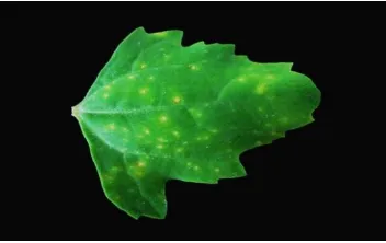
Figure 1. Mosaic symptom of frangipani leaves on the field.
Mechanical inoculation to indicator plant (C. quinoa) and maintenance by three time single lesion showing local lesion (Fig. 2). The local lesion were produced on the inoculated leaves of C. Quinoa [9]. The virus induces chlolotic ringspots or mosaic, and oftendistortion on leaves of frangipani, and severe mosaic symptoms occur on leaves of virus-infected
Nicotiana benthamiana [10].
Figure 2. The local lesions caused by viru isolate Fr-T1 on Chenopodium quinoa leaf
Electron Microscopy
Rigid, rod-shaped particles with average length of 250-300 nm x 18 nm were seen in negatively stained preparations from infected frangipani leaves, infected C. quinoa leaves after three times single lesion and purified virus isolate Fr-T1 (Fig. 3). Electron microscopy test were continued for counting the average size (length and diameter) of 100 virions.
Figure 3. Electron micrograph of negatively stained (2% uranyl acetate) virus particles from crude sap of frangipani leaves showing mosaic symptom (Bar=500nm).
The virus were checked the absorbance value by spectrophotometer and showed the curve spectrum rises rather slowly as the wavelength decreases from 300 to 250 nm, and then rapidly as the wavelength decreases below 250 nm. The purified virus showed the typical UV spectrum of nucleoprotein with A260/280
value is 1.29 and maximum and minimum
absorption at 260 nm and 249 nm, respectively (Fig. 4). There is also evidence that at least at wavelength longer than 250 nm, the virus protein and RNA is partially protected from damage by UV [13].
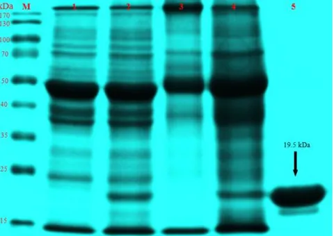
The molecular weight of coat protein of the virus was estimated 19.5 kDa by electrophoresis in sodium dedocyl sulfate polyacrylamide gel (SDS-PAGE) (Fig. 5). Lane 1 and 3 are healthy leaf samples (C. quinoa and field mosaic frangipani leaf) that did not show band on 19.5 kDa, but infected leaf samples (lane 2 and 4) were showing band with identical molecular weight compare to purified virus isolate Fr-T1 (19.5 kDa). This data is slighty different from known molecular weight of tobamoviruses genus, such as the coat protein of TMV has molecular weight of 17–18 kDa [14,15]. The slight difference of coat protein molecular weight between virus isolate Fr-T1 and other tobamoviruses may be as a result when draw horizontal line for comparing marker and samples bands on gel.
Figure 4. The ultraviolet absorption spectrum of purified preparation of virus isolate Fr-T1.
Figure 5. The determination of viral coat proteins molecular weight of virus isolate Fr-T1 by SDS-polyacrylamide gel electrophoresis (SDS-PAGE). (M : Marker, 1 : Healthy Chenopodium quinoa leaf, 2 : Infected Chenopodium quinoa
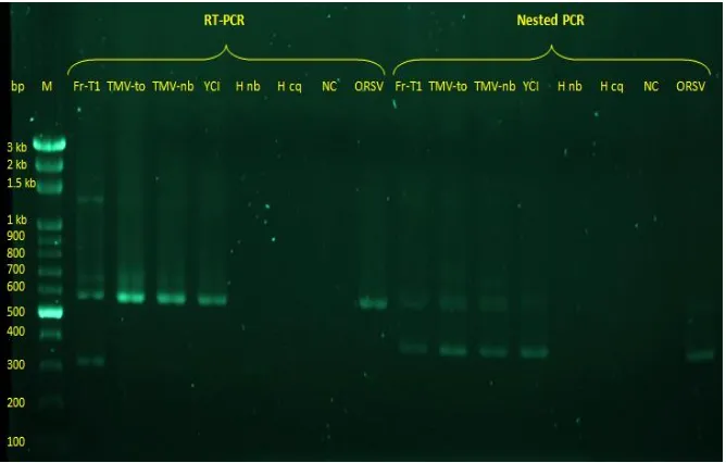
Figure 6. Agarose gel electrophoresis analysis of RT-PCR and Nested PCR products obtained from different tobamovirus isolates. (M : Marker, Fr-T1 : Frangipani virusisolateTaiwan 1, TMV-to : Tobacco mosaic virus infected tomato, TMV-nb : Tobacco mosaic virus infected Nicotiana tabacum, YCI : Yocai virus isolate from company, H nb : Healthy
Nicotiana tabacum, H cq : HealthyChenopodium quinoa, NC : Negative control, ORSV : Odontoglossum ringspot virus).
Agarose gel electrophoresis analysis of RT-PCR (Fig. 6 left) and Nested RT-PCR (Fig. 6 right) products obtained from different tobamovirus isolates were conducted for detection and partial characterization of unknown species. The virus isolate Fr-T1 could be amplified by RT-PCR using degenerate primers TobRT up1 and TobRT do2 and resulted 568 bp. This length are similar comparing to other tobamoviruses isolates (TMV-to,TMV-nb and ORSV) [8].
Nested PCR assays using Fr-TI isolate from RT-PCR product yielded about 400 bp and this amplification products is similar as expected for all tobamovirus isolates tested (TMV-to,TMV-nb and ORSV), but not shown on control (Healthy
Nicotina benthamiana and C. quinoa) (Fig. 7 right). Degenerate primers for detection of tobamoviruses by RT-PCR can be more efficient for the amplification of most members of a gene family, also allowing for the detection of new and unidentified virusspecies, followed by a nested PCR amplification that increased specificity and sensitivity of detection [8].
CONCLUSION
Based on these results, it was confirmed that the rigid rod-shaped virus isolated from mosaic symptom of frangipani leaves has length of 300 nm x 18 nm and could be identified as FrMV, belongs to Tobamovirusupon from its molecular (SDS-PAGE, RT-PCR, Nested PCR and nucleotide
sequence comparison) characterizations. This is the first report that FrMV infecting frangipani (Plumeria sp.) in Taiwan.
REFERENCES
[1] Eggli, U. 2002. Illustrated handbook of succulent plants (Dicotyledons). Springer. Switzerland.
[2] Tao, L.P., A.J.M. Leeuwenberg, D.J. Middle-ton. 1995. Apocynaceae. Flora of China 16. 143-188.
[3] Chung, W.H., C.P. Abe, Y. Yamaoka, T.W. Haung, M. Kakishima. 2005. The first report of Plumeria (Frangipani) rust disease caused by Coleosporium plumeriae in Taiwan. BSPP. New Disease Reports 11: 16.
[4] Chen, T.H., Y.T. Lu. 1995. Partial characteri-zation and ecology of bamboo mosaic Po-texvirus from bamboos in Taiwan. Plant Pa-thol. Bull. 4. 83-90.
[5] Valiunas, D., M. Samuitiene, M.
Navalinskiene, R.E. Davis. 2008. Identifica-tion of viral and phytoplasmal agenta causing disease in Gaillardia Foug. Plants in Lithuania. Agro. Res. 6. 109-118.
[6] Hajibagheri, M.A. 1999. Electron microscopy methods and protocols. Humana Press. Totowa. New Jersey.
[8] Dovas, C.I., K. Efthimiou, N.I. Katis. 2004. Generic detection and differentiation of tobamoviruses by spot nested RT-PCR-RFLP using dI-containing primers along with homologous dG-containing primers. J. Virol. Methods. 117. 137-144.
[9] Francki, R.I.B., M. Zaitlin, C.J. Grivel. 1971. An unusual strain of tobacco mosaic virus from Plumeria acutifolia. Aust. J. Biol. Sci.
24. 815-818.
[10] Lim, M.A., J.S. Hong, Y.S. Song, K.H. Ryu. 2010. The complete genome sequence and genome structure of Frangipani mosaic vi-rus. Arch. Virol. 155. 1543–1546.
[11] VIDE Database. 2011. Plant Viruses Online:
Frangipani mosaic virus. Available at: http://sdb.im.ac.cn/vide/descr345.htm. [12] Varma, A., A.J. Gibbs. 1970. Frangipani
mo-saic virus. Available at: http://www. dpvweb.net/dpv/showdpv.php?dpvno=196. [13] Kleczkowski, A., A.D. McLaren. 1967. Inacti-vation of Infectivity of RNA of Tobacco Mo-saic Virus during ultraviolet irradiation of the whole virus at two wavelengths. J. Gen. Virol. 1. 441-448.
[14] Astier, S., J. Albouy, Y. Maury, C. Robaglia, H. Lecoq. 2007. Principles of plant virology: genome, pathogenicity, virus ecology. Science Publisher. USA.