R E V I E W A R T I C L E
Advanced in Molecular Mechanisms of Atherosclerosis:
From Lipids to Inflammation
Anna Meiliana
1,2,, Nurrani Mustika Dewi
2, Andi Wijaya
1,21Postgraduate Program in Clinical Pharmacy, Padjadjaran University, Jl. Eijkman No.38, Bandung, Indonesia 2Prodia Clinical Laboratory, Jl. Cisangkuy No.2, Bandung, Indonesia
Corresponding author. E-mail: [email protected]
Received date: Jun 21, 2018; Revised date: Jul 24, 2018 ; Accepted date: Jul 31, 2018
B
ACKGROUND:
Atherosclerosis is a leading
cause of vascular disease worldwide. During the
past several decades, landmark discoveries in the
field of vascular biology have evolved our understanding
of the biology of blood vessels and the pathobiology of
local and systemic vascular disease states and have led
to novel disease-modifying therapies for patients. This
review is made to understand the molecular mechanism of
atherosclerosis for these future therapies.
CONTENT:
Advances in molecular biology and -omics
technologies have facilitated
in vitro
and
in vivo
studies
which revealed that blood vessels regulate their own
redox milieu, metabolism, mechanical environment, and
phenotype, in part, through complex interactions between
cellular components of the blood vessel wall and circulating
factors. Dysregulation of these carefully orchestrated
homeostatic interactions has also been implicated as the
mechanism by which risk factors for cardiopulmonary
vascular disease lead to vascular dysfunction, structural
remodeling and, ultimately, adverse clinical events.
SUMMARY:
Atherosclerosis is a heterogeneous disease,
despite a common initiating event of apoB-lipoproteins.
Despite of acute thrombotic complications, an adequate
resolution response is mounted, where efferocytosis
prevents plaque necrosis and a reparative scarring response
(the fibrous cap) prevents plaque disruption. However,
a small percentage of developing atherosclerotic lesions
cannot maintain an adequate resolution response, which
leading to the formation of clinically dangerous plaques that
can trigger acute lumenal thrombosis and tissue ischemia
and infarction.
KEYwORDS:
atherosclerosis, oxidative stress,
inflammation, efferocytosis, foam cells, thrombosis
Indones Biomed J. 2018; 10(2): 104-22
Abstract
Introduction
The burden of cardiovascular disease (CVD) has been risen
to be a leading cause of morbidity and mortality despite the
existence of statin and other preventive strategies to resolve
it.(1,2) Simultaneously, many clinical researches had been
performed to unravel the mechanisms and pathophysiology
of atherosclerosis along with the clinical complications.
These let us discover the development of new therapeutic
approaches to combat CVD which is accompanied by so
many advances and surprises.(3)
Gimbrone and García-Cardeña described the
endothelial cells as a gatekeeper with barrier function,
emerging the defender of vascular homeostasis.(4)
Disturbances in antithrombotic, profibrinolytic,
anti-inflammatory and antioxidant properties of the normal
endothelium lead to endothelial dysfunction and impairment
of its vasodilator capacity.
Lipoproteins, Apolipoproteins and
Atherosclerosis
(Mab) that neutralizes interleukin 1β.(5,6) The point of this
study is about applying anti-inflammatory intervention on
the right spot that will quell the disease without impairing
tumor surveillance or defenses against infection.(7)
Some studies affirmed the dynamic regulation to keep
free cholesterol (FC) in cellular membrane micro domains
and esterified cholesterol in lipid droplets in a balance state,
which was regulated by lipoproteins and cellular cholesterol
exporters. So, increasing FC in immune cells promotes
receptor over sensitization that lead to inflammation,
hematopoietic stem cell proliferation, leukocytosis and
T-cell activation. Thus, these links of cellular cholesterol
and inflammation are affecting not only atherosclerosis but
also influence autoimmune diseases.(3)
The ascertainment of high-density lipoprotein (HDL)
functions (such as cholesterol efflux potential), HDL
particle number and therapeutic approaches that increase
these variables faces challenges from disappointing results
in many clinical studies and Mendelian randomization
analyses.(8) Other approaches focusing on increasing the
HDL function, using HDL mimetics to reverse transporting
cholesterol or apolipoprotein A1 (apo A1) infusion, are also
need to be reconsidered.
Recent studies on triglyceride-rich type lipoproteins
and its pathogenicity have revitalized the enthusiasm for
finding apolipoproteins V, apolipoproteins and C3, Angptl3
and Angptl4 as new therapeutic triglycerides targets
or perhaps more specifically cholesterol-rich remnant
lipoproteins.(8,9) Genome-wide association studies and
Mendelian randomization analyses brought up Lipoprotein
(a) (Lp(a)) as a causative agent in atherothrombosis.(10)
Another study found the role of noncoding small RNAs
(micro-RNAs) as the power switch on atherosclerotic
progression and regression as well as regression and lipid
metabolism, which has opened entirely new vistas on the
molecular pathways that control this disease.(11)
Then any availability of validated genetic markers
proposes the possibility for applied risk stratification
and personalized or precision medicine targeting
therapy mode.(10,11) Paynter,
et al.
, proposed about the
usefulness of genetic information in risk prediction and
pharmacogenomic determinants of the response to therapies
for atherothrombosis.(12) More studies were needed to
bring those hypotheses from basic research to advance
clinical application.(3)
We know that one size fits all approach is nowadays
need to be developed into a smarter design. The goal
of precision medicine in the future management of
atherosclerotic risk in our patients could be achieved by
using biomarkers and genetic information rationally to
classify target therapies toward those who will most likely
to benefit from the treatment.(13,14)
Today, it is no longer a hypothesis, but an established
fact, that increased plasma concentrations of
cholesterol-rich apolipoprotein-B (apoB)-containing lipoproteins are
causatively linked to atherosclerotic CVD and that lowering
low-density lipoprotein (LDL) concentrations with statins
and non-statins reduces atherosclerotic cardiovascular
events in humans.(15-20) After decades of research,
however, there is now a large body of evidence to support
the response-to-retention hypothesis, which proposed that
the key initiating event in atherogenesis is the retention, or
trapping, of cholesterol-rich apoB-containing lipoproteins
within the arterial wall. The retained lipoproteins and their
byproducts provoke a series of strikingly maladaptive
local responses that cause plaque initiation, growth, and
evolution.(21)
The response-to-retention hypothesis, which was drew
on work from the 1940s to the 1980s, shows that lipoproteins
can interact with proteoglycans of the arterial wall.(22-25)
Lipids and apoB are accumulated at lesion-prone sites before
gross morphological changes occur.(26-29) Retention of
apoB-lipoproteins is seen throughout the progression of
atherosclerosis. The consequences of the retention of
apoB-lipoproteins include, not only an accumulation of lipid, but
also prolonged exposure of these particles to local enzymes
and other factors within the vessel wall. The retained and
modified apoB-lipoproteins trigger cellular responses within
the artery wall that accelerate further lipoprotein retention
and lesion development.(30-32)
wall.(28,35) The molecular mediators of this
trans-endothelial movement of lipoproteins remain incompletely
characterized. Recent evidence has suggested roles for
caveolin-1 (36,37) and the scavenger receptor class B type
I (SR-BI) (38). Following retention of cholesterol-rich
apoB-lipoproteins within the artery wall, the lipoproteins
have been shown to undergo several modifications with
important biological consequences (Figure 1).
Aggregated apoB-lipoproteins are avidly taken up
by macrophages (30,39) and by vascular SMCs (40) and
lead to foam-cell formation. These processes stimulate
the release of proatherogenic factors that induce the
synthesis of proteoglycans with enhanced affinity for
atherogenic lipoproteins.(30,39) In addition, monocyte/
macrophages recruited into atheromata secrete proretentive
enzymes, notably lipoprotein lipase, sphingomyelinase and
PLA2, that accelerate further retention of atherogenic
lipoproteins.(21)
Key enzymes implicated in apoB-lipoprotein
retention, aggregation, and atherogenesis include the
secretory sphingomyelinase (S-SMase), lipoprotein lipase,
and the non-pancreatic secretory group V phospholipase-A2
(PLA2-V).(30,41-45) These enzymes, particularly
lipoprotein lipase, are bridging between LDL and
proteoglycans independently of the physical state of apoB
and thereby cause a shift in the retentive mechanism from
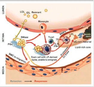
Figure 1. The response-to-retention model of initiation and progression of atherosclerosis.(21) (Adapted with
permission from Wolter Kluwer Health).
a low-affinity process (apoB-GAG binding) in the pristine
arterial wall to a high-affinity process (lipoprotein-GAG
binding) in an established atheroma. This shift in retentive
mechanism has direct clinical consequences. A lifetime
plasma LDL cholesterol concentration of 80 mg/dL will
almost always protects from atherosclerosis (46), but the
pre-existing plaques will grow if the LDL-cholesterol is 80
mg/dL (47).
Key steps in the atherosclerosis process include the
accumulation and oxidation of LDLs by reactive oxygen
species (ROS) within the artery wall accompany with
persistent inflammatory process via infiltration of
monocyte-macrophages, forming foam cells on uptake of
oxidized LDL (ox-LDL). This process sooner or later can
result in rupture or erosion of the arterial wall, forming
thrombus and subsequent platelet aggregation called
atherothrombosis, which resulting in blood flow occlusion
and downstream cellular damage. The involvement of
ROS in atherothrombosis process is including nitric oxide
(NO) inactivation or NO synthase inhibition and platelet
activation via overexpression of platelet eicosanoids and
platelet NO inhibition, which finally contribute to the
arterial dysfunction.(64,65)
Besides LDL, sphingolipids are also proven to have
a potent biological effects in atherosclerotic process.
Ceramides are a group of sphingolipids generated from
hydrolysis of sphingomyelin by sphingomyelinases,
neutral sphingomyelinases, and acidic sphingomyelinases
(A-SMase). By the activation of sphingomyelinases,
ox-LDL increases the production of ceramides.(53,66)
Then sphingomyelinase-dependent hydrolysis of LDL–
sphingomyelin become a potential pathway for ceramide
accumulation in atherosclerotic plaques.(53) Compared
to healthy vascular tissue, glycosphingolipids, such as
glucosylceramide and lactosylceramide, which are generated
from ceramides have also been found higher in human
atherosclerotic plaque.(67,68) Both lactosylceramide and
ceramide through the action of neutral sphingomyelinase
were involved in cell apoptosis (69-71), but lactosylceramide
has been suggested to contribute in plaque formation by
stimulating cell proliferation in human aortic smooth muscle
cells (72,73). Not only associated with, but also suggested
to induce plaque inflammation and instability, sphingolipids
especially glucosylceramide open up opportunities to be a
novel therapeutic targets.
Lipid rafts, microdomains, or nanodomains, different
in sizes from 5- to 500-nm-diameter structures, could be
highly ordered formed regularly from glycerophospholipids,
sphingomyelin and FC, but not cholesteryl ester (CE).
(74-76) Membrane rafts are small heterogeneous, highly
dynamic, sterol- and sphingolipid-enriched domains
that compartmentalize cellular processes.(76) They are
detergent-resistant membrane complexes rich in FC. FC
Oxidative Stress, Inflammation and
Atherosclerosis
serve as the raft stabilizer through hydrophobic binding to
the other components. Lipid raft commonly present in two
types, planar lipid raft and invaginated one, with caveolar or
little cave whose structure depends on the caveolin proteins
that are unique to caveolae. Cholesterol is an essential
component of both lipid rafts and caveolae.(77-81)
To maintain lipid raft, cells require FC. Excess FC for
this purpose will be stored in the cytoplasm as CE. When
the influx of cholesterol is greater than the outflow, the
accumulation of CE droplets and foam cell formation occurs.
Otherwise, loss of foam cell cholesterol will occur. Thus, high
value of EC/total cholesterol ratio implies cell progression,
whereas a low value implies foam cell regression.(82) Lipid
raft must be carefully regulated so the microdomains or
nanodomains are able to provide a platform for organizing
the signaling of many receptors and proteins, including
the B-cell receptor, T-cell receptor (83-85) and major
histocompatibility class receptors (86,87). The cholesterol
used to form and maintain lipid raft can be obtained from
exogenous sources, such as lipoproteins, especially LDL,
or from endogenous cellular synthesis via the mevalonate
pathway in the endoplasmic reticulum followed by transport
to the plasma membrane, or from the intracellular lipid
droplets.(88,89) From lipid droplets, cholesterol will move
out with help of extrinsic signals promoting the hydrolysis
of CE by cholesterol ester hydrolases.(90,91) FC also
can be used for cellular membrane maintenance, unless it
will be moved to a substrate pool for export via ABCA1.
(92,93) The membrane cholesterol that regulates lipid raft
composition, controlled by ABCA1 under the regulation of
the liver X receptor.(94)
Modified lipoproteins, such as ox-LDL, enhance
endothelial damage that trigger leukocyte recruitment,
which eventually fail to clear lipoproteins, undergo cell
death, and contribute to inflammation. Ultimately, growing
lesions will lead to vessel occlusion and subsequent
ischemia or (arterial) thrombosis.(95,96) After long time
been denied, neutrophils and their specific contribution
in atherosclerosis pathophysiology, recent studies present
substantial evidences about the actions of neutrophils in
early and established human and murine atherosclerotic
lesions.
Figure 2. Macrophage apoptosis, autophagy and necroptosis in atherosclerosis.(105) (Adapted with permission from Wolter Kluwer
Health).
of endothelial cells, antigen-presenting cells and platelets.
Thus, NET not only presents in plaques and thrombi but
also plays a causative role in triggering atherosclerotic
plaque formation and arterial thrombosis in any stage of
diseases progression, especially where there are high local
concentration of pro-inflammatory effector molecules.(97)
The present neutrophil-derived proteases and ROS lead to
plaque destabilization.(98-100)
Atherosclerosis is a chronic lipid-driven, maladaptive
and non-resolving inflammatory disease of the vessel
wall. Over several decades, atherosclerosis progresses in
indolence and silence, eventually resulting in the formation
of a life-threatening, rupture-prone atherosclerotic plaque.
(101-103) In brief, the disease is triggered by subendothelial
retention of infiltrated LDL into the intimal space. The
accumulated LDL undergoes oxidation and aggregation
in the intima, presenting a source of chronic stimuli
that instigate and propagate an innate immune reaction.
This includes the recruitment, homing, migration and
differentiation of monocytes into macrophages that avidly
phagocytose modified cholesterol, secrete pro-inflammatory
cytokines, enzymes and ROS and eventually undergo cell
death. Together, these processes and the defective clearance
of the dying cell propagate the inflammatory response,
forming a vulnerable atherosclerotic plaque. The key
characteristics of a vulnerable plaque include a very thin
fibrous cap encapsulating acellular necrotic core regions,
lipid-laden macrophages and T lymphocytes and sometimes,
intraplaque hemorrhage.(104) Figure 2 shows macrophage
apoptosis, autophagy and necroptosis in atherosclerosis.
decrease plaque vulnerability. Otherwise, the canonical
‘don’t eat me’ signal cluster of differentiation (CD)47 plays
a role in impairing efficient efferocytosis then progress the
atherosclerotic lesion.(105) Improving our understanding
of the mechanisms that regulate macrophage death in
atherosclerosis (apoptosis, necroptosis and efferocytosis)
will provide novel therapeutic opportunities to resolve
atherosclerosis and promote plaque stability.(105)
Endothelial, Vascular Smooth Muscle
Cells and Atherosclerosis
Endothelial cell dysfunction (ECD) on the lesion-prone
covering areas of the arterial vasculature contributes in
the pathobiology of atherosclerotic CVD. ECD covers
a constellation of various non-adaptive alterations in
functional phenotype, that regulating hemostasis and
thrombosis, local vascular tone and redox balance, also the
orchestration of acute and chronic inflammatory reactions
within the arterial wall.
ECD manifest as the earliest detectable changes
on lesion-prone areas of the arterial vasculature in
atherosclerotic lesion progression, in form of focal
permeation, trapping, and physicochemical modification
of circulating lipoprotein particles in the subendothelial
space.(106-108) These pathogenic sequence commenced
by selective recruitment of circulating monocytes from
the blood into the intima.(107-113) They differentiate
into macrophages and internally modified lipoproteins
become foam cells, endothelium and macrophages will
be activated in elaboration with multiple chemokines and
growth factors, and induce the nearby smooth muscle cells
(or their precursors) proliferation and extracellular matrix
components synthesis within the intimal compartment,
finally generating a bromuscular plaque.(114)
Developing lesions involve a continuous structural
remodeling, which result in fibrous cap, overlying a
lipid-rich, necrotic core consisting of oxidized lipoproteins,
cholesterol crystals and cellular debris. Matrix remodeling
and calcification varying degrees also play a role in this
complicated plaques, along with a rich population of
inflammatory cells (activated macrophages and T cells,
natural killer T cells and dendritic cells), modulating the
endothelial proinflammatory phenotype and proteolytic
modification of its extracellular matrix component, provide
the plaques’ structural instability.(115-117) When the
plaque is unstable or vulnerable, frank plaque rupture,
with luminal release of the highly thrombogenic contents
of the necrotic core, an atherothrombotic occlusion could
be triggered.(118-121) Evidently, superficial intimal
erosions without plaque rupture also make a consequence
of significant clinical sequelae, suggested due to endothelial
cell apoptosis, with localized endothelial denudation and the
triggering of thrombus formation.(4,122)
By this angle, the vascular endothelium can be
regarded as a dynamically adaptable interface distributed
organ, and at individual cell level, an integrator of the
local pathophysiological milieu. Thus, dysfunction
endothelial certainly alter its normal functional phenotype
non-adaptively, entangle the regulation of hemostasis and
thrombosis (123), local vascular tone (124) and redox
balance (125), also induce the orchestration of acute and
chronic inflammation (124). Thus, the term endothelial
cell dysfunction was proposed into the mainstream of
atherosclerosis research (126,127), and the molecular
manifestations of ECD began to be characterized in detail.
(117,121,124,128)
Several studies documented that Kruppel-like factor
2 (KLF2) expression in endothelial cells promotes an
anti-inflammatory, antithrombotic endothelial phenotype,
due to its antagonism of the nuclear factor kappa-B
(NF-κB) pathway.(129) KLF2 regulates the functions of other
endothelial cells, making it important for atherogenesis,
including endothelial barrier function (130), metabolism
(131) and the release of miRNAs via the shedding of
endothelial microvesicles (132). It also stimulates the
production of several autocoids, including NO and C-type
natriuretic peptide, which were found to be deficient in
dysfunctional endothelium
in vivo
.(133) Study showed that
mice genetically deficient in KLF2 enhanced atherosclerotic
plaque formation compared to wild-type controls.(134)
Nuclear factor erythroid 2-related factor-2 (Nrf2),
activated by atheroprotective flow in cultured endothelial
cells, via the phosphoinositol 3-kinase/ Akt and extracellular
signal-regulated protein kinase 5 pathways, counted to have
an atheroprotector properties. Nrf2 controls a subsequential
of target genes that play a role in the regulation of intracellular
redox balance, as well as resistance to extracellular oxidant
stress.(135-137) Nrf2
in vivo
expressed differentially in
those relatively atherosclerosis-resistant regions of the
vasculature, suggesting its pathophysiological relevance.
(135,138)
vasoprotective endothelial phenotype (4), as they counted
for ≈70% of the atheroprotective flow-induced endothelial
transcriptome activation.(141)
Protein modifications with small ubiquitin-like
modifier (SUMO) have been found to play a key role in
regulating the formation of atherosclerosis.(141-143) SUMO
proteins covalently modify certain residues of specific target
substrates and change the function of these substrates. It has
been well established that the protein inhibitor of activated
signal transducer and activator of transcription (STAT)
family of proteins has not only SUMO E3 ligase activity,
but also transrepression activity.(144) Lerchenmüller,
et al
.,
assume that this may be one of the regulatory mechanisms
by which interferon-inducible transmembrane protein 1
(IFITM1) can inhibit endothelial cell proliferation.(145)
Some studies performed postmortem and/or by imaging
have identified some plaque instability characteristic that
could lead to rupture: 1) a thin or fragmented fibrous cap
with smooth muscle α-actin (ACTA2)–positive cells, guess
to be derived from vascular smooth muscle cells (VSMCs);
2) CD68 or galectin-3 positive cells found in a large mass,
suspect to be macrophages; and 3) necrotic core containing
cells filled with lipid (foam cells) found in a large numbers,
suspect to be macrophages.(146)
These was correlated with the general dogma that
bulks of macrophages and macrophage-derived foam
cells relative to VSMCs found in atherosclerotic plaque,
particularly within the fibrous cap and shoulder regions,
are less stable and more prone to rupture.(107,147) Thus,
VSMCs in advanced lesions could generally regarded as
the atheroprotective plaque-stabilizer, while macrophages
are regarded to promote atherosclerosis and detriment the
plaque stabilization. VSMC can phenotypically switching,
and this is affecting the plaque stability. Inhibiting VSMC
phenotypic switching regulation may be beneficial in
advanced atherosclerosis, suggest this could be a chance
to replace these more conventional antiatherosclerotic
therapies.(146)
MicroRNA and Circadian Control in
Atherosclerosis
Both immune and non-immune cellular constituents
of the vessel wall were involved in many stages of the
pathogenesis of atherosclerotic lesion formation. Over
decades many studies strive to uncover any key signaling
and molecular regulatory pathways involve in the plaque
initiation and progression. MiRNA provides novel
molecular insights on atherosclerosis progressive pathways,
and is reported to have an important role in regulating many
pathophysiological processes, such as cellular adhesion,
proliferation, lipid uptake and efflux, also generation of
inflammatory mediators. The appreciation of miRNAs
detection extracellularly including in circulating blood make
a hike in its potential as new tool for diagnosis, prognosis, or
in response to cardiovascular therapeutics.(11)
MiRNAs are a small (≈18–24 nucleotides),
evolutionary conserved, single-stranded noncoding RNAs
that regulate >60% gene expression, either reducing protein
expression by blocking mRNA translation and by promoting
mRNA degradation at the post-transcriptional level by
typically binding to the 3′-untranslated region (UTR) of
specific target mRNA sequences, using a conserved ≈7–8
nucleotide seed sequence.(148-152) Moreover, one miRNA
can bind to and regulate >1 target, even as a part of the
same signaling pathway, also can harbor several distinct
miRNA-binding sites within its 3′-UTR, adding multiple
levels of regulation. Recent studies reported of LDL and
HDL abundance and function controlled by miRNAs,
have greatly expanded our understanding of the regulatory
circuits governing plasma lipoprotein levels.
MiR-148a is negatively regulating LDL-C plasma
levels by targeting the LDL receptor (LDLR) and is showed
to increase the clearance of circulating labeled LDL and
decrease plasma LDL-C levels.(153,154) Some miRNAs,
such as miR-33 (155-159), miR-758 (160), miR-26 (161),
106 (162), 144 (163,164), 128-118 and
miR-148a (154), play role in regulating HDL biogenesis and
cholesterol efflux by controlling the levels of plasma HDL-C
and the reverse cholesterol transport pathway by targeting
ATP-binding cassette transporter (Figure 3). ABCA1 plays
a central role in these processes by controlling cholesterol
efflux across the cell membrane onto lipid-poor apoA1
(155), and mediate both hepatic HDL biogenesis and the
removal of excess cholesterol from peripheral cell including
macrophages in atherosclerotic plaques.
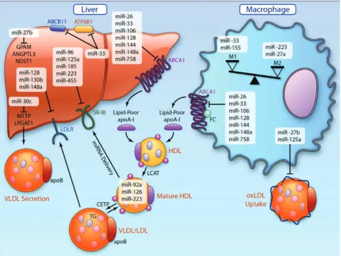
miR-Figure 3. MicroRNA (miRNA) orchestration of cholesterol homeostasis and macrophage activation in atherosclerosis.(11) (Adapted
with permission from American Heart Association).
155 plays a role in macrophage foam cell formation by
negatively regulates macrophage inhibitory factor, via the
transcriptional repressor HMG box-transcriptional protein
1, a protein known to increase the uptake of ox-LDL by
macrophages.(168)
Circulating miRNA can be detected in peripheral
blood, saliva and urine, so their expression may be harbingers
of a range stages of CAD from subclinical atherosclerotic
disease to acute coronary syndromes.(11) miRNAs may
have enormous effects on biological pathways, cell function
and homeostasis in the vessel wall, liver and periphery.
Experimenting a MiRNA mimics or inhibitors may offer an
attractive therapeutic approach for atherosclerotic disease in
specific stages and the management of its complications.
Circadian rhythms refer to 24 hours biological process
that endogenously, entrainable oscillation, which adjust
behavior and physiological activities to environmental
changes. This involves daily rhythmicity such as regulation
of sleep-wake cycles, feeding, body temperature, blood
pressure, heart rate, hormone secretion, metabolism
(including lipid metabolism), and many other biological
functions.(169,170) Our circadian clock is set by the
brain’s suprachiasmic nucleus, which interprets recurring
external stimuli, and autonomous molecular networks in
peripheral cells together. When circadian clock is disrupted
or misaligned, multiple pathologies, including chronic
inflammatory and metabolic diseases such as atherosclerosis
could rise.(171)
Figure 4. Circadian influence on
atherosclerosis.(171) (Adapted with
permission from American Heart Association).
>15 years ago demonstrated the existence of a functional
circadian clock in the vasculature.(173-175) It was found
that among 330 genes, 5% to 10% of the transcriptome,
exhibit circadian expression patterns in mouse aortae.(176)
Circadian patterned genes include those related to the core
molecular clock, lipid and glucose metabolism, protein
folding, and vascular integrity.
VSMC from human carotid plaque exhibits lower
amplitudes of mRNA expression levels of core clock
genes compared to normal carotid smooth muscle cells
from same donors. This rose a notion about the role of
mismatching circadian gene expression patterns toward the
central clocks in atherosclerotic vessels, that might play a
role in plaque stability.(177) Some evidences showed that
brain and muscle Arnt-like protein-1 (BMAL1)-dependent
peripheral circadian clocks in immune cells have a role
in inflammatory markers expression, such as C-C motif
chemokine ligand 2 (CCL2).(178) Studies in atherosclerotic
mouse models indicated the importance of the CCL2
and C-C motif chemokine receptor (CCR2) axis in early
lesion development. CCL2 or its corresponding receptor
CCR2 genetic deficiency leads to a reduction in lesion
development with lower monocyte/macrophage content in
the lesion.(179,180) Indicate that the CCL2-CCR2 axis in
atherogenesis as a result of mobilization and homeostasis
of classical monocytes under steady state rather than their
recruitment.(181) Circadian clock gene perdiod 2 (PER2)
deficiency promotes aortic endothelial dysfunction (182),
which is the initial stage of lesion development.
We can conclude that circadian rhythmicity plays an
important role in atherosclerosis progressive by influencing
atherosclerotic plaque development, either by central clock
or many peripheral clocks existed. Cell-intrinsic molecular
clocks found in leukocytes, endothelial cells, macrophages
and SMC have been identified to be linked to inflammatory
processes underlying atherosclerotic lesion development.
(183) A better understanding of the link between circadian
rhythms and cardiovascular pathologies might help
developing more targeted and personalized therapeutic
strategies for cardiovascular disease patients.
Gut Microbiota in Atherosclerosis
in different anatomic sites such as the gut, skin, respiratory
tract and genitourinary system are characterized by distinct
enzymatic pathways that are adapted to metabolize nutrients
present in the local environment.(185)
To understand the mechanisms by which the
microbiome regulates vascular function, metabolomic
profiling was used to identify specific bacteria-derived
molecules related to energy metabolism and vascular
homeostasis. Further analysis identified trimethylamine as
the gut metabolite and bacteria-derived chemical with the
clearest association with cardiovascular disease.(186) In
clinical studies, unbiased metabolic profiling further revealed
a significant increase in the levels of
trimethylamine-N-oxide (TMAO) and related metabolites in plasma samples
from patients with increased risk for CVD compared
with matched control subjects.(187) TMAO is formed by
bacterial metabolism of choline and phosphatidylcholine in
the gut to yield trimethylamine, which is oxidized in the liver
by the enzyme flavin monooxygenase-3 to form TMAO.
(187) TMAO has become the target of several therapeutic
interventions, ranging from schemes to reduce dietary
intake of trimethylamine precursors to manipulations of the
gut microbiome to reduce trimethylamine synthesis. The
revelation regarding atherosclerosis susceptibility that could
be transmitted from an atherosclerosis-prone strain of mice
to another strain typically resistant to atherosclerosis simply
by the transplantation of gut microbes provided additional
causal evidence to support the role of the gut microbiome in
regulating atherosclerosis (Figure 5).(188)
Studies of the gut microbiome in rodent models show
that in response to changes in dietary intake of carbohydrates,
Figure 5. The gut microbiome and atherosclerosis.(184) (Adapted with permission from American Heart Association).
fat, and fiber, there are changes in the gut microbiota at the
phylum level. Similar studies in humans have shown the
same trend, but there is significant interindividual variability.
There is also regional variability in the microbiome, and
the diversity and composition can reflect an industrialized
versus agrarian diet. Thus, it may be possible to predict
disease risk vis-à-vis diets rich in phosphatidylcholine, a
source of choline, or dietary carnitine, which is ultimately
metabolized to TMAO.(189)
At present, the causal pathways and molecular
mechanisms whereby the gut microbiome initiates and
perpetuates cardiopulmonary vascular disease remain
incompletely characterized. However, early studies in the
field indicate that future
in vitro
and
in vivo
studies of the
blood vessel function must now contend with an additional
layer of complexity in experimental design. These studies
will need to examine local and remote interaction with the
gut microbiota and metabolites. For
in vitro
studies often
done in the presence of antibiotics, this remains a challenge.
Further adding to the experimental complexity, the
microbiome may be altered by diet and by drugs. Thus, in
addition to standardizing diets for
in vivo
studies, the effects
of drugs (and delivery vehicle) will need to be evaluated.
(184)
Gut Microbiota in Atherosclerosis
process underlying normal biological ageing and
aged-related degeneration of multiple tissues. However, recent
studies have identified that cell senescence is also part of
normal tissue remodeling, for example during development,
as a process that regulates tissue mass and architecture.(190)
Cellular senescence induces an evolutionarily conserved
senescence-associated secretory phenotype (SASP),
resulting in release of inflammatory cytokines, chemokines
and proteases.(191) The SASP may be beneficial, for
example attraction of immune cells to clear senescent cells
(192-195) and tumor suppression (196,197). However,
a persistent SASP may be deleterious, promoting cell
proliferation (197), angiogenesis through increased vascular
endothelial growth factor (VEGF) expression (198),
epithelial-to-mesenchymal transformation and invasiveness
of premalignant epithelial cells (199). The role of the SASP
in atherosclerosis remains largely unknown, although the
SASP of human senescent VSMCs requires interleukin
(IL)-1
a
, and causes both endothelial cell dysfunction and
mononuclear cell recruitment (200).
Telomere erosion in endothelial cells is increased in
vessels prone to develop atherosclerosis (201,202), and
the telomeres are shorter in VSMCs in human plaques
compared with non-atherosclerotic vessels from the same
donor (203). However, telomere shortening is not just
a marker of senescence, but also promotes senescence,
plaque development and features of unstable plaques.
For example, telomere uncapping in VSMCs by forced
expression of a dominant-negative telomeric repeat-binding
factor 2 (TRF2) protein accelerates atherogenesis, increases
necrotic core and reduces fibrous cap areas.(204) This
suggests that dysfunctional telomeres predispose to both
initiation and progression of atherosclerosis.(205) The
effects of cell senescence in atherosclerosis clearly depend
upon the cell type being affected. Senescent endothelial
cells also accumulate in vessels with age (202,206) and in
atherosclerosis (207,208), which may importantly contribute
to age-accelerated atherogenesis.
Senescent endothelial cells are dysfunctional and have
reduced and uncoupled endothelial NO synthase (eNOS)
activity, resulting in enhanced superoxide anion production
and decreased NO bioavailability.(208,209) Senescent
endothelial cells also exhibit increasing inflammatory
responses, which include enhanced expression of vascular
cell adhesion molecule-1 (VCAM-1) and intercellular
adhesion molecule-1 (ICAM-1) (208,210), and also
develop a SASP characterized by increased IL-6 and IL-8.
Senescence, eNOS uncoupling and the SASP are regulated
in part by p38a mitogen-activated protein kinase (MAPK),
S6-kinase (S6K) and arginase II (211).
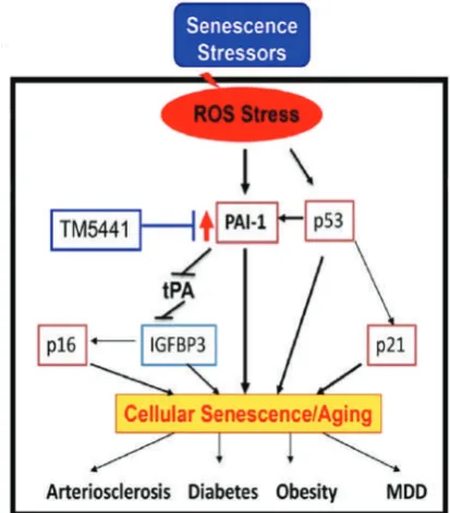
Figure 6. Molecular involvement of PAI-1 (plasminogen activator inhibitor-1) in cellular senescence and associated diseases.(216) (Adapted with permission from American Heart
Association).
The senescence markers include elevated
senescence-associated β-galactosidase (SA β-Gal) activity and
p16Ink4a, p53 and p21 expression.(208,212) However,
whether and how senescent cells contribute to atherogenesis
remains unclear.(213,214) Human plaques contain cells
with shortened telomeres, which predispose cells to
undergo senescence.(204) Consistent with a proatherogenic
role of senescence, the expression of a loss-of-function
TRF2 in VSMCs accelerates plaque growth in the ApoE
−/−mouse model of atherosclerosis, although
in vivo
evidence
for increased senescence in plaques was not provided.
(215) Importantly, senescent cells are metabolically and
synthetically active, producing numerous factors that are
released locally called senescence-messaging secretome or
senescence-associated secretory phenotype. Interestingly,
plasminogen activator inhibitor-1 (PAI-1) has been
identified as a prominent member of the
senescence-messaging secretome.(214) PAI-1 is a validated marker of
cellular senescence (Figure 6).
Genetics and Precision Medicine in
Atherosclerosis
Coronary artery disease (CAD) has important genetic
underpinnings considered equivalent to that of environmental
factors. The heritability of CAD has been estimated between
40% and 60%, on the basis of family and twin studies, a
method that yields high precision despite potential bias.(10)
Precision medicine is an approach to disease treatment and
prevention that seeks to maximize effectiveness by taking
into account individual variability in genes, environment
and lifestyle. One central aim of the recently launched US
Precision Medicine Initiative is the return of genetic results
for clinical utility.(217) The major clinical and biochemical
atherosclerosis risk factors for coronary heart disease
(CHD) and other forms of CVD have been well defined
over the past 50 years by prospective population cohorts
like the Framingham Heart Study and resulting randomized,
controlled treatment trials (RCTs). Genetics for CVD risk
prediction provides the opportunity to more precisely
identify individuals at high risk for developing disease for
whom preventive therapy can be directed.(218)
Our initial understanding regarding genetic risk
for myocardial infarction and other forms of CHD has
focused on rare (<1:100 carrier rate) monogenic etiologies
conferring exceptional risk, such as mutations in genes
LDLR, proprotein convertase subtilisin/kexin type 9
(PCSK9), or ApoB underlying the predisposition for
familial hypercholesterolemia.(219-222) However, because
of the efforts of international consortia over the past decade,
genome wide association studies of hundreds of thousands
of research participants have led to the discovery of more
than 50 common (>1:20 carrier rate) gene variants with
strong evidence for modest increases in CHD risk, and
>150 common genetic variants with strong evidence for
modest alterations of levels of key lipid fractions.(223) An
individual’s CHD genetic risk score (GRS) is an additive
score of the burden of discovered CHD risk alleles that is
often weighted by the estimated disease effect of each allele.
Nevertheless, in a recent post hoc analysis of RCTs
of statin therapy for primary and secondary prevention of
CHD, persons with the highest burden of CHD risk alleles
were not only at increased risk for CHD events, but also,
surprisingly, experienced enhanced absolute and relative
clinical benefit despite similar LDL-cholesterol lowering.
(224) These data suggest that a CHD GRS may identify
people at increased risk who may be more likely to benefit
from preventive interventions.
The evidence of CHD not only affected by
environmental factors, genetics factor play an important
role as rising the risk to 2- until 3-fold in one’s personal
with parental history of premature CHD.(225) Some
observational epidemiological studies showed that plasma
LDL (assessed as LDL-C and TRLs), HDL (assessed as
HDL-C), triglyceride-rich lipoproteins (TRLs), and Lp(a),
all been found to be correlated with CHD risks.(226-228)
About one half of the interindividual variation in plasma
lipid concentrations attributable to genetic variants.(229)
Lp(a) is an LDL-like particle that is covalently linked to a
protein called apolipoprotein(a). Lp(a) level in plasma could
varies up to 1000-fold determined by genetic variation.(230)
Mendelian randomization studies found that genetically
elevated Lp(a) results in increased risk of CHD.(231,232)
Then, it was suggested that decreasing plasma Lp(a) may
have a cardiovascular protective effect.
Lipoprotein-associated phospholipase A2
(Lp-PLA2) is an enzyme that is encoded by the phospholipase
A2 group VII (PLA2G7) gene. Circulating Lp-PLA2 in
plasma primarily associated with LDL particles, and thus
its mass and activity is also associated with CHD risk.(233)
Darapladib, an inhibitor of Lp-PLA2 in Stabilization of
Atherosclerotic Plaque by Initiation of Darapladib Therapy
(STABILITY) trial and The Stabilization Of pLaques usIng
Darapladib-Thrombolysis In Myocardial Infarction 52 Trial
(SOLID-TIMI 52) found that darapladib did not reduce the
risk of CHD (234,235), dubious the Lp-PLA2 as a causal
risk factor for disease.
CHD follow-up studies have demonstrated roles for
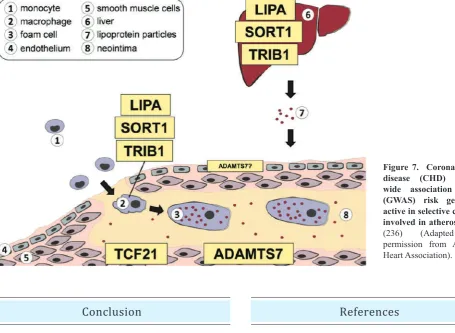
many other genes involved in CHD. Lysosomal acid
lipase A (LIPA), sortilin 1 (SORT1) and tribbles homolog
1 (TRIB1) act as plasma lipid regulators in the liver,
as well as in macrophages biology. Transcription
factor 21 (TCF21) within the vessel wall is upregulated in
dedifferentiated smooth muscle cell, migrate to form fibrous
cap. Adamts7 is also a regulator of smooth muscle
migration but also suggested to role the endothelial cells
(Figure 7).(236)
Figure 7. Coronary heart disease (CHD) genome-wide association studies (GwAS) risk genes are active in selective cell types involved in atherosclerosis.
(236) (Adapted with
permission from American Heart Association).
Cardiovascular system homeostasis is sustained by
the genetic and epigenetic interacting programs. Any
disequilibrium will cause a complex stream of pathogenesis
such as atherosclerosis and its life-threatening complications,
myocardial infarction and stroke. Atherosclerosis is a
chronic inflammatory disease that is initiated by the retention
and accumulation of cholesterol-containing lipoproteins,
particularly low-density lipoprotein, in the artery wall. In the
arterial intima, lipoprotein components that are generated
through oxidative, lipolytic and proteolytic activities lead
to the formation of several danger-associated molecular
patterns, which can activate innate immune cells as well as
vascular cells. Moreover, self- and non-self-antigens, such
as ApoB and heat shock proteins, can contribute to vascular
inflammation by triggering the response of T and B cells
locally. This process can influence the initiation, progression
and stability of plaques. There are several uncertainties and
challenges about the role of genetics in the management
and prevention of chronic disease. The focus on precision
medicine aims to catalyze an effort to better understand
human disease biology, and optimize disease treatment and
prevention by using a combination of clinical, biochemical,
and genetic factors.
Conclusion
References
1. Brown MS, Goldstein JL. Heart attacks: gone with the century? Science. 1996; 272: 629. doi: 10.1126/science.272.5262.629. 2. Mozaffarian D, Benjamin EJ, Go AS, Arnett DK, Blaha MJ, Cushman
M, et al. Heart Disease and Stroke Statistics-2016 Update: A Report
From the American Heart Association. Circulation. 2016; 133: e38-360.
3. Libby P, Bornfelt KE, Tall AR. Atherosclerosis. Success, surprises, and future challenges. Circulation. 2016; 116: 531-4.
4. Gimbrone MA Jr, Garcia-Cardena G. Endothelial cell dysfunction and the pathobiology of atherosclerosis. Circ Res. 2016; 118: 620-36.
5. Ridker PM, Howard CP, Walter V, Everett B, Libby P, Hensen J, et al. Effects of interleukin-1β inhibition with canakinumab
on hemoglobin A1c, lipids, C-reactive protein, interleukin-6, and brinogen: a phase IIb randomized, placebo-controlled trial. Circulation. 2012; 126: 2739-48.
6. Everett BM, Pradhan AD, Solomon DH, Paynter N, Macfadyen J, Zaharris E, et al. Rationale and design of the Cardiovascular In
ammation Reduction Trial: a test of the in ammatory hypothesis of atherothrombosis. Am Heart J. 2013; 166: 199-207.e15.
7. Libby P, Tabas I, Fredman G, Fisher EA. In ammation and its resolution as determinants of acute coronary syndromes. Circ Res. 2014; 114: 1867-79.
8. Musunuru K, Kathiresan S. Surprises from genetic analyses of lipid risk factors for atherosclerosis. Circ Res. 2016; 118: 579-85. 9. Nordestgaard BG. Triglyceride-rich lipoproteins and atherosclerotic
cardiovascular disease: new insights from epidemiology, genetics, and biology. Circ Res. 2016; 118: 547-63.
11. Feinberg MW, Moore KJ. MicroRNA regulation of atherosclerosis. Circ Res. 2016; 118: 703-20.
12. Mega JL, Stitziel NO, Smith JG, Chasman DI, Caulfield MJ, Devlin JJ, et al. Genetic risk, coronary heart disease events, and the clinical
benefit of statin therapy: an analysis of primary and secondary prevention trials. Lancet. 2015; 385: 2264-71.
13. Paynter NP, Ridker PM, Chasman DI. Are genetic tests for atherosclerosis ready for routine clinical use? Circ Res. 2016; 118: 607-19.
14. Herrington W, Lacey B, Sherliker P, Armitage J, Lewington S. Epidemiology of atherosclerosis and the potential to reduce the global burden of atherothrombotic disease. Circ Res. 2016; 118: 535-46.
15. Mihaylova B, Emberson J, Blackwell L, Keech A, Simes J, Barnes EH, et al. The effects of lowering LDL cholesterol with statin
therapy in people at low risk of vascular disease: meta-analysis of individual data from 27 randomised trials. Lancet. 2012; 380: 581-90.
16. Cannon CP, Blazing MA, Giugliano RP, McCagg A, White JA, Theroux P, et al. Ezetimibe added to statin therapy after acute
coronary syndromes. N Engl J Med. 2015; 372: 2387-97.
17. Robinson JG, Farnier M, Krempf M, Bergeron J, Luc G, Averna M, et al. Efficacy and safety of alirocumab in reducing lipids and
cardiovascular events. N Engl J Med. 2015; 372: 1489-99. 18. Sabatine MS, Giugliano RP, Wiviott SD, Raal FJ, Blom DJ, Robinson
J, et al. Efficacy and safety of evolocumab in reducing lipids and
cardiovascular events. N Engl J Med. 2015; 372: 1500-9.
19. Hegele RA, Gidding SS, Ginsberg HN, McPherson R, Raal FJ, Rader DJ, et al. Nonstatin low-density lipoprotein—lowering therapy and
cardiovascular risk reduction—statement from ATVB Council. Arterioscler Thromb Vasc Biol. 2015; 35: 2269-80.
20. Williams KJ, Tabas I, Fisher EA. How an artery heals. Circ Res. 2015; 117: 909-13.
21. Boren J, Williams KJ. The central role of arterial retention of cholesterol-rich apolipoprotein-B-containing lipoproteins in the pathogenesis of atherosclerosis: a triumph of simplicity. Urr Opin Lipidol. 2016; 27: 473-83.
22. Faber M. The human aorta; sulfate-containing polyuronides and the deposition of cholesterol. Arch Pathol. 1949; 48: 342-50.
23. Camejo G, Lopez A, Vegas H, Paoli H. The participation of aortic proteins in the formation of complexes between low density lipoproteins and intima media extracts. Atherosclerosis 1975; 21: 77-91.
24. Iverius PH. The interaction between human plasma lipoproteins and connective tissue glycosaminoglycans. J Biol Chem. 1972; 247: 2607-13.
25. Vijayagopal P, Srinivasan SR, Radhakrishnamurthy B, Berenson GS. Interaction of serum lipoproteins and a proteoglycan from bovine aorta. J Biol Chem 1981; 256: 8234 -41.
26. Smith EB, Slater RS. Lipids and low density lipoproteins in intima in relation to its morphological characteristics. Ciba Found Symp. 1973; 12: 39-62.
27. Tamminen M, Mottino G, Qiao JH, Breslow JL, Frank JS. Ultrastructure of early lipid accumulation in ApoE-deficient mice. Arterioscler Thromb Vasc Biol. 1999; 19: 847-53.
28. Schwenke DC, Carew TE. Initiation of atherosclerotic lesions in cholesterol-fed rabbits. II. Selective retention of LDL vs. selective increases in LDL permeability in susceptible sites of arteries. Arteriosclerosis. 1989; 9: 908-18.
29. Nakashima Y, Fujii H, Sumiyoshi S, Wight TN, Sueishi K. Early human atherosclerosis: accumulation of lipid and proteoglycans in intimal thickenings followed by macrophage infiltration. Arterioscler Thromb Vasc Biol. 2007; 27: 1159-65.
30. Williams KJ, Tabas I. The response-to-retention hypothesis of early atherogenesis. Arterioscler Thromb Vasc Biol. 1995; 15: 551-61.
31. Williams KJ, Tabas I. Lipoprotein retention--and clues for atheroma regression. Arterioscler Thromb Vasc Biol. 2005; 25: 1536-40. 32. Borén J, Gustafsson M, Skålén K, Flood C, Innerarity TL. Role of
extracellular retention of low density lipoproteins in atherosclerosis. Curr Opin Lipidol. 2000; 11: 451-6.
33. Borén J, Olin K, Lee I, Chait A, Wight TN, Innerarity TL. Identification of the principal proteoglycan-binding site in LDL. A single-point mutation in apo-B100 severely affects proteoglycan interaction without affecting LDL receptor binding. J Clin Invest. 1998; 101: 2658-64.
34. Flood C, Gustafsson M, Richardson PE, Harvey SC, Segrest JP, Borén J. Identification of the proteoglycan binding site in apolipoprotein B48. J Biol Chem. 2002; 277: 32228-33.
35. Sloop CH, Dory L, Roheim PS. Interstitial fluid lipoproteins. J Lipid Res. 1987; 28: 225-37.
36. Frank PG, Pavlides S, Cheung MWC, Daumer K, Lisanti MP. Role of caveolin-1 in the regulation of lipoprotein metabolism. Am J Physiol Cell Physiol. 2008; 295: C242-8.
37. Fernández-Hernando C, Yu J, Suárez Y, Rahner C, Dávalos A, Lasunción MA, et al. Genetic evidence supporting a critical role
of endothelial caveolin-1 during the progression of atherosclerosis. Cell Metab. 2009; 10: 48-54.
38. Armstrong SM, Sugiyama MG, Fung KY, Gao Y, Wang C, Levy AS,
et al. A novel assay uncovers an unexpected role for SR-BI in LDL
transcytosis. Cardiovasc Res. 2015; 108: 268-77.
39. Tabas I, Williams KJ, Boren J. Subendothelial lipoprotein retention as the initiating process in atherosclerosis: update and therapeutic implications. Circulation. 2007; 116: 1832-44.
40. Lao KH, Zeng L, Xu Q. Endothelial and smooth muscle cell transformation in atherosclerosis. Curr Opin Lipidol. 2015; 26: 449-56.
41. Bostrom MA, Boyanovsky BB, Jordan CT, Wadsworth MP, Taatjes DJ, de Beer FC, et al. Group V secretory phospholipase A2
promotes atherosclerosis: evidence from genetically altered mice. Arterioscler Thromb Vasc Biol. 2007; 27: 600-6.
42. Oorni K, Kovanen PT. PLA2-V: a real player in atherogenesis. Arterioscler Thromb Vasc Biol. 2007; 27: 445-7
43. Gustafsson M, Levin M, Skalen K, Perman J, Friden V, Jirholt P, et al. Retention of low-density lipoprotein in atherosclerotic lesions of
the mouse: evidence for a role of lipoprotein lipase. Circ Res. 2007; 101: 777-83.
44. Devlin CM, Leventhal AR, Kuriakose G, Schuchman EH, Williams KJ, Tabas I. Acid sphingomyelinase promotes lipoprotein retention within early atheromata and accelerates lesion progression. Arterioscler Thromb Vasc Biol. 2008; 28: 1723-30.
45. Tabas I, Li Y, Brocia RW, Xu SW, Swenson TL, Williams KJ. Lipoprotein lipase and sphingomyelinase synergistically enhance the association of atherogenic lipoproteins with smooth muscle cells and extracellular matrix. A possible mechanism for low density lipoprotein and lipoprotein(a) retention and macrophage foam cell formation. J Biol Chem. 1993; 268: 20419-32.
46. Cohen JC, Boerwinkle E, Mosley TH Jr, Hobbs HH. Sequence variations in PCSK9, low LDL, and protection against coronary heart disease. N Engl J Med. 2006; 354: 1264-72.
47. Nissen SE, Tardif JC, Nicholls SJ, Revkin JH, Shear CL, Duggan WT, et al. Effect of torcetrapib on the progression of coronary
atherosclerosis. N Engl J Med. 2007; 356: 1304-16.
49. Bihari-Varga M. Influence of serum high density lipoprotein on the low density lipoprotein-aortic glycosaminoglycan interactions. Artery. 1978; 4: 504-9.
50. Camejo G, Cortez MM, Lopez F, Starosta R, Mosquera B, Socorro L. Factors modulating the interaction of LDL with an arterial lipoprotein complexing proteoglycan: the effect of HDL. Acta Med Scand Suppl. 1980; 642: 159-64.
51. Umaerus M, Rosengren B, Fagerberg B, Hurt-Camejo E, Camejo G. HDL2 interferes with LDL association with arterial proteoglycans: a possible athero-protective effect. Atherosclerosis. 2012; 225: 115-20.
52. Sneck M, Nguyen SD, Pihlajamaa T, Yohannes G, Riekkola ML, Milne R, et al. Conformational changes of apoB-100 in
SMase-modified LDL mediate formation of large aggregates at acidic pH. J Lipid Res. 2012; 53: 1832-9.
53. Schissel SL, Tweedie-Hardman J, Rapp JH, Graham G, Williams KJ, Tabas I. Rabbit aorta and human atherosclerotic lesions hydrolyze the sphingomyelin of retained low-density lipoprotein. Proposed role for arterial-wall sphingomyelinase in subendothelial retention and aggregation of atherogenic lipoproteins. J Clin Invest. 1996; 98: 1455-64.
54. De Nardo D, Labzin LI, Kono H, Seki R, Schmidt SV, Beyer M, et al. High-density lipoprotein mediates anti-inflammatory
reprogramming of macrophages via the transcriptional regulator ATF3. Nat Immunol. 2014; 15: 152-60.
55. Moore KJ, Fisher EA. High-density lipoproteins put out the fire. Cell Metab. 2014; 19: 175-6.
56. Hewing B, Parathath S, Barrett T, Chung WKK, Astudillo YM, Hamada T, et al. Effects of native and
myeloperoxidase-modified apolipoprotein A-I on reverse cholesterol transport and atherosclerosis in mice. Arterioscler Thromb Vasc Biol. 2014; 34: 779-89.
57. Niyonzima N, Samstad EO, Aune MH, Ryan L, Bakke SS, Rokstad AM, et al. Reconstituted high-density lipoprotein attenuates
cholesterol crystal-induced inflammatory responses by reducing complement activation. J Immunol. 2015; 195: 257-64.
58. Bursill CA, Castro ML, Beattie DT, Nakhla S, van der Vorst E, Heather AK, et al. High-density lipoproteins suppress chemokines
and chemokine receptors in vitro and in vivo. Arterioscler Thromb Vasc Biol. 2010; 30: 1773-8.
59. Nguyen SD, Javanainen M, Rissanen S, Zhao H, Huusko J, Kivelä AM, et al. Apolipoprotein A-I mimetic peptide 4F blocks
sphingomyelinase-induced LDL aggregation. J Lipid Res. 2015; 56: 1206-21.
60. Chiba T, Chang MY, Wang S, Wight TN, McMillen TS, Oram JF,
et al. Serum amyloid A facilitates the binding of high-density
lipoprotein from mice injected with lipopolysaccharide to vascular proteoglycans. Arterioscler Thromb Vasc Biol. 2011; 31: 1326-32. 61. Huang Y, DiDonato JA, Levison BS, Schmitt D, Li L, Wu Y, et al. An
abundant dysfunctional apolipoprotein A1 in human atheroma. Nat Med. 2014; 20: 193-203.
62. Nicholls S, Ray K, Ballantyne C, Beacham L, Miller D, Ruotolo G, et al. Comparative effects of cholesteryl ester transfer protein
inhibition, statin and ezetimibe therapy on atherogenic and protective lipid factors: The accentuate trial. Atherosclerosis. 2016; 252: e237-8.
63. Williams KJ, Fisher EA. Apolipoprotein-B: the crucial protein of atherogenic lipoproteins. In: Wang H, Patterson C, editors. Atherosclerosis: risks, mechanisms, & therapies. Hoboken: John Wiley & Sons; 2015. p.291 – 312.
64. Violi F, Pignatelli P, Basili S. Nutrition, supplements, and vitamins in platelet function and bleeding. Circulation. 2010; 121: 1033-44.
65. Violi F, Carnevale R, Loffredo L, Pignatelli P, Gallin JI. NADPH Oxidase-2 and atherothrombosis. Insight from Chronic Granulamatous Disease. Arterioscler Thromb Vasc Biol. 2017; 37: 218-25.
66. Kinscherf R, Claus R, Deigner HP, Nauen O, Gehrke C, Hermetter A, et al. Modified low density lipoprotein delivers substrate for
ceramide formation and stimulates the sphingomyelin-ceramide pathway in human macrophages. FEBS Lett. 1997; 405: 55-9. 67. Mukhin DN, Chao FF, Kruth HS. Glycosphingolipid accumulation
in the aortic wall is another feature of human atherosclerosis. Arterioscler Thromb Vasc Biol. 1995; 15: 1607-15.
68. Chatterjee SB, Dey S, Shi WY, Thomas K, Hutchins GM. Accumulation of glycosphingolipids in human atherosclerotic plaque and unaffected aorta tissues. Glycobiology. 1997; 7: 57-65. 69. Martin SF, Williams N, Chatterjee S. Lactosylceramide is required in
apoptosis induced by N-Smase. Glycoconj J. 2006; 23: 147-57. 70. Kolmakova A, Kwiterovich P, Virgil D, Alaupovic P, Knight-Gibson
C, Martin SF, et al. Apolipoprotein C-I induces apoptosis in human
aortic smooth muscle cells via recruiting neutral sphingomyelinase. Arterioscler Thromb Vasc Biol. 2004; 24: 264-9.
71. Slowik MR, De Luca LG, Min W, Pober JS. Ceramide is not a signal for tumor necrosis factor-induced gene expression but does cause programmed cell death in human vascular endothelial cells. Circ Res. 1996; 79: 736-47.
72. Mu H, Wang X, Wang H, Lin P, Yao Q, Chen C. Lactosylceramide promotes cell migration and proliferation through activation of ERK1/2 in human aortic smooth muscle cells. Am J Physiol Heart Circ Physiol. 2009; 297: H400-8.
73. Edsfeldt A, Duner P, Stahlman M, Mollet IG, Asciutto G, Grufman H, et al. Sphingolipids contribute to human atheroschlerotic
plaque inflammation. Arterioscler Thromb Vasc Biol. 2016; 36: 1132-40.
74. Orsini F, Cremona A, Arosio P, Corsetto PA, Montorfano G, Lascialfari A, et al. Atomic force microscopy imaging of lipid rafts
of human breast cancer cells. Biochim Biophys Acta. 2012; 1818: 2943-9.
75. Shaw AS. Lipid rafts: now you see them, now you don’t. Nat Immunol. 2006; 7: 1139-42.
76. Pike LJ. Rafts de ned: a report on the Keystone Symposium on Lipid Rafts and Cell Function. J Lipid Res. 2006; 47: 1597-8.
77. Simons K, Ikonen E. Functional rafts in cell membranes. Nature. 1997; 387: 569-72.
78. Anderson RG. The caveolae membrane system. Annu Rev Biochem. 1998; 67: 199-225.
79. Brown DA, London E. Functions of lipid rafts in biological membranes. Annu Rev Cell Dev Biol. 1998; 14: 111-36.
80. Fielding CJ, Fielding PE. Membrane cholesterol and the regulation of signal transduction. Biochem Soc Trans. 2004; 32: 65-9.
81. Chetty PS, Mayne L, Lund-Katz S, Stranz D, Englander SW, Phillips MC. Helical structure and stability in human apolipoprotein A-I by hydrogen exchange and mass spectrometry. Proc Natl Acad Sci USA. 2009; 106: 19005-10.
82. Sorci-Thomas M, Thomas MJ. Microdomains, inflammation and atherosclerosis. Circ Res. 2016; 118: 679-91.
83. Foks AC, Lichtman AH, Kuiper J. Treating atherosclerosis with regulatory T cells. Arterioscler Thromb Vasc Biol. 2015; 35: 280-7. 84. Subramanian M, Thorp E, Hansson GK, Tabas I. Treg-mediated suppression of atherosclerosis requires MYD88 signaling in DCs. J Clin Invest. 2013; 123: 17988.
86. Dixon AM, Drake L, Hughes KT, Sargent E, Hunt D, Harton JA, et al.
Differential transmembrane domain GXXXG motif pairing impacts major histocompatibility complex (MHC) class II structure. J Biol Chem. 2014; 289: 11695-703.
87. Anderson HA, Roche PA. MHC class II association with lipid rafts on the antigen presenting cell surface. Biochim Biophys Acta. 2015; 1853: 775-80.
88. Dubland JA, Francis GA. Lysosomal acid lipase: at the crossroads of normal and atherogenic cholesterol metabolism. Front Cell Dev Biol. 2015; 3: 3. doi: 10.3389/fcell.2015.00003.
89. Jelinek D, Patrick SM, Kitt KN, Chan T, Francis GA, Garver WS. Physiological and coordinate downregulation of the NPC1 and NPC2 genes are associated with the sequestration of LDL-derived cholesterol within endocytic compartments. J Cell Biochem. 2009; 108: 1102-16.
90. Ghosh S. Early steps in reverse cholesterol transport: cholesteryl ester hydrolase and other hydrolases. Curr Opin Endocrinol Diabetes Obes. 2012; 19: 136-41.
91. Ghosh S. Macrophage cholesterol homeostasis and metabolic diseases: critical role of cholesteryl ester mobilization. Expert Rev Cardiovasc Ther. 2011; 9: 329-40.
92. Moore KJ, Sheedy FJ, Fisher EA. Macrophages in atherosclerosis: a dynamic balance. Nat Rev Immunol. 2013; 13: 709-21
93. Neufeld EB, O’Brien K, Walts AD, Stonik JA, Malide D, Combs CA,
et al. The human ABCG1 transporter mobilizes plasma membrane
and late endosomal non-sphingomyelin-associated-cholesterol for efflux and esterification. Biology. 2014; 3: 866-91.
94. Ito A, Hong C, Rong X, Zhu X, Tarling EJ, Hedde PN, et al. Lxrs link
metabolism to in ammation through abca1-dependent regulation of membrane composition and tlr signaling. Elife. 2015; 4: e08009. doi: 10.7554/eLife.08009.
95. Weber C, Noels H. Atherosclerosis: current pathogenesis and therapeutic options. Nat Med. 2011; 17: 1410-22.
96. Lippi G, Franchini M, Targher G. Arterial thrombus formation in cardiovascular disease. Nat Rev Cardiol. 2011; 8: 502-12. 97. Doring Y, Soehnlein O, Weber C. Neutrophil extracellular Traps in
atherosclerosis and atherothrombosis. Circ Res. 2017; 120: 736-43. 98. Döring Y, Drechsler M, Soehnlein O, Weber C. Neutrophils in atherosclerosis: from mice to man. Arterioscler Thromb Vasc Biol. 2015; 35: 288-95.
99. Moreno JA, Ortega-Gómez A, Delbosc S, Beaufort N, Sorbets E, Louedec L, et al. In vitro and in vivo evidence for the role of
elastase shedding of CD163 in human atherothrombosis. Eur Heart J. 2012; 33: 252-63.
100. Soehnlein O. Multiple roles for neutrophils in atherosclerosis. Circ Res. 2012; 110: 875-88.
101. Moore KJ, Tabas I. Macrophages in the pathogenesis of atherosclerosis. Cell. 2011; 145: 341-55.
102. Hansson GK, Libby P, Tabas I. Inflammation and plaque vulnerability. J Intern Med. 2015; 278: 483-93.
103. Sakakura K, Nakano M, Otsuka F, Ladich E, Kolodgie FD, Virmani R. Pathophysiology of atherosclerosis plaque progression. Heart Lung Circ. 2013; 22: 399-411.
104. Yahagi K, Kolodgie FD, Otsuka F, Finn AV, Davis HR, Joner M,
et al. Pathophysiology of native coronary, vein graft, and in-stent
atherosclerosis. Nat Rev Cardiol. 2016; 13: 79-98.
105. Kavuma MM, Rayner KJ, Karunakaran D. The walking dead: macrophage inflammation and death in atherosclerosis. Curr Opin Lipidol 2017; 28: 92-8.
106. Stary HC. Natural history and histological classification of atherosclerotic lesions: an update. Arterioscler Thromb Vasc Biol. 2000; 20: 1177-8.
107. Virmani R, Kolodgie FD, Burke AP, Farb A, Schwartz SM. Lessons from sudden coronary death: a comprehensive morphological classification scheme for atherosclerotic lesions. Arterioscler Thromb Vasc Biol. 2000; 20: 1262-75.
108. Simionescu N, Vasile E, Lupu F, Popescu G, Simionescu M. Prelesional events in atherogenesis. Accumulation of extracellular cholesterol-rich liposomes in the arterial intima and cardiac valves of the hyperlipidemic rabbit. Am J Pathol. 1986; 123: 109-25. 109. Ross R, Glomset JA. Atherosclerosis and the arterial smooth muscle
cell: Proliferation of smooth muscle is a key event in the genesis of the lesions of atherosclerosis. Science. 1973; 180: 1332-9. 110. Ross R, Glomset JA. The pathogenesis of atherosclerosis (first of two
parts). N Engl J Med. 1976; 295: 369-77.
111. Ross R. George Lyman Duff Memorial Lecture. Atherosclerosis: a problem of the biology of arterial wall cells and their interactions with blood components. Arteriosclerosis. 1981; 1: 293-311. 112. Ross R. The pathogenesis of atherosclerosis–an update. N Engl J
Med. 1986; 314: 488-500.
113. Ross R. The pathogenesis of atherosclerosis: a perspective for the 1990s. Nature. 1993; 362: 801-9.
114. Tabas I, García-Cardeña G, Owens GK. Recent insights into the cellular biology of atherosclerosis. J Cell Biol. 2015; 209: 13-22. 115. Libby P. In ammation in atherosclerosis. Nature. 2002; 420: 868-74. 116. Hansson GK. In ammation, atherosclerosis, and coronary artery
disease. N Engl J Med. 2005; 352: 1685-95.
117. Hansson GK, Libby P. The immune response in atherosclerosis: a double-edged sword. Nat Rev Immunol. 2006; 6: 508-19. 118. Davies MJ. Stability and instability: two faces of coronary
atherosclerosis. The Paul Dudley White Lecture 1995. Circulation. 1996; 94: 2013-20.
119. Schwartz SM, Galis ZS, Rosenfeld ME, Falk E. Plaque rupture in humans and mice. Arterioscler Thromb Vasc Biol. 2007; 27: 705-13.
120. Fuster V, Moreno PR, Fayad ZA, Corti R, Badimon JJ. Atherothrombosis and high-risk plaque: part I: evolving concepts. J Am Coll Cardiol. 2005; 46: 937-54.
121. Libby P. Mechanisms of acute coronary syndromes. N Engl J Med. 2013; 369: 883-4.
122. Quillard T, Araújo HA, Franck G, Shvartz E, Sukhova G, Libby P. TLR2 and neutrophils potentiate endothelial stress, apoptosis and detachment: implications for super cial erosion. Eur Heart J. 2015; 36: 1394-404.
123. Gimbrone MA Jr. Vascular Endothelium in Hemostasis & Thrombosis. Edinburgh: Churchill Livingstone; 1986.
124. Gimbrone MA Jr. Vascular endothelium in health & disease. In: Haber E, ed. Molecular Cardiovascular Medicine. New York: Scientific American; 1995. p.49-62.
125. Harrison D, Griendling KK, Landmesser U, Hornig B, Drexler H. Role of oxidative stress in atherosclerosis. Am J Cardiol. 2003; 91: 7-11.
126. Gimbrone MAJ. Endothelial Dysfunction and the Pathogenesis of Atherosclerosis, Proceedings of the Fifth International Symposium. New York: Springer-Verlag; 1980.
127. Gimbrone MA Jr. Vascular endothelium and atherosclerosis. In: Moore S, ed. Vascular Injury and Atherosclerosis. New York: Marcel Dekker; 1981. p.25-52.
128. Gimbrone MA. Atherogenesis: current concepts. In: Schoen FJ, Gimbrone MA Jr, ed. Cardiovascular Pathology: Clincopathologic Correlations and Pathogenic Mechanisms. Baltimore: William & Wilkens; 1995. p.1–11.