Boca Raton London New York Singapore
A CRC title, part of the Taylor & Francis imprint, a member of the Taylor & Francis Group, the academic division of T&F Informa plc.
NEURAL PLASTICITY
IN ADULT SOMATIC
SENSORY- MOTOR SYSTEMS
Edited by
Ford F. Ebner
Vanderbilt University
Department of Psychology
Published in 2005 by CRC Press
Taylor & Francis Group
6000 Broken Sound Parkway NW, Suite 300 Boca Raton, FL 33487-2742
© 2005 by Taylor & Francis Group, LLC CRC Press is an imprint of Taylor & Francis Group No claim to original U.S. Government works
Printed in the United States of America on acid-free paper 10 9 8 7 6 5 4 3 2 1
International Standard Book Number-10: 0-8493-1521-2 (Hardcover) International Standard Book Number-13: 978-0-8493-1521-3 (Hardcover) Library of Congress Card Number 2004058571
This book contains information obtained from authentic and highly regarded sources. Reprinted material is quoted with permission, and sources are indicated. A wide variety of references are listed. Reasonable efforts have been made to publish reliable data and information, but the author and the publisher cannot assume responsibility for the validity of all materials or for the consequences of their use.
No part of this book may be reprinted, reproduced, transmitted, or utilized in any form by any electronic, mechanical, or other means, now known or hereafter invented, including photocopying, microfilming, and recording, or in any information storage or retrieval system, without written permission from the publishers. For permission to photocopy or use material electronically from this work, please access www.copyright.com (http://www.copyright.com/) or contact the Copyright Clearance Center, Inc. (CCC) 222 Rosewood Drive, Danvers, MA 01923, 978-750-8400. CCC is a not-for-profit organization that provides licenses and registration for a variety of users. For organizations that have been granted a photocopy license by the CCC, a separate system of payment has been arranged.
Trademark Notice: Product or corporate names may be trademarks or registered trademarks, and are used only for identification and explanation without intent to infringe.
Library of Congress Cataloging-in-Publication Data
Neural plasticity in adult somatic sensory-motor system / edited by Ford F. Ebner. p. cm. -- (Frontiers in neuroscience)
ISBN 0-8493-1521-2 (alk. paper)
1. Sensorimotor cortex. 2. Neuroplasticity. I. Ebner, Ford F. II. Frontiers in neuroscience (Boca Raton, Fla.)
QP383.15.N475 2005
612.8’252--dc22 2004058571
Visit the Taylor & Francis Web site at
http://www.taylorandfrancis.com
and the CRC Press Web site at
http://www.crcpress.com Taylor & Francis Group
NEURAL PLASTICITY
IN ADULT SOMATIC
FRONTIERS IN NEUROSCIENCE
Series Editors
Sidney A. Simon, Ph.D.
Miguel A.L. Nicolelis, M.D., Ph.D.
Published Titles
Apoptosis in Neurobiology
Yusuf A. Hannun, M.D., Professor of Biomedical Research and Chairman/Department of Biochemistry and Molecular Biology, Medical University of South Carolina
Rose-Mary Boustany, M.D., tenured Associate Professor of Pediatrics and Neurobiology, Duke University Medical Center
Methods for Neural Ensemble Recordings
Miguel A.L. Nicolelis, M.D., Ph.D., Professor of Neurobiology and Biomedical Engineering, Duke University Medical Center
Methods of Behavioral Analysis in Neuroscience
Jerry J. Buccafusco, Ph.D., Alzheimer’s Research Center, Professor of Pharmacology and Toxicology, Professor of Psychiatry and Health Behavior, Medical College of Georgia
Neural Prostheses for Restoration of Sensory and Motor Function
John K. Chapin, Ph.D., Professor of Physiology and Pharmacology, State University of New York Health Science Center
Karen A. Moxon, Ph.D., Assistant Professor/School of Biomedical Engineering, Science, and Health Systems, Drexel University
Computational Neuroscience: Realistic Modeling for Experimentalists
Eric DeSchutter, M.D., Ph.D., Professor/Department of Medicine, University of Antwerp Methods in Pain Research
Lawrence Kruger, Ph.D., Professor of Neurobiology (Emeritus), UCLA School of Medicine and Brain Research Institute
Motor Neurobiology of the Spinal Cord
Timothy C. Cope, Ph.D., Professor of Physiology, Emory University School of Medicine Nicotinic Receptors in the Nervous System
Edward D. Levin, Ph.D., Associate Professor/Department of Psychiatry and Pharmacology and Molecular Cancer Biology and Department of Psychiatry and Behavioral Sciences, Duke University School of Medicine
Methods in Genomic Neuroscience
Helmin R. Chin, Ph.D., Genetics Research Branch, NIMH, NIH
Steven O. Moldin, Ph.D, Genetics Research Branch, NIMH, NIH Methods in Chemosensory Research
Sidney A. Simon, Ph.D., Professor of Neurobiology, Biomedical Engineering, and Anesthesiology, Duke University
The Somatosensory System: Deciphering the Brain’s Own Body Image
Randall J. Nelson, Ph.D., Professor of Anatomy and Neurobiology, University of Tennessee Health Sciences Center
The Superior Colliculus: New Approaches for Studying Sensorimotor Integration
William C. Hall, Ph.D., Department of Neuroscience, Duke University
Adonis Moschovakis, Ph.D., Institute of Applied and Computational Mathematics, Crete New Concepts in Cerebral Ischemia
Rick C. S. Lin, Ph.D., Professor of Anatomy, University of Mississippi Medical Center DNA Arrays: Technologies and Experimental Strategies
Elena Grigorenko, Ph.D., Technology Development Group, Millennium Pharmaceuticals Methods for Alcohol-Related Neuroscience Research
Yuan Liu, Ph.D., National Institute of Neurological Disorders and Stroke, National Institutes of Health
David M. Lovinger, Ph.D., Laboratory of Integrative Neuroscience, NIAAA
In Vivo Optical Imaging of Brain Function
Ron Frostig, Ph.D., Associate Professor/Department of Psychobiology, University of California, Irvine
Primate Audition: Behavior and Neurobiology
Asif A. Ghazanfar, Ph.D., Primate Cognitive Neuroscience Lab, Harvard University Methods in Drug Abuse Research: Cellular and Circuit Level Analyses
Dr. Barry D. Waterhouse, Ph.D., MCP-Hahnemann University Functional and Neural Mechanisms of Interval Timing
Warren H. Meck, Ph.D., Professor of Psychology, Duke University Biomedical Imaging in Experimental Neuroscience
Nick Van Bruggen, Ph.D., Department of Neuroscience Genentech, Inc., South San Francisco
Timothy P.L. Roberts, Ph.D., Associate Professor, University of Toronto The Primate Visual System
John H. Kaas, Department of Psychology, Vanderbilt University
Christine Collins, Department of Psychology, Vanderbilt University Neurosteroid Effects in the Central Nervous System
Sheryl S. Smith, Ph.D., Department of Physiology, SUNY Health Science Center Modern Neurosurgery: Clinical Translation of Neuroscience Advances
Dennis A. Turner, Department of Surgery, Division of Neurosurgery, Duke University Medical Center
Sleep: Circuits and Functions
Pierre-Hervé Luoou, Université Claude Bernard Lyon I, Lyon, France Methods in Insect Sensory Neuroscience
Thomas A. Christensen, Arizona Research Laboratories, Division of Neurobiology, University of Arizona, Tucson, AZ
Motor Cortex in Voluntary Movements
Alexa Riehle, INCM-CNRS, Marseille, France
Preface
Neural plasticity is now well accepted as a universal property of multi-cellular nervous systems. Plasticity has been studied in particular detail in the mammalian cerebral cortex. The word “plasticity” has been applied to a wide variety of cortical changes, so an initial question is always: what metric has been used to conclude that a plastic event has occurred? The chapters in this book illustrate important examples in which the metric for plasticity is physiological alterations in neuronal response properties or changes in behavioral skills. The locus of these changes is in the somatic sensory pathways to and within sensory cortex or motor cortex in response to a variety of challenges. The initial chapters discuss issues relevant to modifications in sensory processing.
Although controversial and easy to ignore, an increasing number of investi-gators are convinced that silent neurons need further study. In somatic sensory cortex the silent neuron idea is linked to a 1988 paper by Robert Dykes and Yves Lamour in which they showed that a large fraction of cortical cells did not fire action potentials in response to tactile stimuli, even though the cells seemed healthy and responded vigorously to locally applied glutamate. Their hypothesis that the silent neurons become wired into cortical circuits during learning was too novel, and arrived too early, to be embraced by other workers in the field without additional lines of evidence. Strong evidence for the existence of silent neurons has since appeared, and the chapter by Michael Brecht and his colleagues in this book poses important questions about the silent neurons’ role in cortical function. The specific contribution of these neurons to cortical plasticity is a particularly important ongoing idea that remains to be clarified.
rep-resentation into isofrequency columns, modular groups of cells that all respond best to the same amplified frequency. These novel findings are considered with regard to classical theories of how resonance facilitates perception in other sensory systems, ranging from the cockroach to the human ear, and also consider how these principles of the biomechanical transduction of information may provide lessons for under-standing the optimal use of tools by humans.
Continuing the coding theme more centrally, Mathew Diamond then discusses the role of modular, maplike cortical organization in the processing of sensory information, including the functional significance of cortical maps, as well as the individual modules that create the topographic framework for spatial coding in primary sensory cortex. These spatial rules for barrel cortex plasticity co-exist with temporal fluctuations in excitability (temporal coding), characterized in anesthetized rats by bursts of spikes that are synchronized across the entire barrel cortex. The bursts appear to briefly open a plasticity gate allowing incoming sensory inputs to modify the efficacy of the activated intracortical circuits. During the time between bursts the plasticity gate is closed and incoming inputs have no long-term effect on intracortical circuits. These modifications by sensory input patterns during discrete intervals provide a theoretical basis for understanding barrel cortex changes in awake, exploring rats because rhythmic oscillations occur in awake rat cortex as well.
was substantial between the two hands, presumably based on interhemispheric connections. In subsequent studies, these findings were extended to a variety of tactile stimuli and tasks leading to the conclusion that transfer of tactile learning appears to be a general rule. It is interesting to speculate that interhemispheric transfer of tactile learning may relate to intermanual referral of tactile sensations following amputation or stroke. The mechanisms of perceptual learning are relevant to the perceptual improvements that are observed in spared modalities following sensory deprivation in a particular modality, such as improved tactile skills in people with very low vision.
Examples of somatic sensory processing after early postnatal sensory deprivation has identified a number of ways in which activity is needed to develop normal sensory processing in cortex. Ford Ebner and Michael Armstrong-James describe the nature of cortical impairments induced by low activity during the early postnatal period in the somatic sensory system in rats and mice after they mature to normal-looking adults. The literature shows that both excitatory and inhibitory processes are affected by sensory deprivation, with the severity of effects depending upon the time of onset, the duration of the deprivation, and the length of the recovery period after deprivation ends. Intracortical circuit dynamics are most severely affected. Neural transmission from cortical layer IV to more superficial layers II/III is a major site of synaptic dysfunction. Trimming all whiskers produces a more uniform down-regulation of sensory transmission than trimming a subset of whiskers presumably because restricted deprivation creates competition between active and inactive inter-connected cell groups. Activity-based changes in function can be induced by altered tactile experience throughout life, but early postnatal deprivation degrades neuronal plasticity, and interferes with the animal’s ability to learn subtle tactile discrimina-tions throughout life.
The remaining chapters deal with the motor side of sensory-motor transforma-tions.
John Chapin and his colleagues discuss the mechanisms by which the brain transforms sensory inputs into motor outputs. The rules for such sensory-motor conversions have proven elusive, and the authors suggest that this is due to the multiplicity of “bridges” between these systems in the CNS. Moreover, while the development and maintenance of the sensorimotor transformation machinery must involve some sort of plasticity, it is not yet clear how or where this plasticity occurs. They then offer specific recommendations for studying these issues in awake animals performing behaviors that involve sensory-motor transformations, an area in which they have made significant contributions.
the task. Further, multijoint responses to ICMS were infrequent before training, but were found in abundance after digit training. The implication is that simultaneous movements may become associated in the cortex through Hebbian synaptic mech-anisms in which horizontal fibers connecting two areas become strengthened through associated repetitive activation. When spontaneous recovery was studied at 3 to 5 months after a hand area motor cortex lesion, skilled use of the hand returned, but roughly half of the digit movement representation was still replaced by shoulder and elbow. However, if squirrel monkeys were trained to retrieve food pellets from food wells, and then re-trained after a motor cortex lesion using the less affected hand (ipsilateral to a small infarct), the monkeys returned to baseline levels on the most difficult food-well task. In this case, motor skill training saved the remaining pre-infarct distal hand representation from the expected takeover by surrounding inputs. The implication of these results is that physical rehabilitation after stroke can drive physiological changes in the cortex associated with recovering skilled hand use, if the conditions are optimized.
Jon Kaas then discusses how motor experience rebalances dynamic systems to reveal latent neural circuit properties. Short term changes emerge over a time period ranging from seconds to hours due to a range of activity-dependent cellular mech-anisms that affect synaptic strengths. Over somewhat longer periods of days to weeks, anatomical circuits may be lost or gained as local circuits grow and rearrange. Over a time period of weeks to months, considerable new growth of axons and synapses can occur that considerably alter the functional organization of sensory and motor systems, sometimes in ways that promote behavioral recovery, and some-times in ways that do not promote such recovery.. One goal of research on sensory-motor plasticity is to understand the mechanisms of change and how to manipulate them in order to maximize recovery after sensory and motor loss. This chapter focuses on changes in the motor system that are the result of a particularly severe type of motor system damage— the loss of an entire forelimb or hindlimb. In humans, badly damaged limbs might require amputation, and it is important to determine what happens to the somatosensory and motor systems as a result of the loss of both the sensory afferents from the limb and the motor neuron outflow to the muscles of that limb.
primary somatosensory cortex has been demonstrated to be strongly correlated with the magnitude of phantom limb pain. Interestingly, phantom pain was more prom-inent in patientsin whom the motor representations of face muscles were displaced medially, possibly reflecting an invasion of the face motor representation in motor cortex.
In the last chapter the behavioral basis of focal hand dystonia is discussed by Nancy Byl as a form of aberrant learning in the somatic sensory cortex. The cause of this disabling movement disorder has remained elusive. It is common in produc-tive, motivated individuals, such as musicians, who perform highly repetiproduc-tive, intensive hand tasks., Their studies document degradation of the cortical somatosen-sory representation of the hand characterized by large receptive fields overlapped across adjacent digits, overlap of glabrous-hairy surfaces, persistence of digital receptive fields across broad cortical distances, high ratio of amplitude to latency in somatic sensory evoked field responses, and abnormal digit representation. Chal-lenging, rewarded, repetitive behavioral tasks that require high speed, high force, precision and intense work cycles with minimal breaks accelerate the onset and severity of dystonia. The development of dystonia may be minimized if individuals use the hands in a functional, mid-range position, take frequent breaks, work at variable speeds for short durations, attend to sensory-motor feedback, and initiate digital movements with the intrinsic muscles. The central theme is that attended, progressive, rewarded, learning-based sensory-motor training consistent with the principles of neuroplasticity, can facilitate recovery of task-specific motor control.
All of the examples in this book suggest that our understanding of neural plasticity and its mechanisms is increasing at a rapid rate, and that the knowledge will modify many of the procedures now in place to improve perceptual and motor skills after brain damage.
Contents
Chapter 1 Silent Neurons in Sensorimotor Cortices: Implications for Cortical Plasticity
Michael Brecht, Miriam Schneider, and Ian D. Manns
Chapter 2 The Vibrissa Resonance Hypothesis
Christopher Moore and Mark L. Andermann
Chapter 3 Spatial and Temporal Rules Underlying Rat Barrel Cortex Plasticity
Mathew E. Diamond
Chapter 4 Probing the Cortical Evidence of Somatosensory Discrimination
Ranulfo Romo, Adrián Hernández, Antonio Zainos, Luis Lemus, Victor de Lafuente, and Rogelio Luna
Chapter 5 Perceptual Learning and Referral in the Tactile System
K. Sathian
Chapter 6 The Effects of Sensory Deprivation on Sensory Function of SI Barrel Cortex
Ford F. Ebner and Michael Armstrong-James
Chapter 7 Role of Plasticity in Sensorimotor Transformations
Linda Hermer-Vazquez, Raymond Hermer-Vazquez, and John K. Chapin
Chapter 8 Neural Plasticity in Adult Motor Cortex
Chapter 9 Reorganization of Motor Cortex after Damage to the Motor System
Jon H. Kaas
Chapter 10 Modulation of Cortical Function and Plasticity in the Human Brain
Friedhelm Hummel, Christian Gerloff, and Leonardo G. Cohen
Chapter 11 Behavioral Basis of Focal Hand Dystonia: Aberrant Learning in the Somatosensory Cortex
Editor
Ford F. Ebner, Ph.D., was raised in the American Pacific Northwest where he
Contributors
Mark L. Andermann
Massachusetts Institute of Technoolgy, McGovern Insitutte of Brain Research and Department of Brain
Adrián Hernandez Department of Neurology and Hertie Institute for Clinical Brain Research
Max Planck Institute for Medical Research Department of Cell Physiology
Christoper I. Moore
Massachusetts Institute of Technology, McGovern Insitute of Brain Research and Department of Brain
-40 -20 0 20 40
220 mm/s 440 mm/s 660 mm/s
-2 A4 Grating Model A4 Grating Experiment
C4 Sandpaper Model C4 Sandpaper Experiment
Frequency (Hz) 130 210 290
NV B2 Vibrissa FSU E2 Vibrissa E2 Vibrissa E2 Vibrissa
FIGURE 2.5 Vibrissa
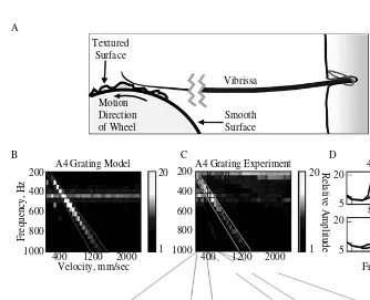
res-the spike in vibrissa velocity (P(f)*f, top) at a wheel speed of 800 mm/s. Neural activity also showed a spike in mean firing rate at this velocity (green line). Neural activity also demon-strated a thresholded sensitivity to the increasing velocity of vibrissa oscillation at higher frequencies (≥ a wheel speed of 2000 mm/sec; see also Figure 2.8). B. Power spectra showing increased velocity of vibrissa motion or increased amplitude of neural activity (bottom) as a function of oscillation frequency and wheel speed. In the top panel, the peak in velocity signal at ~350 Hz (global increase in power) reflects the increased velocity of vibrissa motion generated when the wheel speed drove the predominant spatial frequency present in the texture (shown in the diagonal bands) at the vibrissa resonance (blue box). The increased mean firing rate in the associated neural response is indicated by the vertical band of increased activity observed at a wheel speed of 800 mm/s in the bottom panel. Note also that a peak is present in MUA power spectrum at the vibrissa resonance (~350 Hz), indicating fine temporal fidelity of spiking activity in response to a complex stimulus presentation (see also Figures 2.12–2.14).
Wheel Velocity (mm/s)
800 Velocityof Vibrissa Motion
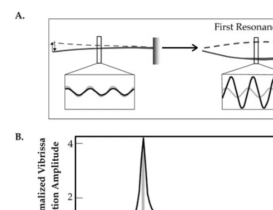
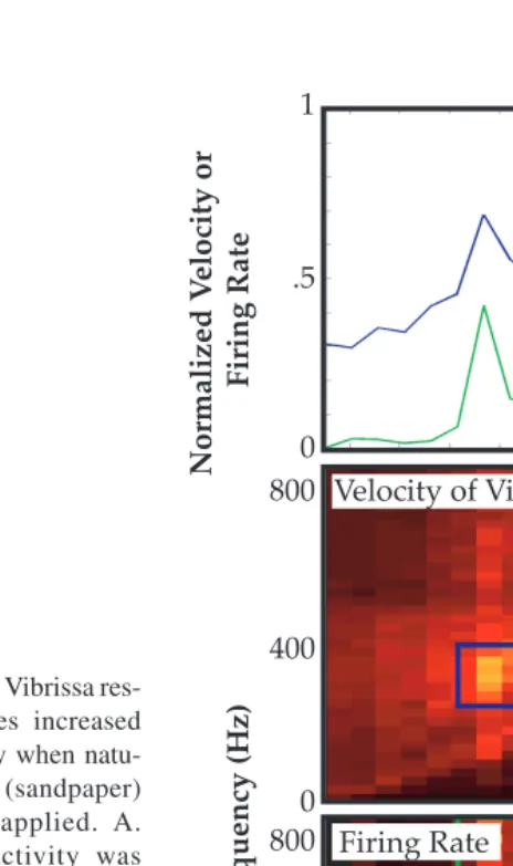
FIGURE 2.6 Vibrissa resonance evokes increased neural activity when synthesized com-plex stimuli are applied. A. White noise stimuli constructed as the sum of phase-shifted sinusoids from 0–600 Hz were presented through a piezoelectric stimulator to the vibrissa. A notched stimulus was also created in which the fundamental resonance and surrounding frequencies (400–500 Hz) were removed from the stimulus and the power adjusted across remaining frequencies. Vibrissa oscillations showed a resonance amplification at ~450 Hz when white noise stimuli were applied (green line) that is not present when notched stimuli were applied (blue line). B. These complex stimuli were presented while recording from a trigeminal ganglion single unit. Average neural activity was summed over the stimulation period (500 msec). As predicted by the differential increase in vibrissa motion, greater mean firing rate was evoked by the non-notched (green bar) than the notched stimulus (blue bar) (N = 37 trials, mean ± SE).
0 200 400 600
0 5 10
WithoutNotching With Notching
S
pi
k
es/s
Frequency (Hz)
N
ormalized
Motion
Amplitude 1
A.i.
200 400 600 200 400 600
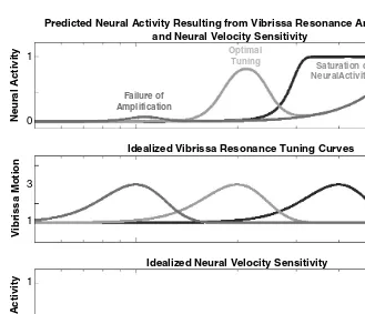
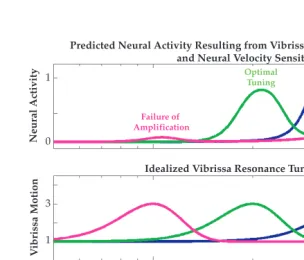
FIGURE 2.8 Neural velocity sensitivity may impact the expression of vibrissa resonance. Bottom panel A model of the neural response to vibrissa stimulation frequency in the absence of resonance amplification. This function was modeled as sin2(pi*f/2000), 0 > f > 1000 Hz, to emulate the neural sensitivity to higher frequency stimulation resulting from velocity sensitivity. Examples of this kind of increase in firing as a thresholded function of vibrissa velocity can be observed in real neural data in Figures 2.4, 2.5, and 2.10 (see also Reference number 63). Middle panel Three idealized examples of vibrissa resonance tuning showing a 3:1 gain in motion amplitude at the fundamental resonance frequency and bandwidth proportional to this frequency. Top panel The predicted neural response to vibrissa stimulation frequency as a function of resonance amplification of peak motion velocity, and intrinsic velocity sensitivity thresholds. For a given amplitude of stimulation, vibrissa resonance amplification that does not drive a neuron near its velocity threshold may fail to be amplified (purple curve, left resonance peak), while resonance amplification that is significantly above the velocity threshold (shown in the bottom panel) may fail to demonstrate tuning due to an upper limit on the range of possible firing rates for a given neuron (blue curve, right resonance peak). A subset of vibrissa resonance tuning curves near to but not above the intrinsic velocity threshold will, in this model, show optimal frequency tuning. Preliminary data suggest that these effects occur in a subset of trigeminal and cortical neurons, and that, within SI, FSU and multi-unit recordings are more susceptible to these impacts of velocity sensitivity.
100 200 400 600
IdealizedNeural VelocitySensitivity Idealized Vibrissa Resonance Tuning Curves
PredictedNeural Activity Resultingfrom Vibrissa Resonance Amplification
andNeural VelocitySensitivity
Failure of
Amplification
Optimal
Tuning Saturation of
Neural Activity
Saturation of Neural Activity
Frequency (Hz)
N
eural
Activity
N
eural
Activity
V
ibrissa
Motion
0 1 0 1
FIGURE 2.10] An example of the temporal evolution of neural frequency tuning. A. Top Peri-stimulus time histograms (PSTHs) are plotted as a function of frequency of stimulation (ordinate) and time (abscissa). Stimuli were applied as 160 µm sinusoids for 500 msec epochs. Resonance tuning can be seen in the selective band of increased firing at ~135 Hz: Intrinsic frequency (velocity) sensitivity can be seen in the increased firing above the threshold of ~ 350 Hz. Bottom Neural tuning curves showing mean firing rate for four different epochs post-stimulus onset. Resonance driven activity was not observed in the first epoch (0–25 msec post-stimulus onset) although robust high frequency responses were present. In later epochs, responses above the intrinsic high frequency threshold diminished in relative prominence while resonance driven neural activity increased. B.Top panel PSTH
of activity evoked at the fundamental resonance frequency (red, 135 Hz) and at a frequency above the intrinsic high frequency threshold (black, 460 Hz). The slower rise time of resonance driven neural activity can be appreciated in this PSTH. Middle panels Traces of vibrissa motion driven by fundamental resonance frequency and high frequency stimuli. The fundamental resonance frequency driven motion shows a gradual increase in motion ampli-tude (red trace). Bottom panel Plots of the peak velocity of vibrissa motion for the funda-mental (red) and high frequency stimuli (black). This time constant for the amplification of vibrissa motion is likely a key factor in the delayed increase in resonance driven activity in this example (see also Figure 2.11).
τ piezo
Evolution of Neural Frequency Tuning
A. B.
Incidence of Neural Frequency Tuning
FIGURE 2.12 Trigeminal ganglion neurons demonstrate neural tuning and an atonal inter-val in the fine timing of their evoked activity. A. The top graph shows a frequency tuning curve for a trigeminal unit, constructed by counting all evoked inter-spike intervals (ISIs), a measure that is functionally equivalent to the mean firing rate. The lower graph shows the count of ISIs at the driving period, indicating fine temporal following of the neuron. Numbers adjacent to each curve indicate the amplitude of vibrissa stimulation applied. Frequency tuning was observed in both the mean firing rate and in the fine timing of neural evoked activity. An increased mean firing rate was observed for stimuli ≥32 µm, while temporal following at the driving frequency was present only for larger amplitudes of stimulation, ≥48 mm. This finding parallels similar observations made in the primate somatosensory system.76 B. A graph of the incidence of ISIs at the fundamental resonance frequency, plotted as a function of the amplitude of stimulation (yellow indicates increased incidence) for the example in A. At larger amplitudes of stimulation, only firing at the fundamental resonance frequency was observed, as shown by the exclusive presence of ISIs at ~7 msec at 80 µm stimulation. In contrast, lower amplitudes of stimulation evoked ISIs at multiples of the driving period. C. A plot from a different single-unit trigeminal recording, showing the mean firing rate (red) and power at the driving frequency (blue). As in the example shown in A and B, temporal following provides a more precise tuning function at frequencies surrounding the vibrissa resonance frequency.
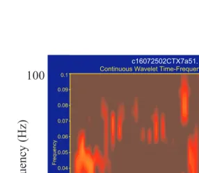
FIGURE 7.6 Sub- and suprathreshold neural activity in the vicinity of one M1 electrode during one trial of skilled reaching. The x-axis shows task-time, the y-axis shows frequency from 0 to 100 Hz, and pixel color represents amount of energy, with hotter colors indicating higher amounts of energy. See text for further description.
c16072502CTX7a51.
Continuous Wavelet Time-Frequency Spectrum
0 200 400 600
Time 0
0.01 0.02 0.03 0.04 0.05 0.06 0.07 0.08 0.09 0.1
F
requency
0 100 50 33.333 25 20 16.667 14.286 12.5 11.111 10
Per
iod
Final Sniff
Contact
Time (ms)
100
Frequency (Hz)
1
Silent Neurons in
Sensorimotor Cortices:
Implications for
Cortical Plasticity
Michael Brecht, Miriam Schneider,
and Ian D. Manns
CONTENTS
I. Evidence for a Predominance of Silent Cortical Neurons in Sensorimotor Cortices
A. Evaluation of Presynaptic Activity in the Somatosensory Cortex
1. Results from Extracellular Unit Recording 2. Results from Sharp Microelectrode Recordings 3. Results from Whole-Cell Recordings in the Vibrissae
Barrel Cortices of Anesthetized Animals
4. Results from Whole-Cell Recordings in the Vibrissae Barrel Cortices of Awake Animals
5. Inconsistencies and Caveats from Whole-Cell Studies 6. Results from Derivatives of the Whole-Cell
Recordings Technique
7. All Techniques Agree that Firing Rates of Cortical Neurons are Very Heterogeneous
B. Evaluation of Postsynaptic Activty in the Somatosensory Cortex
1. Experimental Agreement on the Amplitude of Subthreshold Signals
2. The Synaptic Composition of Postsynaptic Responses Is Controversial
C. Evaluation of the Motor Effects of APs in the Primary Motor Cortex
1. Low Firing Rates Imply a High Efficacy of Cortical APs 2. Small Numbers of APs in Single Cells of the Primary
Motor Cortex Can Evoke Movements
3. Effective Cells Greatly Outnumber Active Cells in M1 II. Silent Neurons and Cortical Plasticity
A. Cortical Plasticity and Alternative Hypotheses for Silent Neurons
1. Silent Cortical Cells as a Corollary of Metabolic Demands
2. Contribution of Silent Cells to Learning and Cortical Plasticity
3. Silent Cells May Signal by Non AP-Dependent Signaling 4. Silent Cells May Function by Sparse AP Activity
B. Data on an Involvement of Silent Cells in Plasticity are Largely Absent
C. Silent Neurons and Synaptic Learning Rules III. Conclusion
References
Silence is music, too.
Miles Davis
Evidence indicates that cortical neurons are mostly silent. Early in the 1970s, Barlow hypothesized that few neurons may be sufficient for a sensory representation.1 This was clarified 15 yr ago by extracellular recordings combined with glutamate appli-cation that indicated a large fraction of cortical cells do not fire spikes in response to tactile stimuli.2 An important implication is that if silent cells, are “desilenced,” they could profoundly contribute to cortical plasticity.
This chapter will review the following: 1. Evidence from new experimental approaches indicates that only a very small fraction of cortical cells do fire APs. 2. While a potential role for these enigmatically silent cortical neurons in cortical plasticity is an attractive hypothesis, very little evidence for this is provided. 3. Examining the contribution of silent cortical neurons to cortical plasticity poses conceptual and experimental challenges.
I. EVIDENCE FOR A PREDOMINANCE OF SILENT CORTICAL NEURONS
IN SENSORIMOTOR CORTICES
review all evidence. Instead, we will mention some landmark studies and point out that very diverse techniques lead to that conclusion. Attention will be paid to our own studies on the quantitative description of neural activity in a rat’s barrel cortex. We will refer to cells as silent neurons, if they discharge less then 0.1 APs for an optimized stimulus (in our case a strong backward deflection of the best whisker).
A. EVALUATION OF PRESYNAPTIC ACTIVITY
IN THE SOMATOSENSORY CORTEX
1. Results from Extracellular Unit Recording
Extracellular unit recording was the first and is still the most common technique to quantify cellular activity in the somatosensory cortex. The general impression of most of these studies is that neurons in the somatosensory cortex discharge APs when the appropriate tactile stimulus is applied.3 Results have been obtained not only in the cat3 and monkey4 primary somatosensory cortex (S1), but also in the vibrissae barrel cortices of rats that were both anesthetized5 and awake.6 In studies like this, when responses to controlled deflections of the (best) principal whisker (PW) are quantified, values around 1 AP per PW stimulus were reported.6-9 These studies also report a considerable level of spontaneous AP activity of around 1Hz.10,11 However, not all studies that analyzed S1 activity by unit recordings came to these conclusions. In a series of influential papers, Dykes and colleagues argued that most neurons in the somatosensory cortex could not be driven by conventional stimuli.2,12 What made these studies so compelling, was the deliberate effort to analyze every AP discharge in order to minimize sampling biases, and even more so, the use of iontophoretic injections of glutamate and other neurotransmitters, which uncovered the existence of previously unresponsive neurons around the recording electrode. Few researchers fully agree to the idea of a majority of unre-sponsive cells in the S1 cortex. Nonetheless the technical elegance of the work of Dykes and colleagues has made it clear that unit recordings result in enormous sampling biases against neurons with low levels of AP activity. Swadlow supported this idea in a series of studies on various cortical areas. In these studies, sampling biases were minimized by antidromic identification of recorded units.13-16 Apart from exceptions like corticofugally projecting layer V neurons, most identified neurons in these studies were found to have very low spontaneous and evoked AP activity. Similarly, some recent unit recording studies on the vibrissae barrel cortex report rather low rates of AP activity.17
2. Results from Sharp Microelectrode Recordings
appear that leaks introduced by the impalement of the cell are responsible for more depolarized membrane potentials and high firing rates observed in sharp microelectrode recordings.20
3. Results from Whole-Cell Recordings in the Vibrissae Barrel Cortices of Anesthetized Animals
The whole-cell recording technique has been used for about ten years for in vivo
recordings.21,22 A substantial number of studies were conducted in the vibrissae barrel cortex of anesthetized rats and most of them came to similar conclusions with respect to AP activity. As first reported, for urethane-anesthetized rats by Moore and Nelson 199823 and confirmed by Zhu and Connors 1999,24 most neurons in the barrel cortex of animals anesthetized with barbiturates do not show evoked AP responses. In our laboratory, we conducted a series of recording studies under urethane anesthesia on identified neurons in the vibrissae region of the ventral posterior medial (VPM) thalamus and the barrel cortex. In the VPM we observed a mean of 0.5 APs per (6˚) PW deflection,25 a result that is only two-fold lower than the findings of unit recordings in the VPM, which report about ~1 AP per PW stimulus.26,27 In contrast, for layer IV barrel cortex neurons we observed a mean of 0.14 APs per PW deflection,28 a response that is five- to ten-fold smaller than what has been reported for layer IV neurons by unit recordings under the same anesthesia.9,29 In layers II/III, we observed evoked AP rates of only 0.031 APs per PW stimulus,30 which is about 40-fold less than what has been reported by unit recordings.29 Thus, the low AP rate estimate of whole-cell recordings versus that of extracellular unit recordings is in line with the idea that unit recordings bias against cells with low firing rates. Indeed, if the firing rate estimates of whole-cell recordings for layer II/III are correct, most of these whole-cells could not possibly be detected by unit recordings because they do not fire APs.
4. Results from Whole-Cell Recordings in the Vibrissae Barrel Cortices of Awake Animals
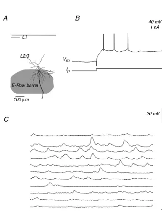
FIGURE 1.1 Activity of a star-shaped pyramidal layer IV neuron in an awake rat A, coronal section through the barrel cortex with topographic position, and the dendritic, axonal arbor of the stimulated pyramid in L4. B, Current injection reveals a regular spiking pattern. C, Ongoing activity of the neuron, while the animal is resting and while it is whisking. Note the absence of APs.
E-Row barrel L1
L2/3
100µm
40 mV
100 ms 1 nA
Ip
resting
whisking Vm
20 mV
100 ms
B
A
5. Inconsistencies and Caveats from Whole-Cell Studies
For obtaining whole-cell recordings, pressure applied to the pipette interior prevents pipette occlusion while cells are approached. As a consequence, intracellular high-potassium solution is pushed into the tissue, and this depolarizes neurons and leads to a transient depression of neuron firing. We therefore compared the results of whole-cell recordings obtained from recordings where we minimized the spill of internal solution (by patching cells with minimal pressure applied to the pipette interior and the first pipette in the experiment) to recordings with massive spillover (patching cells with high pressures after numerous electrode penetrations). With high spillover of potassium, postsynaptic activity can be suppressed during the first 1 to 2 minutes of the recording. This was not the case in recordings without large potassium spillover. However, after less than 5 minutes, recordings under the two conditions were indistinguishable. It is unlikely that spillover of internal solution is a major contributor to the low firing rates observed with whole-cell recordings. Another potentially confounding factor for whole-cell recordings is dialysis of cells by the recording pipette as described below.
Observations in the barrel cortex tend to indicate very low firing rates with whole-cell recordings.23,24,28,30 One study however, reports substantially higher rates of AP activity (spontaneous AP rates of about 1 AP/s,32 The origin of this difference unfortunately is unknown.
6. Results from Derivatives of the Whole-Cell Recordings Technique
a. Cell-Attached Recordings
As already mentioned,, dialysis of recorded neurons with intracellular solution may distort the results of whole-cell recordings. To address this issue we performed sequential cell-attached and whole-cell recordings of AP activity from neurons in the barrel cortices of anesthetized animals.22 To exclude any unintended dialysis, we tested before and after the cell-attached recordings to see whether a giga-seal between pipette and recorded neuron was established. We observed low firing rates (spontaneous AP rates less than 0.1Hz, and less than 0.1 evoked AP per PW stimulus) in both cell-attached and whole-cell recordings and found that AP activity slightly increased in neurons after establishing the whole-cell configuration. Cell-attached recordings are single-cell extracellular recordings, selected for seal formation and not for AP activity, suggesting high firing rates observed with extracellular unit recordings are a result of sampling biases.
b. Targeted Whole-Cell Recordings
Since basically all techniques for recording cellular cortical activity in vivo rely
such interneurons display low levels of evoked AP activity (0.3 APs per PW stimulus). This estimate of AP activity is lower than what was reported from most unit studies of putative interneurons.34-36 As neurons are optically selected by this technique, the possibility of non-representative sampling from a blind approach biased by firing rate is ruled out.
c. Transneuronal Recordings of Spikelet Activity
It has become clear that cortical interneurons are mutually coupled by electric synapses. In the case of strong coupling, presynaptic APs result in an AP-like waveform in postsynaptic interneurons called a spikelet.37,38 As predicted, such spikelets are observed in vivo in recordings from interneurons.33 These recordings reveal that large spikelets (>2mV) occur at low rates between 0.2 to 2.7 Hz (mean ca. 0.5 Hz). Since spikelet-events are likely to reflect APs in one or more electrically coupled presynaptic cells, this infers that most interneurons discharge at low rates <1Hz. It is important to note that AP rates inferred from transneuronal recordings reflect the activity of cells that are not directly recorded and thought to be unaffected by factors such as dialysis of intracellular solution. Thus, a large number of factors that could distort AP counts in conventional recordings can be excluded here. Once again, the AP rates are much lower than those reported from extracellular studies.34-36
7. All Techniques Agree that Firing Rates of Cortical Neurons are Very Heterogeneous
As discussed above, different recording techniques lead to different quantitative assessments of cortical activity. Still most researchers agree that spontaneous and evoked AP rates can be very diverse and may vary by ×10 to ×100 between cells.
B. EVALUATION OF POSTSYNAPTIC ACTIVTY
IN THE SOMATOSENSORY CORTEX
1. Experimental Agreement on the Amplitude of Subthreshold Signals
2. The Synaptic Composition of Postsynaptic Responses Is Controversial
Given general agreement on the amplitude of PSP responses, opinions diverge on the composition of synaptic events that underlie such responses. Some authors favor the idea of a large number of synaptic excitatory and inhibitory inputs that balance each other out to create much smaller net PSPs. Such hypotheses are referred to as high input regimes or synaptic bombardment scenarios.41,42 We have suggested that this may not be the case and that cortical responses are generated by a few carefully selected inputs designated the selective input regime.28
Cortical neurons make and receive a large number of synaptic contacts. For example a layer II/III neuron may receive and allocate about 10,000 (thousand) terminals.43 Since neuron connections between neurons often consist of around five terminals,44,45 each neuron will form connections with a few thousand pre- and postsynaptic cells. In the barrel cortex, it is clear that the average amplitude of single unitary connections is often in the size range of 10% of a sensory evoked PSP. Thus, layer IV PSPs for PW stimuli are on average around 14 mV in size, whereas layer IV to layer IV unitary connections are around 1.6 mV in amplitude.44 In layer II/III neurons, sensory evoked PSPs are around 9 mV in amplitude and layer II/III to layer II/III unitary connections are around 0.8 mV46 in amplitude. In layer V neurons sensory evoked PSPs are on average 5 mV,47 while unitary connections are found to be 0.3 mV.48
From these numbers it is clear that few out of the several thousand presynaptic inputs could underlie sensory responses in the barrel cortex, but a balanced excita-tion-to-inhibition scenario is also possible. This balanced scenario suggests that huge inhibitory and excitatory inputs are hidden in net response. Informal testing of such scenarios by current injection experiments fail to uncover such hidden inputs (Brecht, unpublished data). For slightly suboptimal stimuli, one often observes unitary-response-like synaptic events and total response failures (Brecht and Sakmann, unpublished observations). Such observations are difficult to reconcile with the idea that responses are generated by hundreds or thousands of balanced excitatory and inhibitory synaptic inputs.
3. An Attempt to Quantitatively Determine the Synaptic Composition of a Cortical Sensory Response Suggests Very Low Presynaptic Activity
neuron model49 of a morphologically reconstructed layer II/III neuron. Passive prop-erties of the model, such as membrane resistance, membrane capacitance, and axial resistance were adjusted to appropriate values by comparing the voltage responses of the simulated neuron to those of the recorded neuron.
To simulate the real synapses from layer II/III and layer IV, synaptic conductance changes were placed onto the model neuron. Geometric synaptic distribution and unitary synaptic strength for these synapse-models were taken from in vitro
mea-surements.46,50,51 To mimic realistic temporary synaptic distributions, presynaptic spike times measured in vivo in layer II/III and layer IV were convolved with typical
PSP response time course measured in vitro.
The response analysed here (Figure 1.2B) was small (approximately 100 pA amplitude), but in the range of other PSC amplitudes observed for PW stimulation in these experiments (30pA – 180pA, n = 7). Analysis of this realistic model and comparison of its sensory responses to the recorded sensory response revealed that 10 to 30 active excitatory unitary connections and 1 to 10 inhibitory unitary con-nections best reproduce the clamping behaviour of the recorded sensory responses (Figure 1.2C). More precisely in this scenario, which reproduced the real measure-ment best, 50 excitatory and 7 inhibitory active synaptic terminals corresponding to ~11 excitatory and ~2 inhibitory presynaptic neurons were active (taking 4.5 as the average number of synaptic terminals per unitary connection46). From our simula-tions one can reject a high input scenario (Figure 1.2D), because the voltage clamp
FIGURE 1.2 Voltage clamp behavior of real and simulated synaptic responses of a L2 cortical
pyramidal neuron in barrel cortex A, morphology of the recorded and biocytin filled neuron. B, current response to a 6˚ PW deflection of L2/3 rat barrel cortex neuron for three different holding potentials (arrow: stimulus onset). C, current response of the “best fit” scenario model with 50 excitatory and 7 inhibitory synaptic terminals, which is reproducing the real exper-iment most closely. D, current response of the “synaptic bombardment” scenario model with ~ 500 excitatory and ~ 350 inhibitory synaptic contacts.
behavior of such responses is entirely different from the measured ones (Figure 1.2B). Our analysis was designed to take into account space clamp problems, high access resistance, etc. Nonetheless, it is likely that multiple errors such as mistakes in estimates of the chloride reversal potential, access resistance or geometric distribution of the synapses, will distort our estimate of synaptic inputs. Still, our data seem to rule out the possibility that massive inhibitory inputs mask excitation in layer II/III cells, because such inputs target proximal regions of the neuron and would have undoubtedly been detected in our somatic clamp experiments. In sum-mary, these data suggest that only <100 out of about 10,000 terminals or only <50 unitary connections out of several 1,000 inputs are active during a sensory response to an optimized stimulus (a large PW deflection). Given the experimental uncertain-ties, one should treat these data as an order of magnitude estimate rather than an ultimate count of synaptic inputs. Nevertheless, this evaluation is a further indication that most cortical synapses are silent during whisker stimulation.
C. EVALUATIONOF THE MOTOR EFFECTS OF APSIN THE PRIMARY
MOTOR CORTEX
1. Low Firing Rates Imply a High Efficacy of Cortical APs
If the aforementioned assessments of cortical AP activity by whole-cell recordings were correct, one would conclude that few APs carry out cortical processing. Thus, contrary to mass action views of cortical processing41,42, individual APs might significantly impact on the result of cortical computations. This possibility is difficult to evaluate in sensory cortices. It was demonstrated that microstimulation at very low current levels (5 µA) can bias perceptual judgments.52 Rats,53 mon-keys,54 and humans55 can report intracortical microstimulation in sensory cortices at extremely low current levels (<2 µA). Such currents are thought to stimulate only a few neurons, but the exact number of stimulated cells and APs is unknown in these studies.56
2. Small Numbers of APs in Single Cells of the Primary Motor Cortex Can Evoke Movements
FIGURE 1.3 Whisker movements evoked by intracellular stimulation of an L6 pyramidal neuron. A, dorsal view of a rat’s snout. top: two whisker positions observed in the intracellular stimulation experiment are illustrated. Only the C2 whisker, which carries a reflex foil label, is drawn. B, coronal section through M1 with topographic position, and the morphology of the stimulated pyramid in L6. The cell was recorded close to a site where extracellular stimulation evoked backward movement of C and B whisker rows. C, membrane potential recordings and injection pattern of depolarizing current steps during intracellular stimulation (10 action potentials (APs) at 100 Hz).D, position of whisker C2 during the intracellular stimulation trial. E, movement average of 15 single-cell stimulations. The dashed lines in C–E indicate the onset of AP initation.
2 s 0.4 f
b
E
2 s 0.2 f
b 2 s
20 mV 50 ms
20 mV 2 nA
Vm
Vm Ip
500 µm
L2/3 L5 L6
B A
C
always involved multiple whiskers. The direction of evoked movements depended on the frequency of initiated APs. Thus, it seems that small numbers of APs in single M1 cells could specify motor programs for whisker movements.60
3. Effective Cells Greatly Outnumber Active Cells in M1
The vibrissae motor cortex takes up a large part of the rat’s frontal cortex and based on its surface area, we estimate that it contains 1 to 1.5 million neurons.61 Most of these cells are layer V and layer VI neurons, and in about 20% of these cells we were able to evoke movements by intracellular stimulation.60 A 0.5˚ movement amplitude would seem small in an awake animal, where whisker movements of up 100˚ are observed. However, it is of considerable size if one takes into account that movements are very small under anesthesia. The ongoing whisker movements in the case shown above were only about 1˚ or less in amplitude (see the single trial data in Figure 1.3D).
Thus, 10 APs in one of a million cells can evoke movements with amplitude within the range of the ongoing movements. Intracellular stimulation is even more effective in awake animals.60 Given that intracellularly evoked movement amplitudes were often around 0.5˚ and up to 2 to 3˚, it seems likely if there is a linear relationship, that 100˚ movements could be mediated by a few hundred or a few thousand M1 neurons, i.e., by <1% of M1 neurons. Taken together, the evidence suggests that APs in M1 are highly effective in evoking movements and that only a very small fraction of M1 cells is active during movement generation in the vibrissae motor cortex. The number of cells that are effective in evoking movements, however, is much larger (20%). One would conclude that silent cortical neurons are not simply ineffective.
II. SILENT NEURONS AND CORTICAL PLASTICITY
The second part of this chapter is concerned with the functional significance of silent cortical neurons. If >90% of neurons are silent, the presence of so many silent neurons is a central problem of cortical physiology. The major goal of this section will be to frame questions that could guide research on the significance of silent cortical cells.
A. CORTICAL PLASTICITYAND ALTERNATIVE
HYPOTHESES FOR SILENT NEURONS
1. Silent Cortical Cells as a Corollary of Metabolic Demands
Theory holds that it is implausible that many cortical cells can be active at any given time due to energetic costs of firing APs.63 This theory concludes that less than 5% of cortical neurons could be active even within specifically activated cortical regions (e.g. visual cortex during visual stimulus presentation).
2. Contribution of Silent Cells to Learning and Cortical Plasticity
mechanisms of learning and plasticity. They are conspicuously quiet targets for transformation during plasticity into substrates for nascent neural activity. A con-version of a silent to a spiking neuron is thought to occur as a consequence of a plastic change/learning process and may enhance the ability of the cortical network to adapt to changing demands. A hypothesis like this was expressed (among others) by Dykes and colleagues2 and by Moore and Nelson 199823 which suggested that the large subthreshold RFs of cortical cells serve this purpose. Tenuous support for an idea like this is that silent cortical cells seem to be anatomically integrated into the normal network of cortical neurons and evidence implicates networks in learning and memory. Studies on expression of immediate early genes show that large num-bers of cortical neurons change gene expression during cortical plasticity; these data thus point to activity in (putatively the erstwhile) silent cells.64
3. Silent Cells May Signal by Non AP-Dependent Signaling
Not all neural communication among cortical neurons depends on AP generation. Thus, pre- and postsynaptic neurons exchange complex molecular signals and com-municate electrically via miniature PSPs in the absence of APs. These forms of neural communication deserve special attention and may be important for maintain-ing synaptic strengths in cortical and particularly silent cortical cells.
4. Silent Cells May Function by Sparse AP Activity
Although most cortical cells in sensorimotor cortices fire APs only rarely, even such sparse AP activity, if it is close temporal relation with other sparsely firing neurons (ie. AP rates <0.1 AP per optimal stimulus), could have functional significance.
B. DATAON AN INVOLVEMENTOF SILENT CELLS IN PLASTICITY
ARE LARGELY ABSENT
Critical experiments on the significance of silent cortical neurons have not been done yet. Evidently, recording 0.5 to 2 h from a neuron without AP activity does not reveal the cell’s function. An involvement of silent cells in cortical plasticity would best be revealed by monitoring sub- and suprathreshold activity of identified cells over extended time periods. Chronic recordings of unit activity are possible, but even with tetrode techniques it will be difficult to verify that previously non-spiking cells join the population of active cells. The most promising techniques to confront the problem of silent cells might lie in two photon microscopy-based optical measurements of neural activity. Visualization of neural activity based on dye injection65 or genetic encoded indicators can be done with cellular resolution and individual cells can be identified over extended time periods.66
C. SILENT NEURONS AND SYNAPTIC LEARNING RULES
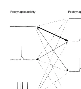
worth recalling.. For optimized stimuli (a strong PW deflection), a large fraction (>90%) of neurons in a cortical column and most neurons in neighboring columns show only subthreshold responses. Thus, in layer II/III of the barrel cortex of an anesthetized rat, we estimate that about 40,000 neurons show subthreshold responses whereas only 200 to 300 APs are evoked in layer II/III.30 So, in a scenario like this what type of information is available to silent cells and their synapses? Figure 1.4 illustrates the most common pre- and postsynaptic activity patterns that are likely to occur under strong sensory stimulation: The most common pre- and postsynaptic activity pattern will be between a silent presynaptic site and a silent postsynaptic site, which is exposed to substantial subthreshold postsynaptic activity. Less fre-quently pre- and postsynaptic activity pattern should consist of a silent presynaptic sites and postsynaptic sites with suprathreshold activity and backpropagating APs. A further pattern of pre- and postsynaptic activity consists of sparse presynaptic
FIGURE 1.4 Pre- and postsynaptic activity patterns during sensory stimulation. Left side,
presynaptic activity patterns: 0 APs, Top; Sparse APs, middle; Bursts of APs bottom. Effects of subthreshold presynaptic activity on axon terminals are neglected. Right side, postsynaptic activity patterns: no subthreshold activity, Top; subthreshold activity, second from top; sub-threshold activity and sparse APs, third from top; subsub-threshold activity and bursts of APs, bottom.
Very common interaction
Common interaction
Rare interaction Presynaptic activity Postsynaptic activity
Domain of the silent neurons
activity and postsynaptic sites with subthreshold activity. Other patterns of pre- and postsynaptic activity would seem to be rare.
From these considerations it becomes clear that many of the classic plasticity protocols that rely on strong pre- and postsynaptic activity cannot predict the types of changes that one may expect in silent cells. Not only in LTP type experiments, but also most paradigms involving spike–timing-dependent plasticity bursts of pre-and postsynaptic activity are applied.67,68 It must be emphasized that most in vitro plasticity experiments are also done in the absence of neuromodulators, which are known to facilitate cortical plasticity in vivo and may be critical in silent cells.
Learning rules which would seem to be important for understanding synaptic change in silent neurons are those uncovered by experiments on homeostatic mechanisms.69 In these experiments, adjustments were observed in not only the AP thresholds but in the synaptic strength of cells in the absence of AP activity.70,71 These synaptic changes may involve heterosynaptic learning rules and could be the mechanism by which silent cells use subthreshold activity to adjust synaptic strength at their largely inactive synapses. This is not to say that conventional forms of synaptic plasticity which involve strong pre- and postsynaptic are irrelevant. Most likely such types of plasticity will occur between the few highly active cortical neurons and will fine tune the properties of these most important cells.
We speculate that spike-based and nonspike-based learning rules set up a con-tinuous competition: a large number of cortical neurons compete to generate a small number of APs that best represent sensory inputs and motor outputs.
III. CONCLUSION
REFERENCES
1. Barlow, H.B., Single units and sensation: a neuron doctrine for perceptual psychol-ogy? Perception, 1, 371, 1972.
2. Dykes, R. and Lamour, Y., Neurons without demonstrable receptive fields outnumber neurons having receptive fields in samples from the somatosensory cortex of anes-thetized or paralyzed cats and rats. Brain Res., 440,133, 1988.
3. Mountcastle, V.B., Modality and topographic properties of single neurons of cat’s somatosensory cortex. J. Neurophysiol., 20, 403, 1957.
4. Mountcastle, V.B., Talbot, W.H., Sakata, H. and Hyvarinen J., Cortical neuronal mechanisms in flutter-vibration studied in unanesthetized monkeys. Neuronal peri-odicity and frequency discrimination. J. Neurophysiol., 32, 452-84, 1969.
5. Welker, C. Receptive fields of barrels in the somatosensory neocortex of the rat. J. Comp. Neurol., 166, 173, 1976.
6. Simons, D.J., Response properties of vibrissa units in rat SI somatosensory neocortex.
J. Neurophysiol., 41, 798, 1978.
7. Armstrong-James, M. and Fox, K. Spatio-temporal divergence and convergence in the rat “barrel” cortex. J. Comp. Neurol., 263, 265, 1987.
8. Armstrong-James, M., Fox, K. and Das-Gupta, A. Flow of excitation within rat barrel cortex on striking a single vibrissa. J. Neurophysiol., 68, 1345, 1992.
9. Diamond, M.E, Armstrong-James, M., and Ebner, F.F., Experience-dependent plas-ticity in the barrel cortex of adult rats. Proc. Natl. Acad. Sci. USA 90, 2602, 1993.
10. Huang, W., Armstrong-James, M., Reama, V., Diamond, M.E. and Ebner, F.F., Con-tribution of supragranular layers to sensory processing and plasticity in adult rat barrel cortex. J. Neurophysiol., 80, 3261, 1998.
11. Armstrong-James, M., Diamond, M. E. and Ebner, F.F., An innocuous bias in whisker use in adult rats modifies receptive fields of barrel cortex neurons. J. Neurosci. 14,
6978-6991, 1994.
12. Warren R.A. and Dykes R.W., Population analysis of single neurons in cat soma-tosensory cortex. Somatosens. Mot. Res., 4, 297, 1992.
13. Swadlow, H.A., Efferent neurons and suspected interneurons in motor cortex of the awake rabbit: axonal properties, sensory receptive fields, and subthreshold synaptic inputs. J. Neurophysiol., 71, 437, 1994.
14. Swadlow, H.A., Efferent neurons and suspected interneurons in second somatosensory cortex of the awake rabbit: receptive fields and axonal properties. J. Neurophysiol.,
66, 1392, 1991.
15. Swadlow, H.A., Efferent neurons and suspected interneurons in S-1 forelimb repre-sentation of the awake rabbit: receptive fields and axonal properties. J. Neurophysiol.,
63, 1477, 1990.
16. Swadlow, H.A., Efferent neurons and suspected interneurons in S-1 vibrissa cortex of the awake rabbit: receptive fields and axonal properties. J. Neurophysiol., 62, 288,
1989.
17. Shimegi S., Ichikawa, T., Akasaki, T., and Sato, H., Temporal characteristics of response integration evoked by multiple whisker stimulations in the barrel cortex of rats. J. Neurosci., 19, 10164, 1999.
18. Carvell, G.E. and Simons, D.J. Membrane potential changes in rat SmI cortical neurons evoked by controlled stimulation of mystacial vibrissae. Brain Res., 448,
19. Timofeev, I., Grenier, F. and Steriade, M., Disfacilitation and active inhibition in the neocortex during the natural sleep-wake cycle: an intracellular study. Proc. Natl. Acad. Sci. USA., 98, 1924, 2001.
20. Li, W.C., Soffe, S.R. and Roberts A. Direct comparison of whole cell patch and sharp electrodes by simultaneous recording from single spinal neurons in frog tadpoles. J. Neurophysiol., 92 380, 2004.
21. Pei, X., Volgushev, M., Vidyasagar, T.R. and Creutzfeldt, O.D., Whole cell recording and conductance measurements in cat visual cortex in vivo,Neuroreport, 2, 485, 1991.
22. Margrie, T. W., Brecht, M. and Sakmann, B., In vivo low resistance whole-cell
recordings from neurons in the anaesthetized and awake mammalian brain. Pflügers Archiv. Eur. J. Physiol. 444,491, 2002.
23. Moore, C.I. andNelson, S.B., Spatio-temporal subthreshold receptive fields in the vibrissa representation of rat primary somatosensory cortex. J. Neurophysiol., 80,
2882, 1998.
24. Zhu, J.J. and Connors, B.W. Intrinsic firing patterns and whisker-evoked synaptic responses of neurons in the rat barrel cortex. J. Neurophysiol., 81, 1171, 1999.
25. Brecht, M. and Sakmann, B., Whisker maps of neuronal subclasses in the rat ventral posterior medial (VPM) thalamus identified by whole-cell voltage recording and morphological reconstruction. J. Physiol., 538, 495, 2002.
26. Armstrong, M. and Callahan, C., Thalamocortical mechanisms in the formation of receptive fieldsof rat barrel cortex neurones. II. spatiotemporal convergence in the thalamic ventroposterior medial nucleus (VPm) and its relevance to generation of receptive fields of S1 cortical "barrel" neurons. J. Comp. Neurol., 303, 211, 1991.
27. Diamond, M.E., Armstrong-James, M., Budway,M.J. and Ebner, F.F., Somatic sensory responses in the rostral sector of the posterior group (POm) and in the ventral posterior medial nucleus (VPM) of the rat thalamus: dependence on the barrel field cortex. J. Comp. Neurol., 319, 66, 1992.
28. Brecht, M. and Sakmann, B., Dynamic representation of whisker deflection by postsynaptic potentials in morphologically reconstructed spiny stellate and pyramidal cells in the barrels and septa of layer IV in rat somatosensory cortex. J. Physiol.,
543, 49, 2002.
29. Armstrong-James, M., The nature and plasticity of sensory processing within adult rat barrel cortex. In The Barrel Cortex of Rodents, Jones, E. G. and Diamond, I. T., Eds., Plenum Press, New York, 1995, pp. 333-374.
30. Brecht, M., Sakmann B., Dynamic receptive fields of reconstructed pyramidal cells in layers I/III of rat somatosensory barrel cortex, J. Physiol., 553:243, 2003.
31. Fanselow, E.E. and Nicolelis, M.A., Behavioral modulation of tactile responses in the rat somatosensory system. J. Neurosci., 19, 7603, 1999.
32. Chung, S., Li, X. and Nelson, S.B., Short-term depression at thalamocortical synapses contributes to rapid adaptation of cortical sensory responses in vivo.Neuron, 34, 437,
2002.
33. Margrie, T.W., Meyer, A.H., Caputi, A., Monyer, H., Hasan M.T., Schaefer, A.T., Denk, W., and Brecht, M., Targeted whole-cell recordings in the mammalian brain
in vivo. Neuron, 39, 911, 2003.
34. Simons, D.J., Temporal and spatial integration in the rat SI vibrissa cortex. J. Neu-rophysiol. 54, 615, 1985.
36. Simons, D.J., Neuronal integration in the somatosensory whisker/barrel cortex. In The Barrel Cortex of Rodents, Jones, E. G. and Diamond, I. T., Eds., Plenum Press, New York, 1995, pp. 262-298.
37. Galarreta, M. and Hestrin, S., Electrical synapses between GABA-releasing interneu-rons. Nat. Rev. Neurosci., 2 425, 2001.
38. Gibson, J.R., Beierlein, M. and Connors, B.W., Two networks of electrically coupled inhibitory neurons in neocortex. Nature, 402, 75, 1999.
39. Petersen, C.C., Grinvald, A. and Sakmann B., Dynamics of sensory responses in layer II/III of rat barrel cortex measured in vivo by voltage-sensitive dye imaging combined
with whole-cell voltage recordings and neuron reconstructions. J. Neurosci., 23, 1298,
2003.
40. Stern, E.A., Maravall, M., Svoboda, K. Rapid development and plasticity of layer II/III maps in rat barrel cortex in vivo. Neuron. 31, 305, 2001.
41. Shadlen, M.N. and Newsome, W.T. The variable discharge of cortical neurons: impli-cations for connectivity, computation, and information coding. J. Neurosci. 18, 3870,
1998.
42. Destexhe, A. and Pare, D., Impact of network activity on the integrative properties of neocortical pyramidal neurons in vivo.J. Neurophysiol. 81, 1531, 1999.
43. DeFelipe, J. and Farinas, I., The pyramidal neuron of the cerebral cortex: morpho-logical and chemical characteristics of the synaptic inputs. Neurobiology, 39, 563,
1992.
44. Feldmeyer, D., Egger, V., Lübke, J. and Sakmann, B., Reliable synaptic connections between pairs of excitatory layer IV neurones within a single ‘barrel’ of developing rat somatosensory cortex. J. Physiol., 521, 169, 1999.
45. Markram, H., Lübke J., Frotscher, M., Roth, A. and Sakmann, B., Physiology and anatomy of synaptic connections between thick tufted pyramidal neurones in the developing rat neocortex. J. Physiol., 500, 409, 1997.
46. Thomson, A.M., Activity-dependent properties of synaptic transmission at two classes of connections made by rat neocortical pyramidal axons in vitro, J. Physiol., 502/1,
131, 1997.
47. Manns, I.D., Sakmann B. and Brecht M., 2004 Sub- and suprathreshold receptive field properties of pyramidal neurons in layers VA and VB of rat somatosensory barrel cortex. J. Physiol., 556, 601, 2004.
48. Reyes, A. and Sakmann, B. Developmental switch in the short-term modification of unitary EPSPs evoked in layer II/III and layer V pyramidal neurons of rat neocortex.
J. Neurosci., 19, 3827, 1999.
49. Hines, M., NEURON — A Program for Simulation of Nerve Equations, aus \newline Eeckman F., Neural Systems: Analysis and Modeling, Kluwer Academic Publishers, 127, 1993.
50. Feldmeyer, D., Lübke, J., Silver, R.A. and Sakmann, B., Synaptic connections between Layer IV spiny neurons — layer II/III pyramidal cell pairs in juvenile rat barrel cortex: physiology and anatomy of inter-laminar signaling within a cortical column, J. Physiol., 538/3, 803, 2002.
51. Gupta, A., Wang, Y. and Markram, H., Organizing principles for a diversity of GABAergic nterneurons and synapses in the neocortex, Science, 287, 273, 2000.
52. Murasugi, C.M., Salzman, C.D., Newsome, W.T., Microstimulation in visual area MT: effects of varying pulse amplitude and frequency. J. Neurosci., 13, 1719, 1993.
![FIGURE 2.10] An example of the temporal evolution of neural frequency tuning. A. TopPeri-stimulus time histograms (PSTHs) are plotted as a function of frequency of stimulation(ordinate) and time (abscissa)](https://thumb-ap.123doks.com/thumbv2/123dok/3941703.1885123/26.612.63.384.76.388/temporal-evolution-frequency-histograms-function-frequency-stimulation-abscissa.webp)