David Publishing Company www.davidpublishing.com P u b l i s h i n g Dav i d
Journal of Life Sciences
Publication Information
Journal of Life Sciences is published monthly in hard copy (ISSN1934-7391) and online (ISSN 1934-7405) by David Publishing Company located at 1840 Industrial Drive, Suite 160, Libertyville, Illinois 60048, USA.
Aims and Scope
Journal of Life Sciences, a monthly professional academic journal, covers all sorts of researches on molecular biology, microbiology, botany, zoology, genetics, bioengineering, ecology, cytobiology, biochemistry, and biophysics, as well as other issues related to life sciences.
Editorial Board Members
Dr. Stefan Hershberger (USA), Dr. Suiyun Chen (China), Dr. Farzana Perveen (Pakistan), Dr. Francisco Torrens (Spain), Dr. Filipa João (Portugal), Dr. Masahiro Yoshida (Japan), Dr. Reyhan Erdogan (Turkey), Dr. Grzegorz Żurek (Poland), Dr. Ali Izadpanah (Canada), Dr. Barbara Wiewióra (Poland), Dr. Valery Lyubimov (Russia), Dr. Amanda de Moraes Narcizo (Brasil), Dr. Marinus Frederik Willem te Pas (The Netherlands), Dr. Anthony Luke Byrne (Australia), Dr. Xingjun Li (China), Dr. Stefania Staibano (Italy), Dr. Wenle Xia (USA).
Manuscripts and correspondence are invited for publication. You can submit your papers via Web Submission, or E-mail to [email protected] or [email protected]. Submission guidelines and Web Submission system are available at http://www.davidpublishing.com.
Editorial Office
1840 Industrial Drive, Suite 160 Libertyville, Illinois 60048 Tel: 1-847-281-9826, Fax: 1-847-281-9855
E-mail:[email protected], [email protected]
Copyright©2011 by David Publishing Company and individual contributors. All rights reserved. David Publishing Company holds the exclusive copyright of all the contents of this journal. In accordance with the international convention, no part of this journal may be reproduced or transmitted by any media or publishing organs (including various websites) without the written permission of the copyright holder. Otherwise, any conduct would be considered as the violation of the copyright. The contents of this journal are available for any citation. However, all the citations should be clearly indicated with the title of this journal, serial number and the name of the author.
Abstracted / Indexed in
Database of EBSCO, Massachusetts, USA Cambridge Scientific Abstracts (CSA), USA
Chinese Database of CEPS, American Federal Computer Library center (OCLC), USA Ulrich’s Periodicals Directory, USA
Chinese Scientific Journals Database, VIP Corporation, Chongqing, China
Subscription Information
Price (per year): Print $420, Online $300, Print and Online $560.
David Publishing Company
1840 Industrial Drive, Suite 160, Libertyville, Illinois 60048 Tel: 1-847-281-9826, Fax: 1-847-281-9855
E-mail: [email protected]
Dav id Publishing Company w ww.davidpublis hing.com
Pu b li sh i ng Dav i d
J LS
Journal of Life Sciences
Volume 5, Number 1, January 2011 (Serial Number 33)
Contents
Research Papers
1 The Human P5B-ATPase ATP13A2 Is Not a Ca 2+
Transporting Pump
Felicitas de Tezanos Pinto, Gerardo Raul Corradi and Hugo Pedro Adamo
7 The Role of Pumpkin Extraction on the Liver and Kidney of Mice Previously Treated with
Haloperidol during Lactation
Samira Omar Balubaid
15 The Sorption Isosteric Heats of Rough Rice in China
Xingjun Li, Zidan Wu and Hui Lu
22 Alterations of Antioxidative Enzymes Activities and Induction of Lipid Peroxidation in
Germinating Wheat Seeds Subjected to Cadmium Stress
Surjendu Kumar Dey
29 Effect of Se-S Cooperated Application on the Mineral Content and Nutrition Quality of Garlic (Allium Sativum L.)
Huanxiu Li, Changquan Wang, Bing Li, Zesheng Yan and Yangxia Zheng
35 Effect of Deficit Irrigation at Different Growth Stages on Wheat Growth and Yield
Seyed Abdolreza Kazemeini and Mohsen Edalat
39 Biological Status of Indus River Dolphin (Platanista Minor Owen) in Indus River, Northern Pakistan
Farzana Perveen, Sardar Azher Mehmood, Shabbir Ahmed and Zia Ur Reman
48 Biochemical Composition and Nutritional Value of the Muscle Tissue of Yellowback Seabream,
Evynnis Tumifrons, from the East China Sea
Lianjun Xia, Qiqun Cheng, Jianxue Lu, Junli Hou, Jian Xin and Min Liu
53 Legionella from Environmental and Clinical Homes for the Mentally Disabled and Comparison of Their Sequence Types
Annalisa Bianchi, Marina Tesauro, Michela Consonni, Fabrizio Pregliasco and Maria Gabriella Galli
Methods and Techniques
59 Detection of the Relationship between Imipenem Susceptible and Non-Susceptible Clinical
Isolates of Acinetobacter Baumannii by Repetitive Element PCR-Mediated DNA Fingerprinting in an Egyptian Hospital
Soheir Helal, Mona M.A. Haleim and Maha Gaafar
66 Assessment of Soil Quality Using Microarthropod Communities Under Different Land System: A Case Study in the Mid-Hills of Central Nepal
Farida Begum, Roshan Man Bajracharya, Subodh Sharma and Bishal K. Sitaula
74 Synthesis and Surface-Active Properties of Carboxymethylcellulose Esters Obtained by
Microwave Assisted Transesterification of Vinyl Laurate
The Human P
5B
-ATPase ATP13A2 Is Not a Ca
2+
Transporting Pump
Felicitas de Tezanos Pinto, Gerardo Raul Corradi and Hugo Pedro Adamo
IQUIFIB-Facultad de Farmacia y Bioquímica, Universidad de Buenos Aires, Junín 956, 1113 Buenos Aires, Argentina
Received: May 30, 2010 / Accepted: July 22, 2010 / Published: January 30, 2011.
Abstract: The human gene ATP13A2 has been proposed to code for an ATP powered ion transporter of the P5B subfamily. Mutations
of the human gene ATP13A2 were found to underlie an autosomal recessive form of early-onset parkinsonism (PD) with pyramidal degeneration and dementia. The ion transported by the ATP13A2 pump is not known, but several studies have shown that the P5-ATPases influence the homeostasis of intracellular Ca2+, and thus it has been suggested that they transport Ca2+. In order to evaluate
this possibility Chinese hamster ovary (CHO) cells stably expressing the human ATP13A2 protein have been obtained and the Ca2+ transport activity of ATP13A2 was assessed by measuring the ATP-dependent uptake of Ca2+ into microsomal vesicles. As a positive control vesicles containing the human plasma membrane Ca2+ pump (PMCA) were used. No significant differences were found between vesicles containing the ATP13A2 protein and the control. Moreover, Ca2+ was unable to induce the formation of the P-ATPase acylphosphate intermediate in vesicles containing the expressed ATP13A2. These results favor the idea that the ATP13A2 does not transport Ca2+.
Key words: P5B-ATP13A2, calcium uptake, CHO cells overexpression.
Abbreviations: PMCA: plasma membrane Ca2+ pump; CHO cells: Chinese hamster ovary cells.
1. Introduction
The P-type superfamily of ion pumps includes transporters energized by the hydrolysis of ATP that transport inorganic cations and other substrates across cell membranes. These P-type ATPases are characterized by the formation of a phosphorylated intermediate during their reaction cycle. They are present in prokaryotes and eukaryotes, and in the basis of their conserved core sequences they have been classified into five subfamilies termed P1-P5 or type I-V. The most poorly understood P-type ATPases are those of the P5 subfamily, which are expressed only in eukaryotes. The P5-ATPases have been divided in two subfamilies termed P5A and P5B based on protein alignments [1]. Both subfamilies harbor the
Corresponding autor: Felicitas de Tezanos Pinto, Ph.D.,
research fields: biochemistry, cellular and molecular biology. E-mail: [email protected].
The Human P5B-ATPase ATP13A2 Is Not a Ca2+ Transporting Pump
2
chromosome 3 in the human gene ATP13A4 was found in patients with autism spectrum disorder (ASD) and specific language impairment (SLI) [4].
The ion specificity of the P5-ATPases is unknown, and recent publications suggest that they affect the intracellular level of different cations [5-8]. The yeast P5A-ATPase Cod1p, in collaboration with the Golgi Ca2+ pump Pmr1p, was suggested to supply calcium to the yeast ER. However, the function of Cod1p in cellular Ca2+ homeostasis is not equivalent to, nor redundant with that of Pmr1p. Moreover, it was shown that the phenotype of mutant yeast cells lacking Cod1p (cod1∆) could be partially suppressed by exogenous calcium [9]. Deletion of Cod1p alone did not affect cellular calcium level, but deletion of both Cod1p and Pmr1p produced a synergistic increase in the intracellular calcium level compared with pmr1∆ alone [5]. In addition Cod1p has been shown to be involved in mechanisms that depend on the ER Ca2+ concentration like glycosylation of proteins in the secretory pathway, protein insertion orientation, and regulation of HMG-CoA reductase degradation [5, 9-11]. The idea of Ca2+ pumping P5-ATPases is favored by a recently publication which showed that the over-expression of human ATP13A4 in COS-7 cells increased the intracellular calcium level [8].
Here, the ability of the human ATP13A2 to transport Ca2+ has been examined under conditions which are optimal for the function of other well known Ca2+ pumps. For this purpose CHO cells were stably transfected with the ATP13A2 cDNA, and clones expressing the ATP13A2 protein were isolated. It was found that the expressed ATP13A2 not only lacks any Ca2+ transport activity but also unable to promote the formation of the catalytic phosphoenzyme from ATP.
2. Material and Methods
2.1 MaterialsReagents were purchased from the following companies: 45Ca and [γ-32P]ATP, PerkinElmer Life Sciences; Immobilon transfer membranes and
nitrocellulose filters, Millipore; immunochemicals, Invitrogen, Molecular Probes, Vector Laboratories and Amersham Biosciences; and reagents for cell culture, thapsigargin, and other chemicals, Sigma. The expression vector pcDNA3.1 carrying the V5-tagged human ATP13A2 cDNA was a generous gift of Drs. Alfredo Ramirez and Christian Kubisch, Institute of Human Genetics, University of Bonn, Germany.
2.2 Protein Expression and Isolation of Cellular Membranes
Stable CHO cell lines expressing the recombinant PMCA were described previously [12]. CHO cells were lipofected with the expression vector pcDNA3.1 carrying the V5-tagged human ATP13A2 cDNA using lipoafectamine 2000 transfection reagent (Invitrogen) according to the manufacturer’s protocol. To express in a stable form the recombinant ATP13A2, the transfected CHO cells were split into dishes of 10 cm in diameter 24 h post-transfection. 24 hours later the transfected cells were cultured in a selective DMEM medium supplemented with antibiotics and 10% of dialyzed fetal calf serum containing the antibiotic G418 at a final concentration of 600 μg/ml. After 3 weeks about 6-8 of the resulting colonies were cloned and expanded, and the expression of the pump was investigated by immunoblotting. The crude microsomal membrane fractions and the erythrocyte inside-out membranes were prepared by the procedure of Enyedi et al. [13] and Sarkadi et al. [14], respectively. Protein concentration was estimated by means of the Bio-Rad protein assay, with bovine serum albumin as a standard. The amount of the expressed protein was estimated by quantitation of the band intensity using the Gel Pro Analyzer 3.0 program (version 3.1 for WindowsTM, Media Cybernetics).
2.3 Detection of the Human ATP13A2 Protein
After three washes with PBS the cells were permeabilized with 0.15% Tween 20 in PBS (PBST) for 5 min on ice. The recombinant ATP13A2 was detected with antibody to V5 (Invitrogen) at a dilution of 1:500 in PBST by incubation over night at 4 ℃. After washing the cells three times with PBST, the anti-V5 was labeled by Zenon Alexa Fluor 568 mouse IgG2a (Molecular Probes) according to the manufacturer’s instructions for two hours at room temperature. Then the cells were washed three times with PBST and after a single wash with PBS the cells were covered by 50 μl of PBS and observed in a confocal microscope FluoView 1000 (Olympus, Japan) with an appropriate Alexa Fluor 568 filter.
SDS-PAGE and immunoblotting were carried out as described previously [15]. Proteins were electrophoresed on a 7.5% acrylamide gel according to Laemmli [16] and subsequently transferred to Millipore Immobilon membranes. The membranes were incubated over night at 4 ℃ with V5 monoclonal antibody (Invitrogen) according to the manufacturer’s protocol. For staining, biotinylated anti-mouse immunoglobulin G and avidin-streptoavidin peroxidase conjugate were used.
2.4 Ca2+ Transport Assay
Ca2+ uptake assays were performed as described previously [12]. The reaction mixture contained 100 mM KCl, 50 mM Tris-HCl (pH 7.3 at 37 ℃), 5 mM NaN3, 0.1 μM thapsigargin, 4 μg/ml oligomycin, 20 mM sodium phosphate, 1.5 mM ATP, 95 μM EGTA, 2.5 mM MgCl2 and CaCl2 (labeled with 45Ca) to give the desired concentration of free Ca2+. The free concentrations of Ca2+ were calculated using the program of Fabiato and Fabiato [17]. Vesicles (10 μg of protein) were preincubated at 37 ℃ for 5 min, and the reaction was initiated by the addition of ATP. The reaction was finished after 5 min by filtering the samples through a 0.45 μm filter. The 45Ca taken up by the vesicles was determined by counting in a scintillation counter.
2.5 Detection of the Phosphorylated Intermediate
The phosphorylation reaction was carried out at 4 ℃ in a medium containing 30 g of microsomal protein, 160 mM KCl, 25 mM Tris-HCl (pH 7.0 at 4 ℃) and 4 μM thapsigargin to obtain a full inhibition of SERCA pump in a reaction volume of 0.25 ml; 0.15 mM CaCl2 was added when indicated. The reaction was initiated by the addition of 1 μM [γ-32P]ATP and terminated after 1 min with 15 μl of a solution containing 100% trichloroacetic acid. The precipitated proteins were dissolved in sample buffer and separated by SDS-PAGE in a 7% acrylamide gel according to Sarkadi et al. [14]. After drying the gel, they were exposed to a storage phosphor screen for 1 day and imaged using a Storm 840 Optical Scanner.
3. Results
3.1 Expression of the Recombinant ATP13A2 Protein
CHO cells were transfected with the pcDNA3.1 expression vector carrying the human ATP13A2 cDNA and stable clones were selected by their resistance to the antibiotic G418. Immunofluorescence experiments showed that the isolated clones successfully expressed the ATP13A2 protein (Fig. 1a). Microsomal membranes from transfected cells were isolated and submitted to SDS-PAGE and immunobloting. The expressed ATP13A2 had the expected migration according to its predicted size of 129 kDa, and judged by the intensity of the bands, it accounted for about 1% of the microsomal protein (Fig. 1b). Thus, the expression level of ATP13A2 was similar to that reached by other P-type ATPases in the same expression system [18].
3.2 Ca2+ Transport Assays
The Human P5B-ATPase ATP13A2 Is Not a Ca2+ Transporting Pump
4
Fig. 1 Expression of the recombinant ATP13A2 pump.
(a) Fluorescence microscopy to visualize ATP13A2 expression in transfected CHO cells. The left panels show CHO cells transfected with the empty vector pcDNA3.1 (upper panels) or with the expression vector pcDNA3.1 carrying the V5-tagged human ATP13A2 cDNA (bottom panels) incubated with an antibody to V5 revealed with Zenon Alexa Fluor 568 mouse IgG2a secondary antibody. The transmittance image of each one is shown in the right panels. (b) Immunoblot of microsomes from CHO cells transfected with cDNA encoding the V5-tagged human ATP13A2. For protein estimation, a V5-tagged Ag+/Cu+ATPase (V5-CopA) of 86 kDa purified from the extremophile organism Archeaglobus fulgidus was used. The amount of protein loaded is indicated at the top of each lane. The proteins were separated by SDS-PAGE and transferred to polyvinylidene difluoride membranes; finally they were detected with the V5 antibody as described under “Material and Methods”.
Ca2+, μM
0.1 1 10
Ca
2+
u
p
ta
k
e
, % o
f m
a
x
im
u
m
0 20 40 60 80 100 120
CHO ATP13A2 h4PMCAxb ePMCA
Fig. 2 Ca2+ dependence of the Ca2+ transport of recombinant ATP13A2 as compared with that of PMCA.
Ca2+ uptake by microsomal vesicles was measured at different free Ca2+ concentrations for 5 min at 37 ℃ as described under “Material and Methods”. The kinetic behavior of the recombinant PMCA (h4PMCAxb, filled triangles) is similar to the PMCA of erythrocytes IOVs (ePMCA, empty triangles). The activity of the recombinant ATP13A2 (empty circles) not differ from that obtained by the endogenous PMCA from CHO cells transfected with the empty vector (filled circles). Data points are the average of three to five experiments. The lines are the best fit to the data given by the Hill equation.
a
measured using either inside out vesicles (IOVs) from erythrocytes, or microsomal vesicles from CHO cells stable expressing the human PMCA isoform 4xb (h4PMCAxb). As shown in Fig. 2, the activity of the PMCA enzyme gradually increased with the concentration of Ca2+, reaching half maximal activity at about 1 μM of free Ca2+
. By contrast, the Ca2+ uptake activity of microsomes expressing the recombinant ATP13A2 was not significantly different from that of microsomal vesicles from CHO control cells transfected with the empty vector indicating that in these conditions no uptake of Ca2+ was associated with the expressed ATP13A2.
3.3 Phosphorylation Reaction
The first step of the catalytic cycle of all well characterized P-type Ca2+ pumps involves the Ca2+ dependent transfer of the γ−phosphate from ATP to the enzyme to form a phosphoenzyme intermediate. As shown in Fig. 3 a strong band was observed when microsomes carrying the recombinant PMCA were phosphorylated with ATP in the presence of Ca2+. Likewise, a band corresponding to the PMCA phosphoenzyme was observed in erythrocyte IOVs. In
Fig. 3 Formation of the phosphorylated intermediate. Phosphoenzyme formation was carried out as described under “Material and Methods”. Lane 1, recombinant ATP13A2 with Ca2+; lane 2, recombinant ATP13A2 without Ca2+; lane 3, PMCA from erythrocyte IOVs with Ca2+ (ePMCA); lane 4, recombinant PMCA with Ca2+ (h4PMCAxb); lane 5, control
empty pcDNA3.1 vector with Ca2+; lane 6, pcDNA3.1 membranes as in lane 5, but the phosphorylation was carried in the absence of thapsigargin allowing the visualization of the SERCA phosphoenzyme; lane 7, control empty pcDNA3.1 vector without Ca2+.
contrast the phosphorylation pattern of membrane vesicles containing the recombinant ATP13A2 was similar to that of control membranes, indicating that no phosphorylation attributable to the ATP13A2 protein had occurred. Thus, the expressed ATP13A2 was not activated by Ca2+ to form the phosphorylated intermediate.
4. Discussion
The P5-ATPases have been associated with the homeostasis of intracellular Ca2+ and this fact has led to the idea that they are Ca2+ transporters. Here, it was investigated calcium as a possible substrate of the human ATP13A2 enzyme by comparing the Ca2+ transport activity and Ca2+-dependent phosphoenzyme formation of ATP13A2 with that of the related Ca2+ specific P2-ATPase PMCA. Neither of the two Ca2+-dependent activities were detected in the recombinant ATP13A2. These results suggest that although P5-ATPases influence many Ca2+ related functions, Ca2+ is not the substrate transported by the human P5-ATP13A2. Mutations in the ATP13A2 gene underlay an autosomal recessive form of early onset parkinsonism [3]. Expression of ATP13A2 in animal models of PD is sufficient to rescue neurodegeneration associated with α-synuclein (α-syn) aggregation [19], which is relevant to this study since it was used the same construct for the expression of ATP13A2 and thus it implies that no additional factors are needed in order to observe the biological response.
The Human P5B-ATPase ATP13A2 Is Not a Ca2+ Transporting Pump
6
excess Mn2+ exposure [19]. On the other hand, it was recently published that the deletion of the P5B-ATPase CATP-5 of Caenorhabditis elegans is responsible for the tolerant phenotype seen in the presence of the toxic spermidine analog norspermidine, raising the possibility that the polyamines are the substrates transported by P5B-ATPases [21].
Acknowledgments
The authors thank Drs. Alfredo Ramirez and Christian Kubisch for supplying vector pcDNA3.1 carrying the V5-tagged human ATP13A2 cDNA and Luis M. Bredeston for the generous gift of the purified V5-tagged Ag+/Cu+ATPase. This work was supported in part by the University of Buenos Aires (UBA, Grant B604), by the Consejo Nacional de Investigaciones Científicas y Técnicas (CONICET, Grant PIP 112-200801-02022), and by Agencia Nacional de Promoción Científica y Tecnológica (ANPCyT, Grant BID PICT 2007-00702).
References
[1] K.B. Axelsen, M.G. Palmgren, Evolution of substrate specificities in the P-type ATPase superfamily,J. Mol. Evol. 46 (1) (1998) 84-101.
[2] P.J. Schultheis, T.T. Hagen, K.K. O’Toole, A. Tachibana, C.R. Burke, D.L. McGill, et al., Characterization of the P5 subfamily of P-type transport ATPases in mice, Biochem. Biophys. Res. Commun. 323 (3) (2004) 731-738.
[3] A. Ramirez, A. Heimbach, J. Grundemann, B. Stiller, D. Hampshire, L.P. Cid, et al., Hereditary parkinsonism with dementia is caused by mutations in ATP13A2, encoding a lysosomal type 5 P-type ATPase, Nat. Genet. 38 (10) (2006) 1184-1191.
[4] D.A. Kwasnicka-Crawford, A.R. Carson,W.Roberts, A.M. Summers, K. Rehnstrom, I. Jarvela, et al., Characterization of a novel cation transporter ATPase gene (ATP13A4) interrupted by 3q25-q29 inversion in an individual with language delay, Genomics 86 (2) (2005) 182-194.
[5] S.R. Cronin, R. Rao, R.Y. Hampton, Cod1p/Spf1p is a P-type ATPase involved in ER function and Ca2+ homeostasis, J. Cell Biol. 157 (2002) 1017-1028.
[6] M.K. Jakobsen, L.R. Poulsen, A. Schulz, P. Fleurat-Lessard, A. Møller, S. Husted, et al., Pollen development and fertilization in Arabidopsis is dependent on the MALE GAMETOGENESIS IMPAIRED ANTHERS gene encoding a Type V P-type ATPase, Genes & Dev. 19 (2005) 2757-2769.
[7] K. Schmidt, D.M. Wolfe, B. Stiller, D.A. Pearce, Cd2+, Mn2+, Ni2+ and Se2+ toxicity to Saccharomyces cerevisiae lacking
YPK9p the orthologue of human ATP13A2, Biochem. Biophys. Res. Commun. 383 (2) (2009) 198-202.
[8] J. Vallipuram, J. Grenville, D.A. Crawford, The E646D-ATP13A4 mutation associated with autism reveals a defect in calcium regulation, Cell. Mol. Neurobiol. 30 (2) (2010) 233-246.
[9] S.R. Cronin, A. Khoury, D.K. Ferry, R.Y. Hampton, Regulation of HMG-CoA reductase degradation requires the P-type ATPase Cod1p/Spf1p, J. Cell Biol. 148 (5) (2000) 915-924.
[10] C. Suzuki, Y.I. Shimma, P-type ATPase spf1 mutants show a novel resistance mechanism for the killer toxin SMKT, Mol. Microbiol. 32 (4) (1999) 813-823.
[11] D.J. Tipper, C.A. Harley, Yeast genes controlling responses to topogenic signals in a model transmembrane protein, Mol. Biol. Cell. 13 (2002) 1158-1174.
[12] H.P. Adamo, M.E. Grimaldi, M.I. García Arguinzonis, Deletions in the N-terminal segment of the plasma membrane Ca2+ pump impair the expression of a correctly folded functional enzyme, Biochemistry 39 (2000) 14893-14899. [13] A. Enyedi, A.K. Verma, A.G. Filoteo, J.T. Penniston, A
highly active 120 kDa truncated mutant of the plasma membrane Ca2+ pump, J. Biol. Chem. 268 (1993) 10621-10626.
[14] B. Sarkadi, A. Enyedi, Z. Foldes-Papp, G. Gardos, Molecular characterization of the in situ red cell membrane calcium pump by limited proteolysis, J. Biol. Chem. 261 (1986) 9552-9557.
[15] F. de Tezanos Pinto, H.P. Adamo, Deletions in the acidic lipid-binding region of the plasma membrane Ca2+ pump, A mutant with high affinity for Ca2+ resembling the acidic lipid-activated enzyme, J. Biol. Chem. 277 (2002) 12784-12789.
[16] U.K. Laemmli, Cleavage of structural proteins during the assembly of the head of bacteriophage T4, Nature 227 (1970) 680-685.
[17] A. Fabiato, F. Fabiato, Calculator programs for computing the composition of the solutions containing multiple metals and ligands used for experiments in skinned muscle cells, J. Physiol. 75 (1979) 463-505.
[18] C.I. Cura, G.R. Corradi, D.E. Rinaldi, H.P. Adamo, High sensibility to reactivation by acidic lipids of the recombinant human plasma membrane Ca2+-ATPase isoform 4xb purified from Saccharomyces cerevisiae, Biochimica et Biophysica Acta (BBA) - Biomembranes 1778 (12) (2008) 2757-2764. [19] A.D. Gitler, A. Chesi, M.L. Geddie, K.E. Strathearn, S.
Hamamichi, K.J. Hill, et al., Alpha-synuclein is part of a diverse and highly conserved interaction network that includes PARK9 and manganese toxicity, Nat. Genet. 41 (3) (2009) 308-315.
[20] A.B. Møller, T. Asp, P. Bach Holm, M.G. Palmgren, Phylogenetic analysis of P5 P-type ATPases, a eukaryotic
lineage of secretory pathway pumps, Mol. Phylogenet. Evol.
46 (2007) 619-634.
[21] A. Heinick, K. Urban, S. Roth, D. Spies, F. Nunes, O. Phanstiel IV, et al., Caenorhabditis elegans P5B-type ATPase
The Role of Pumpkin Extraction on the Liver and Kidney
of Mice Previously Treated with Haloperidol during
Lactation
Samira Omar Balubaid
Department of Biology, King Abdul Aziz University, Jeddah, Saudi Arabia
Received: June 20, 2010 / Accepted: November 03, 2010 / Published: January 30, 2011.
Abstract: The present study was initiated to investigate the effect of haloperidol on the histological structures of the liver and kidney of
mice and their infants. Pumpkin was used to inhibit the toxic effects of this drug. Mothers, directly after delivery from the first day to the weaning age at the 21 day were divided into four groups: the first group (control), the second group supplied with (1 mg haloperidol /kg /B.W./day), the third group was supplied with (1 ml pumpkin extraction /kg /B.W./day), the fourth group was supplied with (1 ml pumpkin /Kg/B.W.). The histological examination of the haloperidol treated mothers revealed its bad condition in compare with the other groups where there was absence of natural architecture of the liver and complete disintegration of the renal tubules. Also, the same symptoms were recorded in the liver and kidney of the infants. An obvious improvement in the structures of liver and kidney in mothers and infants as well as the recovery of most tissues to their normal appearance after the treatment with the mixture of haloperidol and pumpkin in compare with the haloperidol-group and pumpkin-group also showed a normal structure as the control-group.
Key words: Liver, kidney, mice, pumpkin, lactation.
1. Introduction
Depression is a psychological state which is differentiated from the other known disturbances by consistent disordered mode with disturbed sleep, loss of appetite, losing of self confidence, reduced activity and weak memory [1]. Depression is more common in women than men. The different types of depression for ladies were mentioned during ovulation, pregnancy, delivery bleeding and at the post-ovulation time. Haloperidol is the most used drug as anti-depression during pregnancy and lactation, and there is a close relationship between depression and changes in hormones of females [2]. Treatment of pregnant women with antidepressed drug during the last months of pregnancy, may lead to distorted embryos [3] and
Corresponding author: S. Balubaid, Ph.D., associate Prof.,
research field: embryology. E-mail: [email protected].
resulted in an increase in alkaline phosphatase. R. Baldessarini et al. observed that haloperidol had a serious effect on the quantity of milk from mothers through its effect on liver and bile secretion [4]. They also found that the treatment by using this drug for a long period lead to a reduction in the sexual desire in both sexes. While in case of pregnancy and lactation, there were no records for distorted embryos or infants.
The absorption, distribution and secretion of haloperidol-14c in mice after muscular injection or forced feeding, showed that after one hour, the highest level of this drug was recorded in the blood, plasma and in tissues of lung, pancreas, kidney, liver and spleen, also similar elevation in the placenta of mice embryos, while the drug levels in the milk were variable and similar to those levels in the plasma after muscular injection [5].
The Role of Pumpkin Extraction on the Liver and Kidney of Mice Previously Treated with Haloperidol during Lactation
8
spinal cord have been detected in pregnant mice and a significant increase in the prolactine secretion after injection with haloperidol [6].
In a histological study carried by Ref. [7] the testes of mice showed necrosis, decreasing in spermatozoa number and vacuolation of most germ cells. Also clozapine drug caused a disorder on the histological structure of liver, kidney and retina of eyes of the chick embryos [8], an inhibition of CYP2B activity in liver [9] and the drug effect the glomerulus and infiltration process in kidney.
Pumpkin (Cucurbita pepo) is an important medical
plant, which contains a large amount of water (94% of its weight) and carotene which converts to vitamin A in the body [10]. According to the chemical and physical properties of this plant, it is recommended as a good caloric diet [11]. F. Bion et al. recorded that pumpkin usage did not change the mophometric measurements of different organs as liver, kidney, gonads, spleen and brain of mice [12]. The oral daily dose of pumpkin seed extraction to mice did not affect blood glucose level, urea, creatinin, total protein, uric acid and blood cells number [13]. On the other hand [14], revealed that the usage of this plant may inhibit diabetes and it also associated with the reduction of the cellular oxidation potential and blood pressure. This plant contains a higher level of D-chiroinositol which decreases glucose level in the blood, increases the hepatic glycogen, increases insulin level and total hemoglobin of diabetes patients [15]. Therefore, this plant is considered as a good treatment for diabetes, as it composed of polysaccharide and protein [16]. The extracted protein from this plant has an essential role in decreasing the harmful effects of protein malnutrition [17]. Such protein leads to an elevation in the level of hepatic enzymes and a decreasing in the toxic effect of the carbon tetra-oxide because it contains anti-oxidative compound [18]. The extraction from the green leaves of pumpkin were used in treatment of many diseases and due to its higher antioxidative activity it can remove the cellular toxicity of hepatic
tissue and the oxidative potential initiated by garlic [19]. The pumpkin seed oil can prevent changes in plasma lipids and blood pressure associated with inadequate estrogen availability [20]. The pumpkin extracts p5 and p6 had high content of total phenolics and antioxidant activity coupled to moderate to high alpha-glucosidase and angiotensin converting enzyme inhibitory activities and has the potential to reduce hyperglycemia-induced pathogenesis and also associated complication linked to cellular oxidation stress [14].
According to M. Makni et al. [21] who found that pumpkin seed had anti-atherogenic and hepatoprotective effect because pumpkin seed are rich source of an unsaturated fatty acids, antioxidant and fibers.
It is therefore of interest to examine the effect of haloperidol on histological structure of liver and kidney in mothers’ mice during lactation and their infants, also to investigate the role of pumpkin as a protective agent against the haloperidol histopathological alterations.
2. Materials and Methods
Female Swiss albino mice were obtained from experimental animal unit at King Fahd Medical Research Center. The female animals during lactation were housed in individual cages with their infants at a
temperature 20-22 ℃ and were fed a standard Purina
chow and water.
Ten female mice were used in each group. The average weight per experimental animal was 200 g. The female mice during lactation were divided into 4 groups.
(1) The first group was untreated and used as a control.
during Lactation
(3) Animals of group three were given daily the same dose of haloperidol and fed with 1 ml pumpkin extraction /kg B.W. at the same period [15].
(4) The fourth group was given the pumpkin extraction daily at the same dose level for the same period.
At the end of lactation (21 days) liver and kidney specimens from each of the four groups were fixed in neutral formalin. Also, infants were dissected at 7-14-21 days after birth and specimens from liver and kidney were fixed in neutral formalin, embedded in paraffin. The sections were stained with Hematoxylin and Eosin [22].
3. Results
3.1 Changes in Weight
Data in Table 1 showed a highly significant decrease
(P < 0.05) in the infant body weights in the group
treated with Haloperidol drug only as compared with control group. Infant body weights of Haloperidol + pumpkin group showed significant increase than Haloperidol-group. The fourth group which treated with Pumpkin showed a significant increase in body weight as compared with control group during the three weeks of the experiment.
3.2 Histological Changes
3.2.1 Group Treated with Pumpkin Only
Tested sections which treated with pumpkin only in liver and kidney for both mothers and infants showed healthy appearance (Fig. 1) as compared with control group.
3.2.2 Group Treated with the Drug (Haloperidol)
A–Mothers:
(1) Liver
In some regions, it can be seen a dilation and congestion, which is due to the disintegration of the hepatic cells. This disintegration is represented by appearance of fatty vacuoles inside cells (lipid infiltration) and disorder in the liver architecture that could be recognized by the disappearance of the characteristic strands of hepatocyte (Fig. 2). In other regions, disintegration in the wall of the portal and central veins, disintegration of nuclei and cellular membranes of the hepatocyte and fibrosis of the portal regions could be noticed compared with other three groups especially the fourth group (pumpkin).
(2) Kidney
Histological investigation of kidney from mothers treated with the drug 1 mg/kg B.W. for 21 days indicated a complete dissociation of the proximal tubules. This disassociation is represented by disintegration of the lining cells and their nuclei. Some tubules appeared completely vacuolated (degenerated) and most of cells disappeared. Sections also show separation and swelling of the lining cells of the distal tubules. Moreover, separation of the tubular cells from their basement membrane with a partial or complete plugging of the tubular cavities were also recorded. A reduction in the intertubular connective tissues with a large vacuoles, partitioning of glomerulus, reduction of epithelial layer, disorder in the granular layer, dilation of urinary cavity (Fig. 3) and malformation of collecting tubules in the medulla were recorded, compared with other three groups especially the fourth group (pumpkin).
Table 1 Indicated the effect of haloperidol, haloperidol + pumpkin and pumpkin on the infant weights during the experiment in compare with control group.
Groups Weeks
First (Mean ± S.E*) Second (Mean ± S.E*) Third (Mean ± S.E*)
Control Haloperidol Haloperidol + pumpkin Pumpkin
17.52 ± 0.5947 10.04 ± 0.5576 16.22 ± 0.5918 19.72 ±0 .9382
19.01 ± 0.2714 11.71 ± 0.4099 18.65 ± 0.8314 23.50 ± 0.5918
The Role of Pumpkin Extraction on the Liver and Kidney of Mice Previously Treated with Haloperidol during Lactation
10
Fig. 1 Light micrograph of kidney section from infants of 21 days after delivery from pumpkin treated mothers showing normal kidney structure ( H & E 40 x).
Fig. 2 Light micrograph of liver from haloperidol treated mothers showing disappearance of the characteristic strands of hepatocyte deformed hepatocytes (arrows) (E& H 100 x).
Fig. 3 Light micrograph of kidney from haloperidol treated mothers (1 ml/kg B.W.) showing rare intertubular connective tissue (arrow), decayed proximal (PT) and distal (DT) tubules and deformed glomerulus(G) (H &E 100x).
B–Infants (1) Liver
Sections of liver at one week age, showed serious disintegration of the hepatic cells represented by the separation and disruption of these cells in the tissue, rapture of cellular wall, karyolitic nuclei with appearance of lipid filtration and tissue fibrosis (Fig. 4). While at two weeks age, a decrease in the lipid filtration and cellular disintegration were observed. On the other hand, after three weeks, sections of liver appeared pale stained with oedema and a disorder in the structural architecture was recorded. This disorder was due to the separation of hepatic cells which lead to enlargement of the hepatic acini and presence of fatty vacuoles in hepatic cells (Fig. 5), malformation of portal
Fig. 4 Light micrograph of liver section from infants of 7 days after delivery from haloperidol treated mothers showing decayed hepatocytes (H) appearance of fat filtration (Unclear) and tissue fibrosis (arrow) (H & E 100 x).
Fig. 5 Light micrograph of liver section from infants of 21 days after delivery from haloperidol treated mothers showing congestion of blood sinusoid (BV) and oedema (arrow) in the tissue(H & E 100 x).
3 G
DT TP 1
BV
5
2 4
during Lactation
regions, dilation of blood vessels and decreasing in the bile ductules number, compared with other three groups especially the fourth group (pumpkin).
(2) Kidney
Investigation of kidney sections at 7-14 days age showed that the cortex is smaller when compared with medulla, and it have a relatively lower number of glomerulus compared with the control group. Sections also showed a reduction in glomerulus, epithelial disintegration, enlargement of the urinary cavity, a complete disintegration of proximal and distal tubules with cellular infilteration (Fig. 6). After three weeks of delivery, sections of kidney showed congestion inside the tissue, interruption of the inner wall of renal arteries, swelling of proximal and distal tubules cells and adhesion of their nuclei with basement membrane, partitioning and atrophy of glomerulus mainly at cortex (Fig. 7), while these damages were less obvious in the medulla as compared with control or compared with the other three groups especially the fourth group (pumpkin).
3.2.3 Treated Group with the Drug (Haloperidol) and Pumpkin
Generally, there were obvious improvements in the kidney and liver sections of mothers and infants as well as the regain of normal appearance of most tissues. These improvements could be summarized for kidney
Fig. 6 Light micrograph of kidney from infants of 14 days after delivery from haloperidol treated mothers showing shrinked glomerulus (G) decayed proximal (circle) and distal (square) tubules (H & E 40 x).
Fig. 7 Light micrograph of kidney from infants of 21 days after delivery from haloperidol treated mothers showing congestion (BV) and partitioning and atrophy of glomerulus (circle) (H & E40 x).
by the following histological observations, normal appearance of renal tubules, normal appearance, size and number of glomerulus and back to the normal size and structure of intertubular connective tissue (Fig. 8). For liver, sections showed a resistance to the toxicity of the drug and appeared structurally normal. The hepatic strands arranged normally, the portal regions were recovered, oedema disappeared, the fatty filtration was retarded and hepatic cells returned their normal structure (Fig. 9). And also tissues for infants of three weeks’ age. While for one and two weeks age, there were some limited regions still suffered from the harmful influence of the drug, compared with other three groups especially the fourth group (pumpkin).
4. Discussion
The results of the present study indicated that haloperidol administered to mice during lactation decreased the infants’ body weight in comparison with control, while treatment with pumpkin only, showed a marked increase in infant’s body weight. These results were in agreement with those obtained by R. Holson et al. [23], who concluded that all nervous drugs led to retardation of growth due to a reduction in the DNA formation, protein contents. Also, J. Zhang et al. [24] mentioned that injection of pregnant mice with haloperidol lowered the weight of infants of two weeks age in comparison with control.
6
7
The Role of Pumpkin Extraction on the Liver and Kidney of Mice Previously Treated with Haloperidol during Lactation
12
Fig. 8 Light micrograph of kidney from haloperidol and pumpkin treated mothers showing of the normal tissue structure (H & E 40x).
Fig. 9 Light micrograph of liver section from haloperidol and pumpkin treated mothers showing resume normal tissue structure (H & E 10 x).
The present data demonstrated that treatment of mice with haloperidol during lactation can led to changes in liver’s and kidney’s weights in mothers and infants. Before, R. Sommi et al. [25] showed that medium and high doses of hydrochloride alfloxetine, (anti-depression drug) led to an elevation on the cellular hepatic disintegration and an increase in the fatty precipitation in hepatic cells. The toxic impact of the anti-depressed drug resulted in critical morphological and functional changes in the hepatic lipid associated with several disintegrated changes where female mice exposed to antinervous drug during pregnancy especially in time of organs formation or embryonic growth, showed retardation in growth with functional distortion of some organs [26].
The present histological damages in tissues of infants indicated that the drug reaches them from mothers through milk. Haloperidol affects the quantity of milk from mothers through its action on the rate of prolactin hormone secretion [4].
The results obtained in this work showed that haloperidol have a damage effect on mice kidney of mothers and infants and this was supported by A. Patel [27] who mentioned that the anti-nervous drug (haloperidol) to mice during lactation caused embryonic distortion. This effect may be explained by the deficiency in DNA formation [23], delay in the cell division [27] and its direct and indirect effects on the enzymatic activity of cell. This might lead to a disorder in the cellular functional activity and disintegration of cellular cytoplasmic processes in kidney. Another possibility of the effect of the drug (haloperidol) might be through its direct influence on the cell membrane as it had a great ability to dissolve in lipids, and this in turn distorted the nervous impulses through the cell membrane and the cellular metabolism.
Therefore, the damage of epithelium of renal tubules and hepatic cells, dilation of the blood vessels, increasing of the cell size and presence of oedema around cells might refer to the increasing in the permeability of cell membrane and changing in the metabolic mechanisms [8].
The present investigation demonstrated that pumpkin extraction reduced the toxic effect of haloperidol on liver and kidney tissues of female mice and their infants and this might be due to its highly content of B-carotein, which agreed with M. Makni [21] who discussed the antioxidant and hepatoprotective effect of the active groups treated with pumpkin. B-carotene had been proved to be a powerful antioxidant and profound protective actions against tumor [28] because there was increasing interest in the role of antioxidant vitamins like B-carotene in neutralizing free radicals and overtly aggressive oxygen species [29]. B-Carotene was among the most efficient substance known for 9
during Lactation
quenching the excitation energy of single oxygen and also for trapping certain organic free radicals. It had a direct inhibitory effect on liver microsomal enzymes [10], thus offering another mechanism of its anticancer nature. Pumpkin also was considered to be one of the most effective anti-oxidative [18] that increases the rate of hepatic enzymes alkaline phosphatase (ALP), glutamate oxaloacetate transaminase (GOT), and glutamate pyruvate transaminase (GPT) and decrease the toxic effect [17], which agreed with the findings of Ref. [30] in which Nardostachys jatamansi (an aqueous root extract) reversed the haloperidol-induced catalepsy in rats for its antioxidant and anticataleptic effects.
It is concluded that pumpkin extract was effective in preventing the toxic effect of haloperidol on liver and kidney of female mice during lactation, and their infants. Much additional studies were needed before the authors might confidently make recommendations regarding dietary pumpkin in the prevention of toxic effect of the anti-depressive drugs.
Acknowledgments
The author is grateful to Dr. Awatef M. Ali, Assi. Prof. of histology and cell biology, Alexandria University for her kind revision of the manuscript.
References
[1] B. Barentt, Postnatal depression “failure thrive”, Mother Medicine of Australia 33 (3) (1990) 64-72.
[2] M. Sagud, L. Hotujac, A. Mihalijevic-Peles, M. Jacovljevic, Gender difference in depression, Cell Autropol. 26 (2002) 149-154.
[3] H. Nurnberg, Treatment of mania in the last sex monthes of pregnancy, Hosp. Community Psychiatry 31 (2) (1980) 122-126.
[4] R. Baldessarini, F. Tarazi, Drugs and the treatment of psychiatric disorders (Psychosis and Mania), Chapter 19 and 20 in Goodman and Gilmans, The Pharmacological Basis of Therapeutics Mc Graw-Hill, Medical Publishing Division, 10th ed., 2001, pp. 485-520.
[5] H. Miyazaki, Y. Matsunaga, K. Nambu, Y. Oh-e, Y. Yoshida, M. Hashimot, Disposition and metabolism of [14c] haloperidol in rats, Arzneimittelfrschung 36 (3) (1986) 443-452.
[6] A. Jurand, L. Martin, Teratogenic potential of two neurotropic drugs, haloperidol and dextromoramid, tested on mouse embryos, Teratology 42 (1) (1990) 45-54. [7] S. Khalifa, Effect of camels urine and milk, honey bee
with nigela sativa mixture and ginger on the toxic potentials of haloperiol antipsychotie agents on fertility in the male albino rat, J. Toxico. 34 (2006) 119-129. [8] B. Abd El-Magid, F. Ghamdy, Effect of haloperidol
(Hadoldecanoso) anti-depression drug on the development of some organs in chick embryo, Ph.D. Girl’s College of Science, Uni. of King Abdul Aziz, Jeddah, Saudi Arabia, 2007.
[9] R. Tacke, F. Popp, B. Muller, A. Theis, C. Burschka, A. Hamacher, et al., Sila-Haloperidol, asilicon analogue of the dopamine (D(2))-Receptor antagonist Haloperidol: synthesis, pharmacological properties, and metabolic fate, Chem. Med. Chem. 11 (1) (2008) 152-164.
[10] H. Basha, Prophetic Medicine, 2nd ed., Elswady lib., 1993, Jeddah.
[11] J. Salgado, M. Takashima, Chemical and biological characterization of meal and protein isolates from pumpkin seed (Cucurbita moschate), Arch Latinoam Nutr. 42 (4) (1992) 443-450.
[12] F. Bion, D. Pessoa, M. Lapa, A. Campos, N. Antunes, S. Lopes, The use of amultimix as dietary supplement: Study in rats, Arch Latinoam Nutr. 47 (3) (1997) 242-247. [13] A. De Queiroz-Neto, M. Mataqueiro, A. Santana, A.
Alessi, Toxicological evaluation of acute and subacute oral administration of Cucurbita maxima seed extracts to rats and swine, J. Ethnopharmacol. 43 (1) (1994) 45-51. [14] Y. Kwon, E. Apostolidis, Y. Kim, K. Shetty, Eealth
benefits of traditional corn, beans, and pumpkin: In Vitro
studies for hyperglycemia and hypertension management, Med. Food 10 (2) (2007) 226-235.
[15] T. Xia, Q. Wang, D-chiro-inositol found in Cucurbita ficifolia (Cucurbitaceae) fruit extracts plays the hypoglycemia role in streptozocin diabetic rats, Pharm. Pharmacol. 58 (11) (2006) 1527-1532.
[16] L. Quanhong, F. Caili, R. Yukui, H. Guanghui, C. Tongyi, Effect of protein-bound polysaccharide isolated from pumpkin on insulin in diabetic rats, Plant Foods Hum. Nutr. 60 (1) (2005) 13-16.
[17] C. Nkosi, A. Opoku, S. Terblanche, Effect of pumpkin seed (Cucurbita pepo) protein isolate on the activity levels
of certain plasma enzymes in CC14-induced liver injury in low-protein fed rats, Phytother. Res. 19 (4) (2005) 341-345.
The Role of Pumpkin Extraction on the Liver and Kidney of Mice Previously Treated with Haloperidol during Lactation
14
[19] G. Oboh, Hepatoprotective property of ethanolic and aqueous extracts of fluted pumpkin (Telfairia occidentalis) leaves against garlic-induced oxidative stress, J. Med. Food 8 (4) (2005) 560-563.
[20] M. Gossel-Williams, K. Lyttle, T. Clarke, M. Gardner, O. Simon, Supplementation with pumpkin seed oil improves plasma lipid profile and cardiovascular outcomes of female nonovariectomized and ovariectomized Sprague-dawley rats, Phytother. Res. (22) (2008) 873-877. [21] M. Makni, H. Fetoui, N. Gargouri, M. Garoui el, H. Jaber, J. Makni, T. Boudawara, N. Zeghal, Hypolipidemic andhepatoprotective effects of flax and pumpkin seed mixture rich in Omega-3 and Omega-6 fatty acids in hypercholesterolemic rats, Food Chem. Toxicol. Dec. 46 (12) (2008) 3714-3720.
[22] R. Drury, E. Wallington, Carleton’s Histology Technique, 7th Ed., Oxford University Press, New York, Toronto, 1980. [23] R. Holson, P. Webb, T. Grafton, D. Hansen, Prenatal
nuroleptic exposure and growth stunting in rat: In vivo and
vitro examination of sensitive period and possible
mechanisms, Teratology 50 (1994) 125-136.
[24] J. Zhang, L. Jawing, D. Pitts, Prenatal haloperidol reduces the number of active midbrain dopamine neurons in rat
offspring, Neurotoxicol Teratol. (18) (1996) 49-57. [25] R. Sommi, M. CRISMON, C. Bowden, Fluoxetine: A
serotonin specific second generation antidepressant, otology functional effect on the offspring after parental drug exposure, Pharmacotherapy 7 (1987) 1-15.
[26] F. Lu, “Basic Toxicology” Fundamental Target Organs and Riskassessment, 2nd ed, New York, Washington, Philadelphia, London, 1991, pp. 133-144.
[27] A. Patel, Neurobiological aspects of functional teratogenesis: cell proliferation and development of certain nurotransmitters systems, in: T. Fujii, P.M. Adams, (Eds.), Functional Tera, 1987, pp. 133-144.
[28] F. Moreno, M. Rizzi, M. Dagli, M. Penteado, Inhibitory effect of beta-carotene on preneoplastic lesions induced in Wistar rats by the resistant hepatocyte model, Carcinogenesis. 12 (10) (1991) 1817-1822.
[29] G. Burton, Beta-carotene: An unusual type of lipid antioxidant, Science 224 (4649) (1984) 573-579.
The Sorption Isosteric Heats of Rough Rice in China
Xingjun Li, Zidan Wu and Hui Lu
Academy of the State Administration of Grains, Beijing 100037, China
Received: August 30, 2010 / Accepted: November 02, 2010 / Published: January 30, 2011.
Abstract: Equilibrium moisture content (EMC) data for rough rice of thirteen Chinese varieties were obtained by static gravimetric
method at five different temperatures (10, 20, 25, 30, and 35 ℃) and 11% to 96% equilibrium relative humidity (ERH). Six models, i.e., Brunauer-Emmett-Teller, Guggenheim-Anderson-deBoer, Modified Chung-Pfost (MCPE), Modified Henderson, Modified Oswin, and Strohman-Yoerger (STYE) fitted the EMC/ERH data based on the coefficient of determination, residue sum-of-squares, standard error of estimate, and mean relative percent error. The best fitted equations were MCPE and STYE, but MCPE is three-parameter, readily transformed equation and adopted in this study. The isosteric heats for both rough rice desorption and adsorption, and for both the sorption of Japonica and Indica rice, decreased rapidly with an increase in seed moisture content (m.c.) until the m.c. of 20% dry basis (d.b.) reached, and thereafter they decreased smoothly with increasing moisture content. The isosteric heats of rough rice desorption were higher than those of adsorption below the m.c. of 22.5% d.b., but thereafter, there was no significant difference found between desorption and adsorption. The sorption isosteric heats of Indica rice were slightly higher than those of Japonica rice under all moisture contents at a constant temperature. These results provide a sound basis for future work on the drying and storage of rice.
Key words: Equilibrium moisture content, rice (Oryza sativa L.), adsorption, desorption, moisture sorption model, aeration, drying.
Abbreviations: d.b., dry basis; EMC, equilibrium moisture content; ERH, equilibrium relative humidity;
hs, isosteric heat of sorption; hv, latent heat of
vaporization of free water; hw, differential heat of
wetting; m.c., moisture content; MRE, mean relative
percentage error; Ps, saturated vapor pressure; R2,
coefficient of determination; r.h., relative humidity; RSS, residue sum-of-squares; SE, standard error; t, temperature (℃); w.b., wet basis.
1. Introduction
Rice is the major grain in China, with its annual production being around 180 million metric tons in recent years. Due to the large population, a portion of the rough rice grain is stored for a longer period of time (2 years) in China than in developed countries, with deterioration controlled largely through moisture content and temperature. In order to describe the process of drying, the ambient cooling of grain, and
Corresponding author: Xingjun Li, Ph.D., associate
professor, research field: cereal grain biochemistry. E-mail: [email protected].
The Sorption Isosteric Heats of Rough Rice in China
16
recommended the EMC/ERH data of rough rice [5]. However, such suitable equation for Chinese rice varieties is rather deficient.
Knowledge of the heat of sorption is important in understanding the mechanism of grain sorption. It is a valuable tool in designing the drying process [6]. Design of more efficient drying systems could be achieved by stating the correct mathematical models to estimate the heat and mass transfer mechanisms [7-8]. The purpose of the present work is to determine the most suitable ERH/EMC model corresponding to the sorption isotherms of rough rice of thirteen varieties in China, and calculate the isosteric heat of water sorption from the experimental data, providing theoretic basis for rice drying and storage.
2. Materials and Methods
2.1 Materials and Experimental Procedures
Thirteen rice (Oryza sativa L.) varieties in China
were used in the sorption isotherm experiments. These rice varieties, including three classes of Japonica rice and ten classes of Indica rice, were respectively collected from eight provinces of the major rice grain production regions in China during 2007 to 2009. The rough rice grains used for this study were intact, clean and plump. For adsorption experiment, the rice grains were dried to the moisture content (m.c.) of 7-8% wet
basis (w.b.) at 39.0 ℃ in an oven, and then dehydrated
by P2O5 solid in a dessicator to below 5% w.b. as being
samples. For the samples of desorption experiment, the rice varieties were re-moisturized from below 5% w.b. to the m.c. of 22% w.b., and equilibrated at 4 ℃ for two weeks.
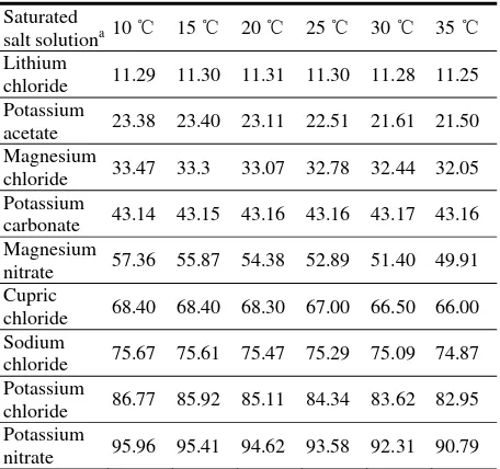
The static gravimetric method, with standard saturated salt solutions in Table 1 to maintain constant vapor pressure [1], was used to obtain nine equilibrium moisture contents at each of five constant temperatures
(10, 20, 25, 30, and 35 ℃). Twenty-seven glass bottles
with a volume of 250 ml each contained 65 ml salt solution, and were kept in one temperature controlled cabinet to maintain nine groups of different relative
Table 1 The equilibrium relative humidity (%) produced by nine saturated salt solutions.
Saturated
salt solutiona10 ℃ 15 ℃ 20 ℃ 25 ℃ 30 ℃ 35 ℃ Lithium
chloride 11.29 11.30 11.31 11.30 11.28 11.25 Potassium
acetate 23.38 23.40 23.11 22.51 21.61 21.50 Magnesium
chloride 33.47 33.3 33.07 32.78 32.44 32.05 Potassium
carbonate 43.14 43.15 43.16 43.16 43.17 43.16 Magnesium
nitrate 57.36 55.87 54.38 52.89 51.40 49.91 Cupric
chloride 68.40 68.40 68.30 67.00 66.50 66.00 Sodium
chloride 75.67 75.61 75.47 75.29 75.09 74.87 Potassium
chloride 86.77 85.92 85.11 84.34 83.62 82.95 Potassium
nitrate 95.96 95.41 94.62 93.58 92.31 90.79
a
Source: Jayas and Mazza [1].
humidity (r.h.) levels ranging from 11.3 to 96%. Every r.h. at one temperature was triplicated and a total of 135 bottles was used in the experiment for five sorption isotherms of a rice variety. The temperature of cabinets was often proofed with a standard thermometer ranging
from 0 to 50 ℃, and controlled to an accuracy of ±
0.5 ℃. The sample of rice seeds (about 4-5 g) was put
into a small bucket (3 cm diameter × 4 cm length) made from copper wire gauze, and hung into the 250 ml bottle on a copper wire pothook under a rubber plug, just 2-3 cm above saturated salt solutions. The rubber plug was tightly plugged into the bottle mouth. From three weeks after exposing the samples in the saturated
vapour at 35 ℃, the copper wire buckets with samples
The sample was dried to constant weight under 103.0
± 0.5 ℃ for 22-28 hours.
2.2 Analysis of the Adsorption and Desorption Data
The adsorption and desorption EMC data of rough rice were fitted to the six moisture sorption isotherm equations given in Table 2, using the non-linear regression procedure in SPSS 13.0 for Windows [10], which minimizes the sum of squares of deviations between experimental and predicted data in a series of iterative steps. The goodness-of-fit of each equation
was evaluated using determination coefficient (R2),
residue sum of squares (RSS), the standard error (SE), and mean relative percentage error (MRE).
The R2 was one of the primary criteria for selecting
the best equation to fit the experimental data. In addition to R2, the other statistical parameters, MRE as a percentage, RSS and SE were used to determine the
quality of the fit. The Eqs. (1)-(4) were used for
calculating R2, RSS, SE, and MRE, respectively.
2
predicated value, mmi is the average of experimental
values, and n is the number of observations. The fit of an equation is good enough for practical purposes when MRE is less than 10% [11].
Table 2 The cited EMC/ERH equations in the study.
Models Equations a
r.h. represents moisture content, M is equilibrium moisture content (% dry basis), t is temperature (℃), and Psis saturated vapor
The Sorption Isosteric Heats of Rough Rice in China
18
2.3 Determination of the Isosteric Heat of Sorption
The total energy required to remove a unit mass of water from grain kernels, the differential heat of
sorption (hs), is conveniently partitioned into two
components, namely the latent heat of vaporization of free water (hv) and the differential heat of wetting (hw).
The hv of adsorption and desorption of rice were
respectively calculated by the following six equations according to Thorpe [7].
. .
dependent on temperature (t, ℃). The saturated vapor
pressure (Ps) can be calculated by Eq. (7). The
derivative of r.h. with respect to t, ∂r h. ./∂Tm c. . depends
on the sorption isotherm equation used, and the Modified Chung-Pfost (MCPE) in Eq. (9) is used in this study.
3. Results
3.1 Fitting of Sorption Equations to Experimental Sorption Data
The results of nonlinear regression analyses of fitting the sorption equations to the experimental data of desorption and adsorption isotherms were respectively evaluated with the indices such as
correlation coefficient (R2), residue sum of squares
(RSS), the standard error (SE), and mean relative percentage error (MRE). Of the six commonly used equations, namely BET, GAB, MCPE, MHE, MOE, and STYE (Table 2), five equations such as STYE,
MCPE, MHE, MOE and GAB gave the better fit to the experimental data of adsorption and desorption isotherms in a wide range of 11.3 to 96.0% ERH, but the BET equation gave the better fit in the range of 11.3 to 49.9% ERH (data not shown). The further comparisons of the sorption equations with a form of
. . ( , )
r h = f M t or M = f r h t( . ., ) for twenty-six sets of isotherm data are given in Table 3. The average
values of R2 and error parameters (RSS, SE, and MRE)
for the twenty-six sets of isotherm data were calculated.
For the form of r h. .= f M t( , ), the equations were ranked for accuracy in an order: STYE, MCPE, MHE, MOE, MHE and GAB, but for that of M =f r h t( . ., ), the order was: BET, MCPE, MHE, MOE, and GAB. However, STYE is four-coefficient, temperature independent equation and can not be explicitly inverted to give EMC as a function of ERH. MCPE, MHE, MOE and GAB equations all are three-coefficient, temperature dependent and easily invertible equations (Table 2). Thus, the MCPE with a form of
. . ( , )
r h = f M t , or with a form of M=f r h t( . ., ) was considered to best describe the equilibrium moisture data of thirteen rice varieties in a wide range of 11.3 to 96.0% ERH, and the best fitted coefficients for both adsorption and desorption isotherms of rough rice data were summarized in Table 4. For MCPE model, the three coefficients C1, C2 and C3 of adsorptive isotherm
equation were different from those of desorptive isotherm equation, and there were some difference in three coefficients C1, C2 and C3 of between Japonica
and Indica rice.
3.2 Isosteric Heat of Sorption
The isosteric heat of sorption (hs) was calculated
from the Eqs. (6) to (9). The coefficients C1, C2, and C3
Table 3 Summary of the results of fitting equations to the data sets of thirteen pairs of rice desorption and adsorption.
Model function Equation Statistical parameters
a
R2 RSS SE MRE %
. .
( , )
r h
=
f M t
GAB 0.99045 0.03092 0.00074 5.90547 MCPE 0.99570 0.01440 0.00020 3.67480 MHE 0.99192 0.02624 0.00042 5.04545 MOE 0.99208 0.02421 0.00085 6.30338 STYE 0.99594 0.01414 0.00044 3.65875
( . ., )
M
=
f r h t
BET 0.98096 1.91840 0.10670 2.98630
GAB 0.97031 47.73069 1.13642 8.46685
MCPE 0.99225 12.24169 0.29150 3.02528
MHE 0.98675 21.32694 0.50775 4.52040
MOE 0.98031 31.34288 0.74628 6.02336
a
The statistical parameter is average of the data sets of thirteen pairs of rough rice desorption and adsorption. R2, correlation coefficient; RSS, residue sum of squares; SE, standard error; MRE means relative percentage error.
Table 4 The best fitted coefficients of MCPE for sorption isotherms of rough rice.
Model Data setsa Model coefficients Statistical parameters
C1 C2 C3 R2 RSS SE MRE %
. . ( , )
r h = f M t
(MCPE)
Desorption 412.543 35.300 0.181 0.9986 0.0046 1.094E-04 1.7654 Adsorption 677.146 110.639 0.184 0.9981 0.0060 1.430E-04 2.1502 Average 483.486 57.569 0.182 0.9985 0.0048 1.131E-04 1.6690 Japonica rice 455.064 63.182 0.175 0.9978 0.0071 1.682E-04 2.5580 Indica rice 504.668 58.354 0.184 0.9985 0.0048 1.136E-04 1.6934
a
Data sets were derivated from the average sorption data of thirteen rice varieties. Desorption, desorption isotherm; Adsorption, adsorption isotherms; Average, the average values obtained from adsorption and desorption isotherms.
adsorption and desorption isotherms.
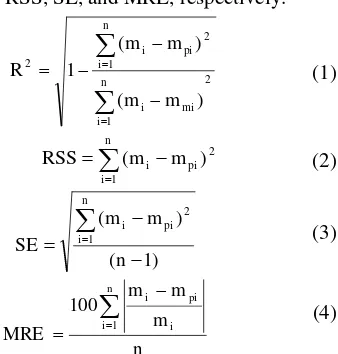
The isosteric heats for both rough rice desorption and adsorption (Fig. 1A), and for both the sorption of Japonica and India rice (Fig. 1B) decreased rapidly with increase in seed moisture content until the moisture content of 20% d.b. was reached, but above 20% d.b. they decreased smoothly with increasing moisture content. At lower moisture contents below 22.5% d.b., the isosteric heats of both desorption and adsorption of rough rice, and of both sorption of Japonica and Indica rice under lower temperatures were higher than those under higher temperatures. The isosteric heats of rough rice desorption were higher than those of adsorption below 22.5% d.b., but thereafter there was no difference found between desorption and adsorption (Fig. 1A). The sorption isosteric heats of Indica rice were slightly higher than
those of Japonica rice at all moisture contents under a constant temperature (Fig. 1B).
4. Discussion
Sun [2] analyzed 17 source sets of rice EMC/ERH data with four commonly cited models such as MCPE, MHE, MOE, and STYE, and considered the STYE as the preferred equation, but three models like MHE, MCPE, and MOE were recommended to fit the EMC/ERH data of rough rice by ASABE [5]. In this study, for the form of r h. .= f M t( , ), the equations were ranked for accuracy in an order: STYE, MCPE, MHE, MOE, MHE and GAB, but for that of
( . ., )
M = f r h t , the order was: BET, MCPE, MHE,
The Sorption Isosteric Heats of Rough Rice in China
Fig. 1 Comparison of adsorption and desorption isosteric heats of rough rice (A), and of the sorption isosteric heats of
Japonica and Indica rice (B) at different temperatures (℃)
predicted by the Modified Chung-Pfost equation.
study to calculate the rough rice isosteric heat of sorption. The isosteric heats for both rough rice desorption and adsorption, and for both the sorption of Japonica and Indica rice, decreased rapidly with an increase in seed moisture content until the m.c. of 20% d.b. was reached, and thereafter they decreased smoothly with increasing moisture content. The isosteric heats of rough rice desorption were higher than those of adsorption below 22.5% d.b. EMC, but thereafter there was no significant difference found between desorption and adsorption. These results show a little difference from the results reported by Öztekin and Soysal [12] that the isosteric heats of rice desorption were higher than those of adsorption below 11% d.b. EMC, but
above 11% d.b. EMC the isosteric heats of desorption were lower than those of adsorption. The sorption isosteric heats of Indica rice were insignificantly higher than those of Japonica rice under all moisture contents at a constant temperature. The rapid increase in the heat of sorption at low m.c. might be due to the existence of highly active polar sites on the surface of rice grains, which were covered with water molecules forming a mono-molecular layer [13]. The decrease in the isosteric heats with higher amounts of sorbed water can be quantitatively explained by considering that sorption initially occurs on the most active available sites giving rise to high interaction energy. As these sites become occupied, sorption occurs on the less active ones, resulting in lower heats of sorption [14]. In low moisture contents, the values of the isosteric heats were higher than the latent heat of vaporization of water, indicating that the energy of binding between the water molecules and the sorption sites was higher than the energy which holds the molecules of pure water together in the liquid phase [15]. At high moisture contents, there was no significant difference between the sorption isosteric heat and the latent heat of vaporization of water over the broad range of moisture contents. In the present study, the heat of sorption of rough rice grains might approach that of pure water at the moisture content of about 22.5% d.b.
It has been noted that hs/hv was calculated to be
dependent on temperature, but the dependence was
small [7]. This temperature dependency of hs/hv was
rather clear in Fig. 1, which might arise from experimental errors in measuring the sorption isotherm, or from the sample properties such as variety, harvest time, pre-treatment, and so on.
The sorption isosteric heats of Indica rice were slightly higher than those of Japonica rice under all moisture contents at a constant temperature. These results could be used for rice drying, storage and aeration.
Acknowledgments
The authors gratefully acknowledge the Scientific
Research Foundation for the Returned Overseas Chinese Scholars, State Human Resources and Social Security Department (CZ1020) and the National Key Technology R&D Program (Project no. 2009BADA0B00-4) for providing financial support. The authors also thank Prof. Digvir S. Jayas from the University of Manitoba in Canada for his kindly suggestions for the experiment, and Dr. Graham Thorpe of Victoria University, Australia for demonstrating how to calculate the isosteric heat of sorption.
References
[1] D.S. Jayas, G. Mazza, Equilibrium moisture characteristics of safflower seeds, Transactions of the ASAE 34 (1991) 2099-2103.
[2] D.W. Sun, Comparison and selection of EMC/ERH isotherm equations for rice, Journal of Stored Products Research 35 (1999) 249-264.
[3] X.J. Li, S.L. Wang, J.S. Wang, Progress in characterization of equilibrium moisture content in grains, Journal of the Chinese Cereals and Oils Association 24 (11) (2009) 137-145. (in Chinese with English abstract) [4] C. Chen, R.V. Morey, Comparison of four EMC/ERH
equations, Transactions of the ASAE 32 (1989) 983-990.
[5] ASABE Standards, ASAE D245.5 OCT1995, Moisture relationships of plant-based agricultural products, American Society of Agricultural Engineers, St. Joseph, Michigan, USA, 2006, 49085-49659.
[6] H.A. Iglesias, J. Chirife, P. Viollaz, Thermodynamics of water vapour sorption by sugar beet root, Journal of Food Technology 11 (1976) 91-101.
[7] G.R. Thorpe, Physical basis of aeration, in: S. Navarro, R. Noyes (Eds.), The Mechanics and Physical of Modern Grain Aeration Management, CRC Press, Boca Raton, 2001, pp. 135-144, 186.
[8] X.J. Li, Z.Y. Wei, Z.Y. Cao, Q.Y. Feng, J.S. Wang, Equilibrium moisture content and sorption isosteric heats of five wheat varieties in China, Journal of Stored Products Research 47 (2011) 39-47.
[9] AOAC, Official Methods of Analysis, 13th edition, Washington DC, Assoc. Off. Anal. Chem., 1980.
[10] SPSS Inc., SPSS for Windows, Release 13.0.1., SPSS Inc., Chicago, USA, 2006.
[11] R.J. Aguerre, C. Suarez, P.E. Viollaz, New BET type multilayer sorption isotherms: Part II. Modelling water sorption in foods, Lebensmittel-Wissenschaft und-Technologie 22 (1989) 192-195.
[12] S. Öztekin, Y. Soysal, Comparison of adsorption and desorption isosteric heats for some grains, Agricultural Engineering International: the CIGR Journal of Scientific Research and Development2 (2000)1-17.
[13]E. Tsami, Net isosteric heat of sorption in dried fruits, Journal of Food Engineering 14 (1991) 327-335.
[14] N. Wang, J.G. Brennan, Moisture sorption isotherm characteristics of potatoes at four temperatures, Journal of Food Engineering 14 (1991) 269-282.