Effect of the chemical state of uranium atom on the energy
of spin-orbital splitting of its inner orbitals
By Yuri F. Batrakov, Andrey G. Krivitsky∗, Oleg V. Pospelov and Elena V. Puchkova
Section of Radiochemistry, Faculty of Chemistry, St. Petersburg State University, St. Petersburg, 199034, Russia (Received August 7, 2003; accepted in revised form October 10, 2003)
Uranium / Uranium compound / Relativistic effects / Chemical bond / X-ray emission / Chemical shift
Summary. Chemical shifts (ChSh) of nine emission lines
of the uranium L-series in uranium oxides UO2+x (x=0–1)
with respect to UO2 were studied by using a precise
crystal-diffraction X-ray spectrometer. ChSh of Lα1, α2 uranium and
thorium lines in solid solutions yUO2·(1−y)ThO2 (y=0÷1)
were measured with respect to UO2 and ThO2, respectively.
The changes in energy of spin-orbital splitting (SOS) –∆δnl± of innernl-orbitals of the uranium atom were calculated from
the data of ChSh of spin-doublet lines. For UO2+x oxides,
a linear decrease in ∆δnl± values with increasing degree
of uranium oxidation was found. Sign inversion of ∆δnl±
for uranium levels was found on passing to solid solutions. No change in the SOS energy of inner thorium levels was detected. The values of ∆δnl± were found to correlate with the experimental values of the effective magnetic moment of uranium in oxides.
On the basis of the comparison of experimental ∆δnl±
values with Dirac–Hartree–Fock atomic calculations, it was concluded that the observed variations in ∆δnl± values are due to the redistribution of electron and spin density between the 5f7/2- and 5f5/2-levels of the fine structure of the uranium
atom without changes in atomic charge state. On the basis of the hypothesis of intraatomic relativistic U 5f7/2↔U 5f5/2
transition, a model of paramagnetic moment formation on the uranium atom in uranium dioxide was proposed.
1. Introduction
The chemistry of heavy element atoms was the sub-ject of many theoretical and experimental studies in the past decades. In fundamental reviews by Pyykkö and De-sclaux [1] and Pitzer [2], it is noted that the most outstanding chemical behavior anomalies of compounds of heavy atoms are due to relativistic effects (RE). The RE in chemical term is understood as all effects in the electron structure of an atom which appears passing to the finite value of the velocity of light c=137.0359895(61)a.u.1 as compared
to c= ∞ [3]. In chemistry, three main REs are usually considered:
*Author for correspondence (E-mail: [email protected]). 1Atomic units, the system of units where e≡me≡h≡1.
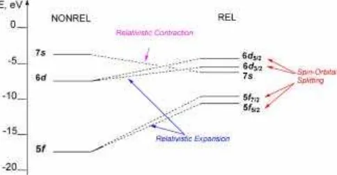
1. Direct RE of contraction and energetic stabilization of s1/2- andp1/2-atomic orbitals (AOs).
2. Indirect RE of self-consistent expansion and energetic destabilization ofd- and f-AOs.
3. Effect of spin-orbital splitting (SOS) of AOs having a non-zero orbital angular momentum l into two sub-shellsnl+andnl−with total angular momentum j=l±
1/2, respectively.
For the case of uranium valence orbitals, these effects are schematically shown in Fig. 1.
With increasing atomic number Z, the relativistic com-ponent of total energy increases proportionally to Zn
(n>1). The SOS energy of valence orbitals is
propor-tional to Z4 and in the case of light actinides according to the data of atomic calculations in the Dirac–Hartree– Fock (DHF) approximation, it is several electron-volts (eV, 1 eV≈1.60×10⁻¹⁹J). In other words, it is comparable to
chemical bond energy. Hence, there are prerequisites for the relatively independent participation in chemical bond-ing of wave functions correspondbond-ing to split nl± valence
levels [4]. However, at present the question whether it is ne-cessary to take into account RE (in particular the effect of SOS of valence orbitals) in modeling the chemical bond-ing of light actinide atoms is still unsolved [5–7]. This is mainly caused by the lack of experimental data on the ba-sis of which qualitative and quantitative evaluation could be made about the participation of valence levels of the fine structure of the heavy atom in the process of chemical bonding.
In the case of the uranium atom, the problem of the degree of participation of 5f-electrons in chemical bond-ing is also unsolved [7, 8]. In X-ray photoelectron spectra (XPS), a regularly decreasing intensity of the narrow line of 5f-electrons (with an energy Eb=2.0±0.1 eV) is
ob-served in a series of UO2+xoxides with increasingx[9–11].
The explanations of this phenomenon are very contradic-tory [9–13]. This is probably due to insufficient XPS sensi-tivity to fine energetic changes of the atomic electron struc-ture (state). It is known that the error in the determination of the binding energy of electrons and of the line width in the XPS method is about 100 meV and that in the determination of relative line intensity is 10% [9].
A possibility to introduce clarity into these problems is to use the method of chemical shift (ChSh) of hard X-ray emission lines. ChSh is defined as a change in the energy of an X-ray emission line due to modification of bonds of the atom. This method gives information about the chemical state of the atom in the sample. The aim of our work is to search for an experimental parameter making it possible to characterize the chemical effects of SOS of valence orbitals in the uranium atoms.
Compound Crystal structure Provided by Synthesis
references UO2.02 fluorite type, St. Petersburg University, [14]a
cubic UO2 Dept. of Radiochemistry
ThO2.0 fluorite type, Commercial
cubic ThO2
U4O9(UO2.25) fluorite type, V.G. Khlopin Radium [15, 16] cubic U4O9 Institute, St. Petersburg
U2O5(UO2.50) hexagonal U2O5 V.G. Khlopin Radium [17] Institute, St. Petersburg
U3O8(UO2.67) orthorhombic St. Petersburg University, [18]
α-U3O8 Dept. of Radiochemistry
UO3.0 orthorhombic St. Petersburg University, [19, 20]
γ-UO3 Dept. of Radiochemistry
yUO2·(1−y)ThO2 fluorite type, St. Petersburg University, [21, 22]
cubicb Dept. of Radiochemistry
UO2(NO3)2·6H2O see ref. St. Petersburg University, [23] Dept. of Radiochemistry
UO2(NO3)2·2H2O see ref. St. Petersburg University, [24] Dept. of Radiochemistry
UO2SO4 see ref. Samara State University, [25]
Inorganic Chem. Dept.
UO2SO4·2.5H2O see ref. Samara State University, [25]
Inorganic Chem. Dept.
UO2SeO4 see ref. Samara State University, [25, 26] Inorganic Chem. Dept.
UO2SeO4·2.5H2O see ref. Samara State University, [25, 26]
Inorganic Chem. Dept. a: The reduction of commercial UO2.14with carbon monoxide at 400–500◦C.
b: The solid solutions of UO2·ThO2obey Vegard’s law.
Table 1.The characterization of measured
samples.
2. Experimental
Twenty-one samples were measured, which were uranium oxides, uranium-thorium solid solutions, and uranyl com-pounds, as listed in Table 1. The structure of the samples was confirmed by our X-ray powder diagram investigations.
Chemical shifts were measured for:
a) Nine lines (composing two multiplets: 2p-3dand 2p-4d, two doublets: 2s-3pand 2s-4p) of the L-uranium series for uranium oxides UO2+x(x=0–1) with respect to UO2
(Table 2).
b) Lα1, α2-uranium and thorium lines for solid solutions
yUO2·(1−y)ThO2 (y=0÷1) with respect to UO2 and
ThO2, respectively (Table 3).
c) Lα1, α2- uranium lines for uranyl compounds:
UO2(NO3)2·2H2O, UO2(NO3)2·6H2O, UO2SO4,
UO2SO4·2.5H2O, UO2SeO4and UO2SeO4·2.5H2O with
respect to UO2(Table 4).
focus-Table 2.Chemical shifts (in meV) of theL-uranium series in oxides UO2+x.
Multiplet/ 2p-3da 2p-4d 2s-3p 2s-4p
Doublet
Line Lα1 Lα2 Lβ2 Lβ15 Lγ1 Lβ4 Lβ3 Lγ3 Lγ2
Transition 3d+→2p+ 3d−→2p+ 4d+→2p+ 4d−→2p+ 4d−→2p− 3p−→2s 3p+→2s 4p+→2s 4p−→2s
Exp. error ±3 ±4 ±3 ±6 ±4 ±6 ±6 ±6 ±4
UO2 0 0 0 0 0 0 0 0 0
U4O9 −51 3 −37 −8 −23 −7 −118 n/m n/m
U2O5 −101 +19 −67 −10 −34 n/mb n/m n/m n/m
U3O8 −118 +46 −82 +9 −33 −31 −192 n/m n/m
UO3 −152 +88 −85 +44 −1 +15 −169 −97 +5
a: 2p-3dmultiplet consists of three lines:Lα1,Lα2, andLβ1(3d−→2p−transition);
b: Not measured because of low line intensity (50 times lower than the intensity of the most marked ULα1-line).
Composition δULα1 δULα2 ∆δU 3d± δThLα1 δThLα2 ∆δTh 3d±
Exp. error ±3 ±5 ±6 ±3 ±5 ±6
UO2 0 0 0 − − −
0.9UO2-0.1ThO2 +2 +4 −2 −30 −33 +3
0.85UO2-0.15ThO2 −2 −3 +1 −29 −30 +1
0.8UO2-0.2ThO2 2 −9 +11 −23 −29 +5
0.7UO2-0.3ThO2 8 −5 +13 −13 −27 +14
0.5UO2-0.5ThO2 6 −12 +18 −5 −13 +8
0.3UO2-0.7ThO2 3 −27 +30 −3 −12 +9
0.2UO2-0.8ThO2 8 −24 +32 −5 −10 +5
0.15UO2-0.85ThO2 14 −28 +42 −3 −8 +5
0.1UO2-0.9ThO2 18 −39 +57 +1 −2 +3
ThO2 − − − 0 0 0
Table 3. Chemical shifts (δ, in meV) of
Lα1- andLα2-lines of uranium and
tho-rium and changes in SOS energy (∆δ3d±,
in meV) of 3d-orbital of uranium and tho-rium in solid solutionsyUO2·(1−y)ThO2.
Table 4.Chemical shifts (δ, in meV) ofLα1- and Lα2-uranium lines
and changes in SOS energy (∆δ3d±, in meV) of 3d-uranium orbital in
uranyl compounds.
Compound δULα1 δULα2 ∆δU 3d±
Exp. error ±3 ±5 ±6
UO2 0 0 0
UO2(NO3)2·6H2O −153 +116 −269 UO2(NO3)2·2H2O −139 +101 −240
UO2SO4 −139 +125 −264
UO2SO4·2.5H2O −151 +108 −259
UO2SeO4 −131 +131 −262
UO2SeO4·2.5H2O −134 +119 −253
UO3 −152 +88 −240
ing according Couchois’ technique [29]. A detailed scheme of the instrument is shown in Fig. 2.
A 0.3 mm thick quartz crystal curved in the shape of a part of the cylinder with a 2000 mm radius was used as a monochromator. Adjustment to a certain line with a wavelength λwas done with the aid of a theodolit (3) by counting the required angleθaccording to the Wulf–Braggs’ equation:
nλ=2dsinθ , (1)
where n is the reflection order (n=1), d is the interpla-nar spacing of the reflecting surfaces of the monochromator (d=1.81674 Å), andθis the diffraction angle.
Fig. 2. Scheme of X-ray spectrometer: 1. a focal point; 2.
mirror-equivalence position; 3. optical angle meter; 4. quartz monochromator; 5. sample; 6. rotating cassette; 7. Roentgen tube; 8. collimator; 9. step-motor-driven micrometer; 10. the lever; 11. scintillation detector.
Each sample was powdered, mixed with polystyrene powder and pressed into a tablet, 20 mm in diameter. The tablet was then placed in a metal holder and mounted in a special rotating cassette (6), which can carry six such samples (5). Fluorescent (secondary) X-ray emissionhν1of
and 44 mA. After collimation (8), the radiation was decom-posed with a quartz monochromator (4), adjusted to the selected X-ray line, and was recorded with the aid of a scin-tillation detector (11) on the basis of a NaI(Tl) crystal.
Preliminary adjustment to the appropriate X-ray emis-sion line was carried out with the help of optical angle meter (3). Line scanning was carried out discretely (18–32 points per line) with different pitch for each line by a turning monochromator on 2–10 angle seconds with a step-motor-driven micrometer (9). Line intensity measurements at each point were carried out successively for all samples by their successive introduction into the primary beam. To compen-sate for optical aberration effects and the effects of external factors, the experiment was repeated many times. The ob-tained results were averaged.
The mathematical processing of the spectra (determin-ation of the position of the line maximum and of its natural width) included the approximation of experimental data by the convolution of the Lorentzian function describing the true form of the X-ray line and the Gaussian function de-scribing the widening of this line due to the mosaic (block) structure of the monochromator [30, 31].
The absolute error in the determination of the line shift attains 1 meV, and the relative precision in its determination is σE
E ≈10
−8(whereσE is the mean-square error in the
de-termination of line energy E). The spectrometer resolution calculated by using theKα1-line of tin as the ratio of
exper-imentally observed width of the emission line at half-height Γ to its energy is 10−5. We emphasize this point because the
photon energy region between 10 and 20 keV is rather un-favorable for crystal diffractometry measurements and the lack of accurate experimental data concerning L X-rays of heavy elements results.
3. Results and discussion
A physical value that should be chosen as the parameter making it possible to judge about the participation of split components of uranium valence orbitals in chemical bond-ing must provides information about changes in the SOS energy of the innernl-orbital when the chemical state of the atom is varied [32]. It can be easily shown that this parame-ter is the difference (∆δnl±) in the ChSh (δ) of spin-doublet lines having one common level. For instance, such a level for ULα1- and ULα2-lines is the 2p+-level. Then the difference
in chemical shifts with respect to reference sample is
δLα1−δLα2= the innernl j-electron. Hence, if the ChSh of spin-doublet lines for various uranium compounds are known it is pos-sible to calculate the change in the SOS energy (∆δnl±) of innernl-levels and to draw generalizing conclusions about RE of SOS.
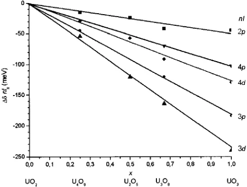
Fig. 3.Changes in SOS energy of inner uranium nl-orbitals∆δnl±
(respect to UO2) in oxides UO2+x vs.x.
It is necessary to study first the behavior of the parame-ter∆δnl±depending on theredox stateof the atom. For this purpose, on the basis of the measured values of ChSh (with respect to UO2) of spin-doublet lines (Table 2) the changes
of the SOS energy of inner 2p-, 3p-, 3d-, 4p- and 4d-orbitals of uranium were obtained (Table 5) in a series of oxides with the composition of UO2+x (x=0–1). It is evident that with
changing index at oxygen x a whole set of uranium atom characteristics also changes: partial charge on this atom as well asmagnetic stateandvalence statefrom U(IV) in UO2
to U(VI) in UO3.
Fig. 3 shows the linear character of the dependence of the parameter∆δnl± onxfor all inner uranium AOs. With increasing degree of uranium oxidation, a monotonic de-crease in the SOS of all inner uranium orbitals is observed. Therefore, a conclusion can be drawn that the effect of pro-cesses in the valence shell at the atom on the change in the SOS energy of core AOs is of a monotonic character. The fact that the inner uranium levels respond linearly to changes in the chemical state of the atom (i.e. in the per-turbation of the valence shell of the atom) is not trivial be-cause the potential of inner electrons screening by valence ones is of a more complex character. Moreover, it should be noted that the greatest response (maximal changes in the SOS energy) are observed for the 3d-uranium orbital. The SOS energy of the U 3d-orbital in uranium trioxide is almost 250 meV lower than that in uranium dioxide. There-fore, it is the uranium 3d-orbital that is convenient for study-ing the effect of the chemical state of the atom on RE of SOS.
To study the behavior of the parameter ∆δ3d± in com-pounds of the same valency, the ChSh of ULα1-, and ULα2
-lines were measured with respect to UO2for two groups of
isovalent compounds:
1) solid solutionsyUO2·(1−y)ThO2(y=0–1) (Table 3) in
which uranium is in the tetravalent state;
2) uranyl compounds UO2(NO3)2·2H2O, UO2(NO3)2·
6H2O, UO2SO4, UO2SO4·2.5H2O, UO2SeO4 and
UO2SeO4·2.5H2O of hexavalent uranium (Table 4). For
solid solutions, ChSh of ThLα1-, and ThLα2-lines with
nl Formula ∆δnl±
UO2 U4O9 U2O5 U3O8 UO3 Error
2p ∆δ2p±=δLβ15−δLγ1 0 −15 −24 −42 −45 ±5 3p ∆δ3p±=δLβ3−δLβ4 0 −45 n/ma −120 −184 ±8 3d ∆δ3d±=δLα1−δLα2 0 −54 −120 −164 −240 ±5 4p ∆δ4p±=δLγ3−δLγ2 0 n/m n/m −70 −102 ±7 4d ∆δ4d±=δLβ2−δLβ15 0 −29 −57 −91 −129 ±7 a: Not measured.
Table 5.Changes in SOS energy (∆δnl±,
in meV) of inner nl-uranium orbitals in oxides UO2+x.
Fig. 4.Changes in SOS energy of 3d-orbitals∆δ3d±of uranium
(re-spect to UO2) and thorium (re(re-spect to ThO2)vs. composition of solid solutionyUO2·(1−y)ThO2.
Fig. 4 shows the dependence of changes in the SOS en-ergy of U 3d- and Th 3d-orbitals on the composition (y) of the solid solution yUO2·(1−y)ThO2. With increasing
con-tent of thorium dioxide in solid solution, the energy dif-ference between the split components (±) of the uranium 3d-orbital increases. This is probably due to the dilution of the paramagnetic matrix of uranium dioxide by diamagnetic thorium dioxide. The fact that such changes are not observed in the thorium system confirms the above suggestion about the effect of paramagnetic properties of UO2.
Table 4 gives the ChSh of Lα1-, and ULα2-lines with
respect to UO2, and ∆δ3d± values calculated from their
basis for diamagnetic uranyl compounds. It is quite clear that ChSh values for the compounds with uranyl group vary over a wide range. However, their differences (∆δ3d± values) are in a narrow range (240–270)±6 meV. It can be suggested that this is caused by the uranyl structure of these compounds. Slight variation in the SOS energy is due to effect of ligands of the second coordination sphere. It should be noted that the observed effect, the constant value of ∆δ3d± can serve as a distinguishing feature of uranyl compounds.
From the viewpoint of the role of the uranium atom mag-netic state in chemical bonding effects it is of interest to examine the dependence of SOS energy of U 3d-orbitals on the effective magnetic moment on the uranium atom (µeffect).
Fig. 5 shows the correlation of the∆δ3d±with experimental values ofµeffecttaken from Refs. [21, 22, 33]. In a system of
coordinatesµeffect−∆δ3d±, uranium oxides UO2+xand solid
Fig. 5.Correlation of experimental values [21, 33] of effective
mag-netic moment on uranium atom (µeffect) with the value of changes in
SOS energy of uranium 3d-orbitals (∆δ3d±).
solutionsyUO2·(1−y)ThO2are located on straight lines
cor-responding to different linear functions. This indicates that the formation mechanisms of magnetic properties in these two systems differ greatly. Uranyl compounds are localized on the abscissa (µeffect=0), as has been shown above, in
a narrow range of ∆δ3d± values. This correlation can be used for evaluating the uranium atom magnetic state in the investigated compound.
The above facts show that RE of SOS of inner atomic or-bital levels directly depend on the uranium magnetic state which is determined by the state of valence shell of the atom. This conclusion is confirmed by the data of isotopic ef-fects [34, 35] in twosomes233U–238U and235U–238U for the
compounds: U3O8, UO2and Cs[UO2(NO3)2]. The value of
∆δnl± (nl=2p, 3p, 3d, and 4p) for chemically identical compounds with different isotopic composition is lower by one order then in changing the chemical state of the atom (see Table 6). It should be noted that the235U–238U isotopic
shifts ofLβ3-,Lβ4-,Lγ2-, andLγ3-lines attain the values of
+250±20 meV [35]. Even if relative large errors in the re-sults of measurements of isotopic effects of the parameter ∆δnl± (Table 6) are taken into account, it can be suggested that the change in the SOS energy of inner orbitals of the atom is not greatly affected by changes in the isotopic state of its nucleus.
nl ∆δnl±(233U–238U) ∆δnl±(235U–238U)
(in meV, respect to238U) in SOS energy changes of
NonRel Rel Rel Rel Rela Rel Rel
5f→ ∞ 5f+→5f− 5f+→ ∞ 5f−→ ∞ 5f+→5f− 5f+→ ∞ 5f−→ ∞
Table 7. Theoretical (DHF) values of
changes in SOS energy (∆δnl±, in meV)
of innernl-uranium orbitals.
1) nonrelativistic removal of one U 5f-electron (NonRel 5f →8);
2) relativistic removal of one U 5f5/2-electron (Rel 5f−
→8);
3) relativistic removal of one U 5f7/2-electron (Rel 5f+
→8);
4) relativistic transmission of one electron from U 5f7/2- to
U 5f5/2-orbital (Rel 5f+→5f−).
Table 7 lists the results of our calculation by the Dirac– Hartree–Fock (DHF) method (the applicability of DHF-calculations for the purposes of X-ray emission spec-troscopy, in particular, for the chemical shifts method has been shown in Ref. [36]) of changes in SOS energy on inner nluranium AOs (nl=2p, 3p, 3d, 4p, and 4d) for the four above transitions from two initial configurations of uranium: neutral atom U [Rn] 5f−1.55f
cancy on the innernl j-shell.
The data in this table show that the∆δnl±values almost
do not depend on the number of electrons (occupancy) on uranium 6d- and 7s-levels. Hence, all energetics of RE of SOS is determined by 5f-levels.
In Fig. 6 the theoretical ∆δnl± values are compared
with experimental values of the SOS energy of inner nl-uranium orbitals in UO3with respect to UO2(Table 5). For
all four theoretical and experimental cases maximum values of∆δnl± are observed on the 3d-orbital. Only the model of
a relativistic electron transition from 5f7/2- to 5f5/2-orbital
of uranium (Rel 5f+→5f−) describes the experiment
sat-isfactorily. In other words, a conclusion can be drawn that anintraatomic relativisticU 5f+→U 5f−transitionis
pos-sible. Its analogue is to some extent the nonrelativistic tran-sition Th 6d→Th 5f in thorium compounds from which the filling of 5f-levels in the actinide series starts [37]. The change in the SOS energy of inner AOs is due to electron redistribution between 5f+- and 5f−-levels of the fine
struc-Fig. 6.Experimental and theoretical values of changes in SOS energy
of inner uraniumnl-orbitals (∆δnl±).
ture of the uranium atom without a change in atomic charge state:
∆δnl±=Q5f±·C nl5
f±, (4)
where Q5f± is the number of electrons redistributed
be-tween 5f+- and 5f−-levels of the uranium atom,Cnl5f± is
the change in the SOS energy of thenl-level on transition of one electron 5f+→5f−. Then the appearance of
param-agnetic moment on uranium in UO2from diamagnetic UO3
(the unpaired electron density formation) can be illustrated by the following scheme:
Fig. 7.Correlation of experimental values [21, 33] of effective
mag-netic moment on uranium atom (µeffect) with the number of electrons
(Q5f±) participating in the redistribution between 5f+- and 5f−
-uranium orbitals. Line – theoretical function (5), points – experiment.
effective magnetic moment:
µseffect= [N(N+2)]1/2=2
[Q5f±(Q5f±+1)]1/2. (5)
On the basis of Eq. (4) from experimental values of ∆δnl±(see Table 5) and calculated coefficients Cnl5
f±(see Table 7) one can find the number of redistributed electrons Q5f± for uranium oxides UO2+x (with respect to UO2). In
other words, one can ascribe to each compound in addition to the experimental value of effective magnetic moment on uranium atom µeffect the number of electron Q5f±
partici-pating in the redistribution between 5f+- and 5f−-levels of uranium. In Fig. 7 the theoretical function (5) is compared with the dependence of experimental values [21, 33] ofµeffect
on the number Q5f± for uranium oxides. The correlation between theoretical and experimental dependencies makes it possible to draw the conclusion that suggestion about the mechanism of the appearance of paramagnetic moment on uranium in UO2 because of redistribution of 5f-electrons
has a right to existence.
It may be suggested that the intraatomic relativistic 5f+→5f− transition is a fine energetic tuning to a spe-cific chemical state. As a result of this transition unique chemical properties of uranium atom are probably formed in the uranium-oxygen system [38]: polyvalence of the oxide series, numerous structural modifications, and structural-chemical compromise which consists in the preserva-tion of oxide structure with increasing oxygen content (UO2.00→UO2.25).
It should be emphasized that these conclusions confirm the theoretical concept of core orbitals [39–41] developing recently. This concept is based on Clausius’ virial theorem and reflects a very important role of inner atomic orbitals in chemical bonds formation.
4. Conclusion
On the basis of experiments and calculations, it is possible to draw the conclusion that the SOS effect of inner atomic orbitals strongly depends on the chemical state of the
ura-nium atom. This dependence is a direct consequence of the unique intra-atomic electron redistribution between 5f+ -and 5f−-split levels of the uranium atom fine structure with-out changes in atomic charge state.
Acknowledgment.The authors wish to express appreciations to Dr. Lev L. Makarov who contributed much to this work in its early stage, Dr. Ilja I. Tupitsyn for his kind help with DHF programs, and Dr. Roman V. Bogdanov for stimulating discussions of the paper.
This work was carried out with the financial support of the Russian Federation Ministry of Education- grant PD 02-1.3-306.
References
1. Pyykkö, P., Desclaux, J. P.: Relativity and the Periodic System of
Elements. Acc. Chem. Res.12, 276 (1979).
2. Pitzer, K. S.: Relativistic effects on chemical properties. Acc.
Chem. Res.12, 271 (1979).
3. Pyykkö, P.: Relativistic effects in structural chemistry. Chem. Rev.
88, 563 (1988).
4. Schwarz, W. H. E., van Wezenbeek, E. M., Baerends, E. J., Snij-ders, J. C.: The origin of relativistic effects of atomic orbitals.
J. Phys. B.: At. Mol. Opt. Phys.22, 1515 (1989).
5. Ionova, G.: Relativistic Effects in Properties of Heavy Elements: Theory and Experiment. In: 5th Intern. Conf. Nucl. Radiochem., Extended Abstracts, Vol. 1. Pontresina, Switzerland (2000) p. 213. 6. Balasubramanian, K.: Relativistic effects and electronic structure
of lanthanide and actinide molecules. In:Handbook of Physics
and Chemistry of Rare Earth. (Gschneidner, K. A., Eyring, L., Choppin, G. R., Lander, G. H., eds.) Elsevier, Amsterdam (1994) Vol. 18, Chapt. 119, p. 29.
7. Schwarz, W. H. E.: The Concept of the Chemical Bond. In:
Theor-etical Models of Chemical Bonding. (Maksic, Z. B., ed.) Springer, Berlin (1990) Vol. 2, p. 593.
8. Pepper, M., Bursten, B. E.: The electronic structure of actinide-containing molecules: a challenge to applied quantum chemistry.
Chem. Rev.91, 719 (1991).
9. Teterin, Yu. A., Terechov, V. A., Ryzhkov, M. V., Utkin, I. O.,
Ivanov, K. E., Teterin, A. Yu., Nikitin, A. S.: The role of the U 6p,
5f electrons in chemical bonding of uranil and uranium
fluo-rides: X-ray photoelectron and X-ray emission studies. J. Electron
Spectrosc. Relat. Phenom.114, 915 (2001).
10. Moser, H. R., Delley, B., Schneider, W. D., Baer, Y.:
Characteri-zation of f electrons in light lanthanide and actinide metals by
electron-energy-loss and X-ray photoelectron spectroscopy. Phys.
Rev. B29, 2947 (1984).
11. Veal, B. W., Lam, D. J.: X-ray photoelectron studies of thorium,
uranium, and their dioxides. Phys. Rev. B10, 4902 (1974).
12. Batrakov, Yu. F., Makarov, L. L.: On the role of 5f-electrons in
the chemical bonding of the light actinides by theirLX-ray
emis-sion spectra. Vestnik SPbGU. Ser. 44, 40 (1995).
13. Makarov, L. L.: X-ray emission effects as a tool to study light
actinides. Czech. J. Phys.49, Suppl. S1, Part 2, 610 (1999).
14. Crosswhite, H. M.: Gmelin Handbook of Inorganic Chemistry.
8thedn., Springer, Berlin (1984), Uranium Suppl. Vol. C4
Ura-nium Dioxide, Sect. 1.2., p. 11.
15. Gronvold, F. J.: High-temperature X-ray study of uranium oxides
in the UO2-U3O8region. Inorg. Nucl. Chem.1, 357 (1955).
16. Shaner, B. E.: Metallographic determination of the UO2-U4O9
phase diagram. J. Nucl. Mater.2, 110 (1960).
17. Blinova, N. I., Kurbatov, V. V., Solncev, V. M.: X-ray investigation
of the system U3O8-U2O5. Radiokhimija (Radiochemistry)6, 463
(1964).
18. Kovba, L. M.: The rectification of the homogenous regions for
U3O8±x and U8O21±x oxides. Radiokhimija (Radiochemistry)9,
134 (1967).
19. Sheft, I., Fried, S., Davidson, N.: Preparation of uranium trioxide.
J. Am. Chem. Soc.72, 2172 (1950).
20. Hoekstra, H. R., Siegel, S.: The uranium-oxygen system:
U3O8-UO3. Inorg. Nucl. Chem.18, 154 (1961).
21. Trizebiatowski, W., Selwood, P. W.: Magnetic susceptibilities of
urania-thoria solid solutions. J. Am. Chem. Soc. 72, 4504
22. Hinatsu, Y., Fujino, T.: Magnetic susceptibilities of UO2-ThO2
solid solutions. J. Solid State Chem.60, 195 (1985).
23. Vdovenko, V. M., Stroganov, E. V., Sokolov, A. P., Zandin, V. N.: The structure of the uranyl nitrate hexahydrate. Radiokhimija
(Radiochemistry)2, 24 (1960).
24. Vdovenko, V. M., Stroganov, E. V., Sokolov, A. P.: The structure investigating of the crystals of uranyl nitrate trihydrate and
uranyl nitrate dihydrate. Radiokhimija (Radiochemistry) 3, 19
(1961).
25. Brandenburg, N. P., Loopstra, B. O.:β-uranyl sulphate and uranyl
selenate. Acta Cryst. B34, 3734 (1978).
26. Serezhkin, V. N., Tabachenko, V. V., Serezhkina, L. B.: Synthesis and investigating of the uranyl selenate. Radiokhimija
(Radio-chemistry)20, 214 (1978).
27. Sumbaev, O. I.: The effect of the chemical shift of the X-ray
Klines in heavy atoms. Phys. Lett. A30, 129 (1969).
28. Sumbaev, O. I.: Crystal-diffraction gamma-spectrometers. Gos
AtomIzdat, Moscow (1963) pp. 110–111.
29. Cauchois, Y.: Spectrographie des rayonsxpar transmission d’un
faisceau noncanalise’ a travers un cristal courbe’ (I). J. Phys. Rad.
3, 320 (1932).
30. Lee, P. L., Boehm, F., Vogel, P.: Gen. Phys.9, 616 (1974).
31. Tupitsyn, I. I., Makarov, L. L., Batrakov, J. F.: Sign of the
spin-polarized effects in the chemical shifts of the X-ray CuKα1,2
emission transitions. J. Phys. Chem. Solids59, 809 (1998).
32. Batrakov, Yu. F., Krivitsky, A. G., Puchkova, E. V.: Relativistic Ef-fects on Light Actinides Chemical Properties. In: 2nd Russian Youth Science Conference on the Fundamental Problems of
Ra-diochemistry and Atomic Energy. Abstract, N. Novgorod (2002) p. 26.
33. Selwod, P. W.: Bull. Soc. Chim.D, 122 (1949).
34. Makarov, L. L., Suglobov, D. N., Batrakov, Yu. F.: The isotope
shifts in UL X-ray emission lines. Radiokhimija
(Radiochem-istry)38, 206 (1996).
35. Makarov, L. L., Batrakov, Ju. F., Solomennikov, A. A.: ULX-ray
emission isotope effects: 238–235–233. In: 5th Intern. Conf. Nucl. Radiochem., Extended Abstracts, Vol. 1. Pontresina, Switzerland (2000) p. 346.
36. Bratcev, V. F., Deyneka, G. B., Tupitsyn, I. I.: Application of Hartree–Fock method to calculation of relativistic wave functions.
Izvestija AN SSSR. Ser. Fizicheskaja41, 2656 (1977).
37. Makarov, L. L., Karazija, R. I., Batrakov, Yu. F., Chibisov, N. P., Mosevich, A. N., Zaytsev, Yu. M., Udris, A. I., Shishkunova, L. V.:
Chemical effects in the ThL-spectra. Radiokhimija
(Radiochem-istry)20, 116 (1978).
38. Katz, J. J., Seaborg, G. T., Morss, L. R.:The chemistry of the
ac-tinide elements. 2nd edn., Chapman and Hall, New York (1986) pp. 1131–1165.
39. Korolkov, D. V.:Theory of valency in Progress. (Kuznetsov, V. I.,
ed.) Mir, Moscow (1980) p. 210.
40. Korolkov, D. V.: Core and Valence Atomic Orbitals in Chemical
Bond Formation. In: Series Sov. Sci. Rev. B. Chem. Harwood
Academic Publishers GmbH, UK (1992) Vol. 17, p. 103.
41. Korolkov, D. V.: Electronic Structure and Properties of

![Fig. 5. Correlation of experimental values [21,33] of effective mag-SOS energy of uranium 3netic moment on uranium atom (µeffect) with the value of changes ind-orbitals (∆δ3d±).](https://thumb-ap.123doks.com/thumbv2/123dok/2796873.1686309/5.595.307.550.153.359/correlation-experimental-effective-uranium-uranium-ueffect-changes-orbitals.webp)

![Fig. 7. Correlation of experimental values [21,33] of effective mag-(netic moment on uranium atom (µeffect) with the number of electronsQ5 f±) participating in the redistribution between 5 f+- and 5 f−-uranium orbitals](https://thumb-ap.123doks.com/thumbv2/123dok/2796873.1686309/7.595.43.282.41.227/correlation-experimental-effective-ueffect-electronsq-participating-redistribution-orbitals.webp)
