NOTE Bacteriology
Two Genotypes among ‘
Candidatus
Mycoplasma haemobos’ Strains Based on the
16S-23S rRNA Intergenic Spacer Sequences
Fumina SASAOKA
1), Jin SUZUKI
1), Yusaku WATANABE
1,4), Masatoshi FUJIHARA
1,4), Kazuya NAGAI
2),
Toh-ichi HIRATA
3)and Ryô HARASAWA
1,4)*
1)Department of Veterinary Microbiology, Faculty of Agriculture, Iwate University, Morioka 020–8550, Japan 2)Cryobiofrontier Research Center, Faculty of Agriculture, Iwate University, Morioka 020–8550, Japan 3)Omyojin Research Farm, Faculty of Agriculture, Iwate University, Morioka 020–8550, Japan 4)The United Graduate School of Veterinary Science, Gifu University, Gifu 501–1193, Japan
(Received 6 August 2012/Accepted 1 October 2012/Published online in J-STAGE 15 October 2012)
ABSTRACT. ‘Candidatus Mycoplasma haemobos’, sometimes causative of bovine infectious anemia at various extents, has been demonstrated
throughout the world. Here, we show two distinct types of ‘Ca. M. haemobos’ are distributed among cattle in Japan, by examining the primary and secondary structures of the 16S-23S rRNA intergenic spacer region that has been shown to be a stable genetic marker for mycoplasma species. Our results may explain differences in severity of anemic condition as well as provide a genetic marker for an epidemiological study of bovine hemoplasma infections.
KEY WORDS: hemoplasma, mycoplasma, rRNA.
doi: 10.1292/jvms.12-0349; J. Vet. Med. Sci. 75(3): 361–364, 2013
‘
Candidatus
Mycoplasma haemobos (synonym of ‘
Ca.
M.
haemobovis’)’ is a trivial name of hemotropic mycoplasma
that may cause anemia at various degrees in cattle [11, 12,
16, 18]. Reason of different severity in anemic condition
caused by ‘
Ca
. M. haemobos’ infection has yet been
un-known. Although this alleged species has provisionally been
identiied by only nucleotide sequence of the 16S rRNA
gene because of uncultivable trait [10, 17, 20], genetic
varia-tion in this species remained unexplored. In our previous
study, the 16S-23S rRNA intergenic spacer (ITS) region of
hemoplasma was found to be a useful tool for determination
of taxonomic status of this particular species [15] as well
as other mycoplasma species [2, 4–6]. Here, we show the
‘
Ca.
M. haemobos’ strains are divided into two distinct types
according to the ITS sequences.
EDTA-anticoagulated blood samples were collected from
25 Japanese black cattle bred on an experimental farm at
Omyojin (latitude 39.7N and longitude 141.1E) of Iwate
University during October 2011 to March 2012. Total DNA
was extracted from 200
µl
of the whole blood samples by
using the QIAamp DNA Blood Mini Kit (QIAGEN, Hilden,
Germany) according to the manufacturer’s instructions,
eluting into 200
µl
of buffer AE, and stored at −20°C until
examination in the PCR assay. No clinical symptom was
reported on the cattle examined, despite infection by
Bovine
leukemia virus
.
Hemoplasma infection was found in all the 25 cattle
diag-nosed by real-time PCR by using forward primer Hemo-F1,
5′-ATATTCCTACGGGAAGCAGC-3′, equivalent to nucle
-otide numbers 328 to 347 of
M. wenyonii
and reverse primer
Hemo-R1, 5′-ACCGCAGCTGCTGGCACATA-3′, equiva
-lent to nucleotide numbers 503 to 522 of
M. wenyonii
as
de-scribed previously [13]. Real-time PCR was performed in a
SmartCycler instrument (Cepheid, Sunnyvale, CA, U.S.A.)
with SYBR Premix Ex
Taq
(Code #RR041A, TaKaRa Bio.,
Otsu, Japan). The reaction mixture contained 1
µl
of each
primer (10 pmol/
µl
), 12.5
µl
of 2X premix reaction buffer
and water to volume of 23
µl
. Finally, 2
µl
of DNA samples
as templates were added to this mixture. Ampliication was
carried out 40 cycles of denaturation at 95°C for 5 sec, re
-naturation at 57°C for 20 sec and elongation at 72°C for 15
sec, after the initial denaturation at 94°C for 30 sec. After
real-time PCR, melting experiment was performed from 60
to 95°C at 0.2°C/sec with smooth curve setting averaging
one point. Melting peaks were visualized by plotting the
irst derivative against the melting temperature as described
previously [7].
Of all the cattle examined, eight cattle were found
in-fected with ‘
Ca
. M. haemobos’ alone based on the 16S rRNA
gene analysis, and they were subjected to analysis of the ITS
region by end-point PCR. Briely, PCR ampliication was
carried out at 94°C for 30 sec, 55°C for 2 min and 72°C for 2
min for 30 cycles using forward (5′-GTTCCCAGGTCTTG
-TACACA-3′) and reverse (5′-CAGTACTTGTTCACTATC
-GGTA-3′) primers as described previously [1]. The PCR
products were then fractionated on horizontal, submerged
1.0% SeaKem ME agarose gels (FMC Bioproducts,
Rock-land, ME, U.S.A.) in TAE (40 mM Tris, pH8.0, 5 mM sodium
acetate and 1 mM disodium ethylenediaminetetra
cetate)
buffer at 50 volts for 60 min. After electrophoresis, the gels
were stained in ethidium bromide solution (0.4
µ
g/m
l
) for
*CorrespondenCe to: Harasawa, R, Department of Veterinary Microbiology, Faculty of Agriculture, Iwate University, Morioka 020–8550, Japan.
e-mail: [email protected]
F. SASAOKA ET AL.
362
15 min and visualized under UV transilluminator. DNA in
a clearly visible band was extracted by using NucleoSpin
Extract II kit (Macherey-Nagel, Düren, Germany) and was
subjected to direct sequencing in a 3500 Genetic Analyzer
(Applied Biosystems, Foster City, CA, U.S.A.). The
Gen-Bank/EMBL/DDBJ accession numbers for ITS sequences of
‘
Ca.
M. haemobos’ strains used in this study are AB740009
through AB740016.
The nucleotide sequences of ITS regions of the ‘
Ca
.
M. haemobos’ strains were compared with authentic
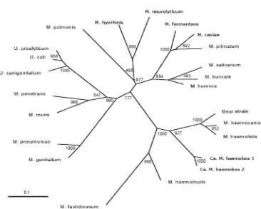
my-coplasma species in phylogenetic analysis. In the present
study, we examined the phylogenetic relatedness among 23
mycoplasma species including not only 3 ureaplasmas but
also 6 hemoplasmas,
M. haemomuris
,
M. haemofelis
, ‘
Ca.
M. haemominutum’, bear strain and 2 types of ‘
Ca
. M.
hae-mobos’ (Fig. 1). In addition to our previous illustration of
ITS from ‘
Ca
. M. haemobos’ type 1 [15], the present
analy-sis revealed existence of another type of ITS among ‘
Ca
.
M. haemobos’ strains. Alignment of nucleotide sequences of
ITS regions deined so far indicated 94% similarity between
these two genotypes (Table 1). Of the eight strains, six were
identical to the ‘
Ca
. M. haemobos’ type 1, and the remaining
two strains showed an identical but were distinct from type 1
sequence. Although we examined only Japanese black cattle
without clinical symptom, this variation can be used for an
epidemiological marker of ‘
Ca
. M. haemobos’ infections in
cattle population, since nucleotide sequences in ITS region
have been conserved within a mycoplasma species or
sub-species [2, 6, 8].
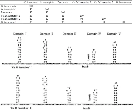
The secondary structures of the ITS were predicted
accord-ing to the algorithm of Zuker and Stiegler [21]. Five
stem-loop domains were allocated in ITS of ‘
Ca
. M. haemobos’
(Fig. 2). Domains III and V were well conservative, despite
several deletions in others domains. Secondary structures in
ITS region have sometime provided a key character to
dis-tinguish closely related species of mycoplasmas [3, 6, 7, 9].
In the present study, ITS regions of the two genotypes of
‘
Ca
. M. haemobos’ showed characteristic features of
myco-Fig. 1. Unrooted phylogenetic tree based on the ITS comparison, suggesting a monophyletic relationship among hemoplasmas and M. fastidiosum. Following nucleotide sequences obtained from the DNA databases
are shown with an accession number in parenthesis. They are M. salivarium PG20 (X58558), M. pulmonis
m53 (X58554), M. neurolyticum Sabin Type A (X58552), M. buccale CH20247 (D89504), M. primatum
HRC292 (D89509), M. caviae G122 (D89505), M. hominis PG21 (X58559), M. fermentans PG18 (X58553),
M. hyorhinis BTS-7 (X58555), M. penetrans GTU54 (D89508), M. muris RIII4 (D89507), M. pneumoniae
BOVINE HEMOPLASMAS 363
plasmas, lacking spacer tRNA genes and having boxA and
boxB motifs [15].
In conclusion, 2 genotypes of ‘
Ca
. M. haemobos’ were
demonstrated by analyzing the primary and secondary
struc-tures of ITS regions. This may provide a clue to elucidate
differences in severity of anemia in cattle, though virulence
of these 2 genotypes remained unexplored. These genotypes
can also be used for a genetic marker for bovine hemoplasma
infections.
ACKNOWLEDGMENTS. This study was partly supported
by a Grant-in-Aid (No. 23658251) for Scientiic Research
of the Japan Society for the Promotion of Science from the
Ministry of Education, Culture, Sports, Science, and
Tech-nology of Japan.
REFERENCES
1. Harasawa, R. 1996. PCR: application of nested PCR to detec-tion of mycoplasmas. pp.75–79. In: Molecular and Diagnostic Procedures in Mycoplasmology, vol. 2.(Razin, S. and Tully, J.G. eds.), Academic Press, New York.
2. Harasawa, R. 1999. Genetic relationships among mycoplasmas
based on the 16S-23S rRNA spacer sequence. Microbiol. Immu-nol.43: 127–132. [Medline]
3. Harasawa, R., Hotzel, H. and Sachse, K. 2000. Comparison of the 16S-23S rRNA intergenic spacer regions among strains of the Mycoplasma mycoides cluster, and reassessment of the taxo-nomic position of Mycoplasma sp. bovine group 7. Int. J. Syst. Evol. Microbiol.50: 1325–1329. [Medline] [CrossRef]
4. Harasawa, R. and Kanamoto, Y. 1999. Differentiation of two biovars of Ureaplasma urealyticum based on the 16S-23S rRNA intergenic spacer region. J. Clin. Microbiol. 37: 4135–4138.
[Medline]
5. Harasawa, R., Kawahara, M. and Rikihisa, Y. 2002.
Character-Table 1. Similarity matrix showing nucleotide sequence homology among the ITS regions of hemoplasmas. Nucleotide sequences of ITS regions have been determined on only four species. Numbers indicate homology percentage between two ITS sequences.
M. haemocanis M. haemofelis Bear strain Ca. M. haemobos 1 Ca. M. haemobos 2 M. haemomuris M. haemocanis 100
M. haemofelis 95 100
Bear strain 93 93 100
Ca. M. haemobos 1 80 81 81 100
Ca. M. haemobos 2 82 82 83 94 100
M. haemomuris 44 44 44 43 44 100
F. SASAOKA ET AL.
364
istics of the 16S-23S rRNA intergenic spacer region of Myco-plasma haemomuris, previously classiied as ‘Haemobartonella muris’. J. Vet. Med. Sci.64: 1161–1164. [Medline] [CrossRef]
6. Harasawa, R., Lefkowitz, E. J., Glass, J. I. and Cassell, G. H. 1996. Phylogenetic analysis of the 16S-23S rRNA intergenic spacer region of the genus Ureaplasma. J. Vet. Med. Sci.58: 191–195. [Medline] [CrossRef]
7. Harasawa, R., Mizusawa, H., Fuji, M., Yamamoto, J., Mukai, H., Uemori, T., Asada, K. and Kato, I. 2005. Rapid detection and differentiation of the major mycoplasma contaminants in cell cultures using real-time PCR with SYBR Green I and melting curve analysis. Microbiol. Immunol.49: 859–863. [Medline]
8. Harasawa, R., Pitcher, D. G., Ramirez, A. S. and Bradbury, J. M. 2004. A putative transposase gene in the 16S-23S rRNA inter-genic spacer region of Mycoplasma imitans. Microbiology150: 1023–1029. [Medline] [CrossRef]
9. Harasawa, R., Uemori, T., Asada, K., Kato, I. and Shiragami, N.
1992. ‘boxA’-like sequence between the 16S/23S spacer in the
rRNA operon. FEBS Lett.297: 209–211. [Medline] [CrossRef]
10. Hoelzle, K., Hofmann-Lehmann, R. and Hoelzle, L. E. 2010. ‘Candidatus Mycoplasma haemobos’, a new bovine haemot-rophic Mycoplasma species? Vet. Microbiol. 144: 525–526.
[Medline] [CrossRef]
11. Hoelzle, K., Winkler, M., Kramer, M. M., Wittenbrink, M. M., Dieckmann, S. M. and Hoelze, L. E. 2011. Detection of Candi-datus Mycoplasma haemobos in cattle with anemia. Vet. J.187: 408–410. [Medline] [CrossRef]
12. Hofmann-Lehmann, R., Meli, M. L., Dreher, U. M., Gönczi, E., Deplazes, P., Braun, U., Engels, M., Schüpbach, J., Jörger, K., Thoma, R., Griot, C., Stark, K. D. C., Willi, B., Schmidt, J., Kocan, K. M. and Lutz, H. 2004. Concurrent infections with vector-borne pathogens associated with fetal hemolytic anemia in a cattle herd in Switerland. J. Clin. Microbiol.42: 3775–3780.
[Medline] [CrossRef]
13. Nishizawa, I., Sato, M., Fujihara, M., Sato, S. and Harasawa, R.
2010. Differential detection of hemotropic Mycoplasma species in cattle by melting curve analysis of PCR products. J. Vet. Med. Sci.72: 77–79. (Erratum. J. Vet. Med. Sci. 72: 1704.) [Medline]
[CrossRef]
14. Saitou, N. and Nei, M. 1987. The neighbor-joining method: a new method for reconstructing phylogenetic trees. Mol. Biol. Evol.4: 406–425. [Medline]
15. Sasaoka, F., Suzuki, J., Fujihara, M., Watanabe, Y., Nagai, K. and Harasawa, R. 2012. Examination of the 16S-23S rRNA
intergenic spacer sequences of ‘Cadidatus Mycoplasma haemo-bos’ and Mycoplasma haemofelis. J. Vet. Med. Sci.74: 83–87.
[Medline] [CrossRef]
16. Su, Q. L., Song, H. Q., Lin, R. Q., Yuan, Z. G., Yang, J. F., Zhao, W. Y. and Zhu, X. Q. 2010. The detection of ‘Candidatus My-coplasma haemobos’ in cattle and buffalo in China. Trop. Anim. Health Prod.42: 1805–1808. [Medline] [CrossRef]
17. Tagawa, M., Matsumoto, K. and Inokuma, H. 2008. Molecular detection of Mycoplasma wenyonii and ‘Candidatus Myco-plasma haemobos’ in cattle in Hokkaido, Japan. Vet. Microbiol. 132: 177–180. [Medline] [CrossRef]
18. Tagawa, M., Matsumoto, K., Yokoyama, N. and Inokuma, H. 2010. Comparison of the effect of two hemoplasma species on hematological parameters in cattle. J. Vet. Med. Sci.72: 113–115.
[Medline] [CrossRef]
19. Thompson, J. D., Higgins, D. G. and Gibson, T. J. 1994. CLUST-AL W: improving the sensitivity of progressive multiple
se-quence alignment through sese-quence weighting, position-speciic
gap penalties and weight matrix choice. Nucleic Acids Res.22: 4673–4680. [Medline] [CrossRef]
20. Uilenberg, G. 2009. ‘Candidatus Mycoplasma haemobos’. Vet. Microbiol.138: 200–201. [Medline] [CrossRef]
21. Zuker, M. and Stiegler, P. 1981. Optimal computer folding of