CORRELATION BETWEEN MAGNESIUM SERUM LEVELS AND PEAK
EXPIRATORY FLOW IN CHRONIC ASTHMA PATIENTS
1 2 3*
Grendi Faneri Yonarko , Suhardi DA. , Barmawi Hisyam 1. Faculty of Medicine, Universitas Gadjah Mada, Yogyakarta
2. Division of Nephrology, Department of Internal Medicine, Dr. Sardjito General Hospital, Yogyakarta 3. Division of Pulmonology, Department of Internal Medicine, Dr. Sardjito General Hospital, Yogyakarta
*Corresponding Author: [email protected]
ABSTRACT
Background: Magnesium Serum in some previous studies had been related to asthma events and severity levels. Correlation between magnesium serum levels and severity of asthma was controversies. It might be influenced by race, genetic pattern, diet and demography factor.
Objective: The objective of this study was to know relationship between magnesium serum levels and predictive PEF (Peak Expiratory Flow) in chronic asthma patients in Yogyakarta.
Method: This was a cross-sectional study; subjects were asthma patients who visited Pulmonology Outpatient Department in Dr. Sardjito General Hospital Yogyakarta, from September 2004 - March 2005. We performed clinical evaluation, magnesium serum levels (normal 0.65 mmol/L-1.04 mmol/L), predictive PEF and medications. Correlation between magnesium serum levels and PEF were analyzed by Pearson correlation test. This study used analysis of variance (anova) to analyze mean difference among more than 2 groups and multiple regressions to know the variables which influenced serum magnesium levels.
Result: There were 62 asthma patients in this study. There was no hypomagnesaemia. The mean of magnesium was 0.89 ± 0.08 mmol/L. Results
INTRODUCTION
Asthma is a serious problem. The prevalence of asthma increased in the developing
1
country during the last 20 years . The increasing number of asthma patients registered in Indonesia is
2
9
population . Some studies and meta-analysis reported that magnesium intravenous effective to treat intermediate and severe acute exacerbation of
10,11,12,13
asthma .
Correlation between serum magnesium and asthma is controversy. The concentration of serum magnesium in asthma population in Yogyakarta is unknown. The prevalence of hypomagnesaemia and evidence of the correlation can support of the prevention and therapeutic of asthma. Status of magnesium was measured by serum magnesium
14
level . Peak Expiratory Flow (PEF) was used to 15,16
measure lung function .
The hypothesis was the serum magnesium had the correlation with Peak Expiratory Flow (PEF) in chronic asthma patients.
SUBJECTS AND METHODS
It was a cross-sectional study. Subjects were chronic asthma patients who visited to Pulmonology Outpatient Department Dr. Sardjito General Hospital Yogyakarta, from September 2004 - March 2005.
Eligibility criteria for entry in this study were as follows: diagnosed as chronic asthma patients; more than 18 years old; signs informed consent. The exclusion criteria were as follows: used diuretic, thiazid, aminoglicoside, siclosporine, cisplatin, foscarnet, laxative, oral magnesium, enema, antacide, magnesium intravena; alcohol abuse; history of diabetes mellitus, heart and renal diseases; current diarhhoea; and current pregnancy.
The independent variable is magnesium serum. The dependent variable is PEF. The study used consecutive sampling. Chronic asthmatics, who agreed to take apart in the study were interviewed and clinically evaluated. A questionnaire that included the patients age, sex,
duration of asthma, number of hospitalization was obtained from each subject. PEF was measured 3 times to get the best value. Measurements of serum magnesium level were carried out using colorimetric from Randox.
STATISTICAL ANALYSIS
The statistical analysis was done using SPSS 11. Characteristic features were showed by mean and standard deviation. Distribution was tested by Kolmogorov Smirnov. Correlation between serum magnesium levels and PEF were analyzed by Pearson correlation test. In all tests, a p value of <0.05 was considered significant. The value of power was 90%. We used anova to compare mean when analyze more than 2 group.
RESULTS
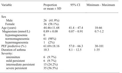
Total 62 chronic asthma patients were studied. They were 26 male subjects (41.9%) and 46 female subjects (58.1%). Mean of ages were 44.46±11.68 years. Mean of magnesium was 0.89 ± 0.08 mmol/L. There was no prevalence of hypomagnesemia. The study found 1 (2%) subject h a d h y p e r m a g n e s a e m i a a n d 6 1 ( 9 8 % ) normomagnesaemia subjects.
Table 1. Characteristic features
Variable Proportion
or mean ± SD
95% CI Minimum - Maximum
Sex
Male
Female Age (years)
Magnesium (mmol/L)
hypomagnesemia
normomagnesemia
hypermagnesemia PEF predictive (% ) Duration of asthma Severity:
intermitten mild persistent
intermediate persistent severe persistent
26 (41.9%) 36 (58.1%). 44.46±11.68 0.89 ± 0.08 0
61 (98%) 1 (2%) 61.69±18.16 10.3
6 (9.7%) 6 (9.7%) 15 (24.2%) 35 (56.5%)
41.4 – 47.4 0.87 – 0.91
57.0 – 66.3 8.1 – 12.5
18-66 0.7-1.2
30-101 1-35
Table 2. Correlation between Magnesium and PEF
Variabel r p
Magnesium
0.281
0.027*
*significant (<0,05), Pearson test
Table 3. Correlation between magnesium serum and admission to emergency room or hospitalization
Graphic 1. Scatter Plot of correlation of predictive PEF (%) and magnesium serum (mmol/L).
Serum magnesium (mmol/dL)
1.3 1.2
1.1 1.0
.9 .8
.7 .6
PEF predictive (%)
120
100
80
60
40
20
DISCUSSION
This study did not find hypomagnesemia. 8,17 This result was different with previous study . This result might be interfered by some factors including diet and genetic pattern. The type of diet of Yogyakarta population and the method of cooking may influence magnesium status. Some impairment and variant of genetic pattern could also influence magnesium absorption from intestinal and
18,19,20 magnesium excretion through renal . This factor could bias of the study. The tight exclusion criteria might be excluding the population of hypomagnesemia. But the same exclusion criteria was used on Alamoudi study in Arab Saudi and found prevalence of hypomagnesemia at
8,17 approximately 27% .
The serum magnesium may not reflect a true status of magnesium. Magnesium predominantly was in intracellular and only 1% in
21
serum . The correlation between concentration of serum magnesium and intracellular magnesium is still unclear. Zervas reported that the asthmatic acute attack patients had erythrocytes magnesium level lower than healthy people or chronic asthma
argument. We suggested a larger sample in next extended study to achieve a more conclusive result.
CONCLUSION
The study showed a weak correlation of serum magnesium and PEF(r=0.28 p<0.05). This result could be influenced by some factors. The strong correlation was higher in hypomagnesemia population than in normomagnesaemia population. This study did not have a hypomagnesaemia population so that we cannot analyze this probability.
The uncontrolled factor like allergen factor, air pollution infection, weather, emotion, food and additive might cause bias in this study.
REFERENCES
1. Global Initiative for Asthma (GINA), 2005, Global Strategy For Asthma Management and Prevention, National Instititute of Health. www.Ginasthma.com,
4. Spivey, W. H., Skobelpff, E. M, Levin R.M., 1984, Effect of Magnesium Chloride on Rabbit Bronchial smooth Muscle, Annual Emergency Medicine 19:1107-1112
5. Dominguez L.J, Barbagallo, M., Lorenzo, G. D., Drago A. et al., 1998, Bronchial Reactivity and Intracellular Magnesium: a Possible Mechanism for the Bronchodilating Effects of Magnesium in Asthma, Clinical Science 95: 137-142.
6. Fassler, C.A., Rodriguez, M., Badesch, D.B., Stone, W.J, Marini J.J , 1985, Magnesium toxicity as a Cause of Hypotension and Hypoventilation. Arch Internal Medicine 145, 1604-1606
7. Britton, J., Pavord, I., Richards, K., 1994, Dietary Magnesium, Lung function, Wheezing, and Airway Hypereactivity in a Random Adult Population Sample, Lancet 344: 357-62
8. Alamoudi, O. S, 2000, Hipomagnesemia in Chronic Stable Asthmatics: Prevalence, Correlation with Severity and Hospitality, Europe Respiratory Journal, 16: 427- 431
9. Alter H. J., Thomas D. K. Hilty W. M., 2000, Intravenous Magnesium as an Adjuvant in Acute Bronkospasme: A Meta Analysis, Annals of Emergency Medicine vol 36:191-197.
10. Ciarallo, L., Broisseau, D. Reinert, S., 2000, Higher Dose Intravenous Magnesium Therapy for Children with Moderate to Severe Acute Asthma, Arch Pediatric Adolescence Med ; 154 (10):979-83
11. Silverman R. A., Osborn H. Runge J., Gallagher E. J., 2002, IV Magnesium Sulfate in the Treatment of Acute Severe Asthma, a
Multicenter Randomized Controlled Trial, CHEST, 22 : 489-497.
12. Ian J , Johnson K , Montelpare , McAuliffe, 2002, Variability within individuals of plasma ionic magnesium concentrations, BMC Physiology, 2:6 ,www.biomedcentral.com
13. National Asthma Education an Prevention Program.Expert (NAEPP) ,1997, Panel Report 2: guidelines for Diagnosis and Management of Asthma, Bethesda, MD: National Institutes of Health, in B.D. Rose (Eds), UpToDate version 14.1 14. Prasson J.,Mani S.K.,2003, Peak Expiratory Flow vs
s p i r o m e t r y i n P a t i e n t s w i t h A s t h m a , www.rcjournal.com
15. Alamoudi, O.S., 2001, Electrolyte Disturbance in Patients With Chronic, Stable Asthma, Chest 120:431-436
1
16. Agus, Z. A.,2005 , Causes of Hypomagnesemia, dalam B.D Rose, UpToDate ed 14.1
2
17. Agus, Z. A.,2005 , Causes and Treatment of Hypermagnesemia, dalam B.D Rose UpToDate ed 14.1
3
18. Agus, Z. A.,2005 , Regulation of Magnesium Balance, dalam B.D Rose UpToDate ed 14.1
19. Zervas, E., Papatheodorou G., 2003, Reduced Intracelluler Mg Concentration in Patients With Acute Asthma, Chest:123:113-118
20. Cohen J, 1977, Statistical Power Analysis for the Behavioral Science, Academy Press, p. 93 21. Zervas E., Loukides S., Papatheodorou G., Psathakis
PROGNOSTIC FACTORS OF LEPTOSPIROSIS PATIENTS IN DR.
SARDJITO GENERAL HOSPITAL, YOGYAKARTA, INDONESIA
1 2 2* 2
Kurniaatmaja, E.R , Priambodo D , Humardewayanti R , Loehoeri S 1. Faculty of Medicine, Universitas Gadjah Mada, Yogyakarta
2. Subdivision of Tropical Medicine and Infectious Diseases Department of Internal Medicine Universitas Gadjah Mada / Dr. Sardjito General Hospital, Yogyakarta, Indonesia
*Corresponding Author:[email protected]
ABSTRACT
Background:
. Severe disease can be fatal, although majority of cases are mild and self-limited.
Objective: To determine the prognostic factors for leptospirosis that associated with mortality in patients with leptospirosis in Dr. Sardjito General Hospital, Yogyakarta.
e conducted a retrospective study of data collected in our hospital between Jan 2010 until May 2011, from whom the diagnosis of leptospirosis was confirmed based on pertinent clinical and epidemiological data and positive serology.
Result: Thirty two patients were included in this study, including 29 survivors (90.62%) and 3 non-survivors (9.38%). Of these 32 patients, 26 patients (81.25%) were admitted to the medical ward and 6 patients (18.75 %) were admitted to the ICU. Multivariate logistic regression demonstrated that three factors were independently associated with mortality: higher level of potassium (OR 10.8; CI 1.194-97.728; p<0.01) on admission and neurological dysfunction (altered mentation or seizure) (OR 30; CI 4.367–206.07; p<0.01)
Conclusion: The mortality of leptospirosis remains high despite improvements in patients care. In order to improve the early treatment of high-risk
Leptospirosis, an infectious disease that affects humans and animals, is a common zoonosis with a variety of clinical manifestations. Yogyakarta is one of the cities with a high incidence of leptospirosis. It is important to recognize the clinical features and prognostic factors of this disease
Methods: W
Keywords : Leptospirosis, prognostic factors, mortality
Background
Leptospirosis is the most widespread zoonosis with recent outbreaks in several Asian, 1 Central, and South American countries. Leptospirosis is a zoonotic disease, which is caused by Leptospira and transmitted to human by contact with Leptospira contaminated animal urine or
Leptospira contaminated environment. Thirty-two patients with Leptospirosis had admitted in Dr. Sardjito General Hospital from January 2010 until
2
May 2011. In Indonesia, the spread of leptospirosis is in the Province of West Java, Central Java, Yogyakarta, Lampung, South Sumatra, Bengkulu, Riau, West Sumatra, North Sumatra, Bali, NTB, South Sulawesi, North Sulawesi, East Kalimantan and West Kalimantan. The mortality rate of leptospirosis in Indonesia is high, reaching 2.5 to 16.45%. At the age of over 50 years mortality rate can be up to 56%. When Leptospirosis patient is accompanied by a yellow lining of the eye indicating damage to liver tissue, the risk of death will be higher. Several publications reported mortality rates between 3% -54% depending on the infected organ
3 system.
We believe that early evaluation of disease severity at the time of admission might be useful in improving the care of patients with leptopsirosis.
Table 1. The mean hospital stay ± SD for the survivors
Variabel Survivors Non-survivors p
Gender, n (%)
Man Woman
21 8
3
0 0.55
Age (years ), mean ± SD
42.41± 13.44 57.33± 14.43 0.2
20-46years
47-76 years
17 12
0
3 0.092
At risk occupation, n (%)
Yes No
12 17
2
1 0.57
Rodent exposure, n (%)
Yes No
21 8
2
1 1
Hemodialisis, n
Yes No
10 19
1
2 1
ICU, n
Yes No
4 25
2
1 0.083
The mean age ± SD for the survivors was 42.41 years ± 13.44 years, and for the non-survivors was 57.33 years ± 14.43 years. In age variable there was no differences between the survivors and non survivors (p = 0.092)
The gender ratio between male and female patients with leptospirosis was 3:1, man 75% and women 25%. There was no differences in gender between the survivors and non survivors (p = 0.55)
Table 2. Clinical sign and symptoms in survivors and non survivors among patients with Leptospirosis
Sign and symptoms
Survivors(n =29) Non survivors(n =3) pvalue
Fever
2 0 1.000
Dyspnea 15 2 1.000
Jaundice 19 3 0.534
Cardiovascular collapse
16 1 0.589
Conjunctival Suffusion
21 2 1.000
Oliguria 9 3 0.044*
Respiratory symptoms
7 2 0.184
Neurological dysfunction
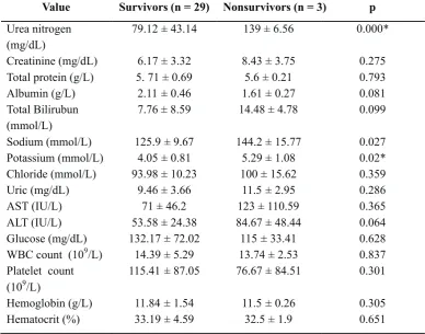
Table 3. Result of univariate analysis of laboratory values between survivors and non-survivors among patients with leptospirosis.
Value Survivors (n = 29) Nonsurvivors (n = 3) p
Urea nitrogen (mg/dL)
79.12 ±43.14 139 ±6.56 0.000*
Creatinine (mg/dL) 6.17 ±3.32 8.43 ±3.75 0.275 Total protein (g/L) 5. 71 ±0.69 5.6 ±0.21 0.793
Albumin (g/L) 2.11 ±0.46 1.61 ±0.27 0.081
Total Bilirubun (mmol/L)
7.76 ± 8.59 14.48 ± 4.78 0.099
Sodium (mmol/L) 125.9 ±9.67 144.2 ±15.77 0.027 Potassium (mmol/L) 4.05 ±0.81 5.29 ±1.08 0.02* Chloride (mmol/L) 93.98 ±10.23 100 ±15.62 0.359
Uric (mg/dL) 9.46 ±3.66 11.5 ±2.95 0.286
AST (IU/L) 71 ±46.2 123 ±110.59 0.365
ALT (IU/L) 53.58 ±24.38 84.67 ±48.44 0.064
Glucose (mg/dL) 132.17 ±72.02 115 ±33.41 0.628 WBC count (109/L) 14.39 ±5.29 13.74 ±2.53 0.837 Platelet count
(109/L)
115.41 ±87.05 76.67 ±84.51 0.301
Hemoglobin (g/L) 11.84 ±1.54 11.5 ±0.26 0.305
Hematocrit (%) 33.19 ± 4.59 32.5 ± 1.9 0.651
*statistically significant
Table 4. Result of multivariate stepwise logistic regression analysis of risk factors for patients with Leptospirosis.
Risk Factor OR 95% CI P value
Oliguria 0.75 0.541 –1.04 0.018
Urea nitrogen (mg/dL)
0.813 0.642 -1.028 0.025
Potassium (mmol/L)
10.8 1.194 –97.728 0.009
Neurological symptoms
10 with a worse prognosis for oliguric renal failure. Ninety-seven percent of the patients had renal failure and all patients who died were oliguric. In our study, oliguria was a very sensitive criterion of the severity of the disease. Similarly, Seguro et al noted that the mortality rate for oliguric patients with acute renal failure were higher than that for patients with persistent dieresis. Again, 90% of leptospirosis is anicteric with a lower mortality compared with icteric forms. In our group, 68% of patients were icteric and there was no difference in mortality.
Neurologic dysfunction (altered mentation or seizures) was the most significant predictor of mortality; most patients with neurologic dysfunction also had significant renal and hepatic disease contributing to encephalopathy. Altered mental status was the strongest independent predictor of death in urban leptospirosis in Brazil; other poor prognostic factors were oliguria, advanced age, renal and respiratory insufficiency.In this study, there was an association between altered
12 mental status and mortality.
Conclusions
The mortality of leptospirosis remains high despite improvements in patients care. In order to improve the early treatment of high-risk patients, these two clinically and two laboratory criteria, independently associated with mortality, could be used at the time of admission.
References
1. Vinetz JM. Leptospirosis. Curr Opin Infect Dis. 2001; 14:527–538.
2. Tunissea A. Anlisis Spasial Faktor Risiko Lingkungan pada Kejadian Leptospirosis di Kota Semarang. 2008; 1-4.
3. Murtiningsih B, Budiharta S, Supardi S. Faktor Risiko Leptospirosis di Provinsi Yogyakarta dan sekitarnya. 2005; BKM; 21:1-8.
4. Dupont H, Perdrizet DD, Perie JL, et al. Leptospirosis: Prognostic Factors Associated with Mortality. Clinical Infectious Diseases 1997; 25:720–4.
5. Unnikrishnan D, Pisharody R, Vijayalakshmy N. Prognostic Factors in Leptospirosis. A study from Kerala, India. Infectious Diseases in Clinical Practice 2005; 13:104-107.
6. Riwidikno H. Uji Beda. In Statistik Kesehatan : Belajar mudah teknik analisis data dalam Penelitian Kesehatan. Jogjakarta: Mitra Cendikia Press. 2007; 4-70.
7. Dahlan, M. Sopiyudin. Uji Hipotesis. In statistika untuk Kedokteran dan Kesehatan. Jakarta : PT. Arkans. 2006; 1-135.
8. Lopes AA, Costa E, Costa YA, et al. The association between serum potassium at hospital admission and the case-fatality rate of leptospirosis in men. Rev Inst Med Trop Sao Paulo. 2001; 43:217–220.
9. Daher E, Zanetta DM, Cavalcante MB, et al. Risk factors for death and changing patterns in leptospirosis acute renal failure. Am J Trop Med Hyg. 1999; 61:630–634
10. Levett PN. Leptospirosis. Clin Microbiol Rev. 2001;14:296–326
11. Muthusethupathi MA, Shivakumar S, Vijayakumar R, et al. Renal involvement in leptospirosis—our experience in Madras City. J Postgrad Med. 1994; 40:127–131.