SEA-H LM -332
Distribution: General
Guidelines on
Standard O perating Procedures
for Laboratory Diagnosis of
H IV-O pportunistic Infections
Edited by:
D r Sudarshan Kumari
Regional Adviser
Blood Safety and Clinical Technology
WHO, SEARO
New D elhi – 110 002
W orld Health O rganization
© W ord Health O rganization 2001
This document is not a formal publication of the W orld Health O rganization (W HO ), and all rights are reserved by the O rganization. The document may, however, by freely reviewed, abstracted, reproduced or translated, in part or in whole, but not for sale or for use in conjunction with commercial purposes.
Contents
Page
Foreword ... iii
Preface ... v
Acknowledgements... vii
1. Standard Operating Procedures for Diagnosis of HIV Infection ...1
Introduction ... 1
Clinical manifestations... 1
Laboratory diagnosis... 1
Safety considerations... 2
Specimen collection ... 2
Specimen transport and storage ... 3
Specimen processing... 3
Reporting procedure ... 5
References... 5
2. Standard Operating Procedures for Laboratory Diagnosis of Viral Opportunistic Infections in HIV-Infected Patients ...7
Introduction ... 7
Clinical manifestations... 7
Safety considerations... 8
Specimen collection ... 8
Specimen transport and storage ... 9
Specimen processing... 9
Reporting procedure ... 9
Giemsa staining... 10
Reference ... 10
3. Standard Operating Procedures for Laboratory Diagnosis of Tuberculosis and M. avium Complex Disease in HIV-Positive Patients...11
Introduction ... 11
Laboratory diagnosis... 11
Collection of sputum for diagnosis of AIDS and M AC ... 12
Sputum smear M icroscopy ... 14
Collection of other specimens(1)... 14
Smear examination ... 14
Staining procedure ... 15
Smear reading... 15
Sputum culture (M odified Petroff’s M ethod) ... 15
Drug susceptibility tests... 16
Identification... 18
M ycobacterium avium complex (M AC)... 21
Q uality assurance programme ... 23
Grade of TB laboratory diagnostic procedures to be performed at different levels ... 24
References... 24
Appendix ... 26
4. Standard Operating Procedures for the Laboratory Diagnosis of other Bacterial Infections in HIV/AIDS Patients...31
Introduction ... 31
Clinical association and diagnosis... 31
Isolation and identification of Salmonella ... 33
Nocardiosis... 34
Isolation and identification of Nocardia... 36
References... 36
5. Standard Operating Procedures for the Laboratory Diagnosis of Common Fungal Opportunistic Infections in HIV/AIDS Patients...37
Introduction ... 37
Common opportunistic infections in AIDS patients... 38
Serology Test... 51
References... 53
Appendix ... 54
6. Standard Operating Procedures for the Laboratory Diagnosis of Common Parasitic Opportunistic Infections in HIV/AIDS Patients...59
Introduction ... 59
Clinical association and diagnosis... 59
Identification of parasites... 71
Protozoa in wet mounts... 71
References... 75
Foreword
Standard O perating Procedures (SO Ps) are an essential part of good laboratory practices. These provide a stable pattern of function by the laboratory staff and ensure consistency of quality in laboratory results. SO Ps are also a prerequisite for accreditation of laboratories. WH O has been providing technical support in various laboratory technologies by providing guidelines for the development of SO Ps. Guidelines on Standard O perating Procedures for Laboratory D iagnosis of H IV-O pportunistic Infections is another step in this direction.
H IV/AIDS is perhaps the most important challenge being faced by the human race. In spite of considerable efforts and inputs, specific antiviral therapy that is accessible and affordable to the grow ing number of H IV/AID S cases in developing countries could not be provided so far. These patients w ith a w eakened immune system contract opportunistic infections. It is estimated that the problem w ill confront M ember Countries of the South-East Asia Region w ith greater vengeance in the days to come. An early diagnosis of these infections is of vital importance for better management and initiation of preventive measures.
O pportunistic infections differ from conventional communicable diseases and have assumed importance only after the advent of H IV/AID S. Understandably, the expertise for the diagnosis of these infections is sub-optimal in the developing countries. Guidelines on Standard O perating Procedures for Laboratory D iagnosis of H IV-O pportunistic Infections is aimed at providing technical support to laboratories that are involved in diagnosing opportunistic infections. O n the basis of these guidelines, the laboratories need to develop their SO Ps and train their staff in their effective use. Periodic review and updating of these SO Ps in the light of new know ledge and technology are also suggested.
I am sure this publication w ill meet its intended objectives and help mankind in its fight against the menace of H IV/AID S.
Preface
O pportunistic infections (O Is) are caused either by organisms of low or no virulence w hich are non-pathogenic in individuals w ith an intact immune system, or by know n pathogens w ho present in a different w ay than usual in immunodeficient individuals in the form of increased virulence, recurrence, multi-drug resistance or atypical presentation.
The spectrum of opportunistic infections has been found to vary from continent to continent and region to region. With the unprecedented increase in the number of AID S cases, O Is are also increasing. The early diagnosis of these infections is vital for better management and preventive measures. There are misconceptions about the diagnosis of these infections. A consultative group meeting w as organized at the WH O Collaborating Centre for Training and Research on H IV/AID S, Clinical M anagement and Counselling, in the D epartment of Communicable D isease Control, M inistry of Public H ealth, Thailand, from 27-30 September 1999 to finalize Standard O perating Procedures (SO Ps) for commonly encountered O Is in the South-East Asia Region.
Guidelines for preparation of SO Ps are presented in six chapters dealing w ith the diagnosis of H IV infection and bacterial, viral, parasitic and fungal O Is commonly associated w ith AID S. An exclusive chapter has been included for the laboratory diagnosis of tuberculosis and M ycobacterium avium complex disease in H IV-infected patients, since tuberculosis has been found to be the most commonly encountered O I in AID S patients in the South-East Asia Region.
SO Ps for these diseases focus on specimen collection, storage and transportation, specimen processing, examination, identification, recording and reporting, interpretation of results, quality assurance procedures and safety precautions. Tests and methods, w hich are either in use or can be easily adopted in most of the laboratories in the M ember Countries have been included. References have been provided for further reading on the subject.
It is hoped that this publication w ill benefit immensely laboratories in the M ember Countries in establishing facilities for diagnosis of O Is. There is a continual need to update and periodically review the SO Ps in each laboratory based on its local requirements.
Acknowledgements
The drafts on Guidelines for Preparation of Standard O perating Procedures w ere finalized during a Consultative M eeting on Standard O perating Procedures for D iagnosis of H IV-O pportunistic Infections organized by D r Pikul M oolasart at the WH IV-O Collaborating Centre for Training and Research on H IV/AID S, Bamrasnaradura H ospital, Nonthaburi, Thailand from 27-30 September 1999.
1. Standard O perating Procedures for
Diagnosis of HIV Infection
Introduction
This SO P is confined to the standard techniques of anti-H IV testing as per W H O testing strategies.
Clinical manifestations
Human immunodeficiency virus (HIV) infection can lead to a variety of diseases. In primary infection, some adolescents or individuals may experience the acute retroviral syndrome at or near the time of seroconversion, then recover and remain asymptomatic for years before progression to the full-blown disease of Acquired Immunodeficiency Syndrome (AIDS).
Laboratory diagnosis
The standard test for diagnosis of HIV infection is the serology for antibody detection. HIV antibody assays are now commercially available in various formats. The adverse economic, social and psychological cost of false positive and false negative assays for HIV infection have pushed investigators and manufacturers to develop diagnostic anti-H IV test kits with high sensitivity and specificity.
There are two types of HIV: HIV-1 and H IV-2. H IV-1 is divided into distinct genetic subtypes. The HIV assay should include the antigen for detection of H IV-1, anti-H IV-2 and anti-H IV-1 subtype O antibodies.
ELISA is the most widely used anti-HIV antibody test. The principles of ELISA are classified as indirect, competitive and sandwich test. The competitive principle is not popular because of the low sensitivity. The antigens used are prepared from viral lysate or recombinant proteins and/or synthetic peptides. The ELISA techniques require an ELISA Reader and are suitable for use in the laboratories where more than 30 samples are tested at each time. Gelatin particle agglutination (GPA) is a simple test and can be used as an alternative or supplemental test to ELISA.
W estern blot which is the standard confirmatory test should only be used to resolve indeterminate results and diagnosis of H IV-2.
The specimen of choice for anti-H IV testing is serum or plasma. H owever, assays for detection of anti-HIV in whole blood, saliva/oral fluid, urine and dried blood spot have also been developed. These specimens have been utilized for surveillance purposes.
In primary infection, the virus in the blood could be demonstrated by nucleic acid-based test (PCR for proviral DNA and RT-PCR for viral RNA), p24 antigen testing or culture. In most cases, the period of viremia is approximately less than one week prior to the appearance of antibody. Antibodies to HIV are detectable within four to six weeks of infection by commonly employed tests and in virtually all infected individuals within six months and it persists for life. The level of viremia is a predictive marker of HIV disease progression.
Diagnosis of H IV infection in babies born to H IV-infected mothers cannot be established by conventional antibody tests. This is because the presence of anti-H IV antibody in the newborn may not necessarily indicate primary infection, but may also be due to passive transmission of anti-HIV antibody from mother to the uninfected child that may persist even up to 18 months. Detection of viral RNA, or viral DNA, viral culture, or detection of the IgA class of antibodies to HIV (IgA does not pass placenta) are more appropriate methods for diagnosis of HIV infection at the early stage (i.e. less than 18 months).
Safety considerations
Laboratory safety must be strictly followed according to good laboratory practice (GLP) guidelines and universal precautions. All laboratory accidents should be listed and documented. Laboratory personnel should not be allowed to work alone and must be familiar with, and use at all times aseptic or sterile techniques.
Specimen collection • Specimens should be collected with universal precautions and put in sterile leak proof unbreakable containers.
Specimen transport • Specimens should be sent to the laboratory as soon as possible.
Specimen processing • Specimens should be separated in a closed system centrifuge.
Specimen collection
O ptimal time of specimen collection
• Blood specimens could be collected at any convenient time.
Correct specimen type and method of collection
• W hole blood or anticoagulated blood
Adequate quantity and appropriate number of specimens
Specimen transport and storage
Time between specimen collection and processing
• Specimens should be transported and processed as soon as possible or within 24 - 48 hours.
Special considerations to minimize Deterioration
• If serum/plasma has been separated, it can be stored in a refrigerator for a week or in a freezer at –20°C or lower temperature for a longer period.
Specimen processing
Test selection • High quality reagents (ELISA or other simple/rapid assays) should be selected as appropriate.
Appearance • The appearance of the sample should be checked for evidence of heemolysis, jaundice or lipemia.
Strategy for anti-H IV testing
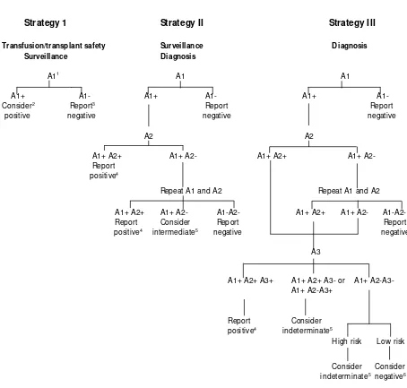
• The strategy of H IV testing is based on the objective of testing the prevalence of HIV in the population. For details see Table 1 andFigure 1.
Confirmatory Test • In populations where the prevalence of HIV infection is greater than 10%, the initial reactive specimen will be tested by a different screening test (e.g. ELISA or simple/rapid) based on different antigen preparation (e.g. viral lysate and recombinant antigen) or different test principles (e.g. indirect ELISA and sandwich ELISA). Any serum that is reactive on the first test but non-reactive on the second test, should be retested with the two assays. Concordant results after repeat testing will indicate a positive or negative result. If the results of the two assays remain discordant the serum is considered indeterminate.
• In populations in which the prevalence of HIV infection is less than 10%, after the results of the two assays are concordantly positive, the third test is employed. For the sera found to be repeatedly discordant i.e. positive by first and negative by second test the third test is also employed. The three tests must be based on different antigen preparations or test principles. Serum that is reactive by all the three tests is considered positive. Serum that remains discordant in the second test or is reactive in the first and second test but non reactive in the third test is considered indeterminate.
Table 1: UNAID S and WH O recommendations for H IV testing strategies according to test objective and prevalence of infection in the sample population.
O bjective of testing Prevalence of
infection Testing strategy
Transfusion/Transplant safety All prevalence I
Figure 1: Schematic representation of the UN AID S and WH O H IV testing strategies
Strategy 1
Strategy II
Strategy III
Transfusion/transplant safety Surveillance D iagnosis Surveillance D iagnosis
A11 A1 A1
A1+ A1- A1+ A1- A1+ A1-
Consider2 Report3 Report Report
positive negative negative negative
A2 A2
A1+ A2+ A1+ A2- A1+ A2+ A1+ A2-
Report positive4
Repeat A1 and A2 Repeat A1 and A2
A1+ A2+ A1+ A2- A1-A2- A1+ A2+ A1+ A2- A1-A2-
Report Consider Report Report
positive4
intermediate5
negative negative
A3
A1+ A2+ A3+ A1+ A2+ A3- or A1+ A2-A3- A1+ A2-A3+
Report Consider
positive4 indeterminate5
H igh risk Low risk
Consider Consider
indeterminate5 negative6
1Assay A1, A2 and A3 represent three different assays
2 Such a result is not adequate for diagnostic purposes: use strategy II or III. W hatever the diagnosis, donations which were earlier
reactive should not be used for transfusion or transplant
3Report: result may be reported
4For newly diagnosed individuals a positive result should be confirmed on a second specimen 5Testing should be repeated on a second specimen taken after 14 days
The follow-up sample from patients with indeterminate result should be collected after two weeks. If the second sample also shows indeterminate result, it should be tested by a confirmatory assay (e.g. W estern Blot). However, if the confirmatory test fails to resolve the serodiagnosis, follow up testing should be undertaken at four weeks, three months, six months and 12 month-intervals. After 12 months, such indeterminate results should be considered as negative.
Reporting procedure
The results are kept confidential.
Report • Negative - If the initial/screening test shows non-reactive result.
• Positive - If the sample shows reactive results concordantly by three screening tests. The subsequent sample is requested to retest before the result is reported to the patient.
• Indeterminate - If the sample shows discordant results by the three screening tests, the follow up samples are required to retest at two weeks and at three, six and 12 months before the result is reported to the individual. If the results remain indeterminate after one year, the person is considered to be HIV antibody negative.
Special considerations • Pre test-, post test-counselling service should be provided and the result should be kept confidential.
Reference
2. Standard O perating Procedures for
Laboratory Diagnosis of Viral
O pportunistic Infections in
HIV-Infected Patients
Introduction
This SO P is confined to simple techniques for diagnosis of herpes simplex virus (H SV), varicella-zoster virus (VZV) and cytomegalovirus (CM V) which are the common opportunistic viral infections in HIV-infected persons.
Clinical manifestations
H erpes simplex virus type 1 (H SV 1) and type 2 (H SV 2) infections cause primary and recurrent oral, genital and rectal ulceration and occasionally disseminated visceral and central nervous system disease.
VZV causes varicella in primary infection, and herpes zoster when reinfected later in lifetime. In H IV-infected persons, reactivation of VZV cause prolonged and severe manifestation of herpes zoster and occasionally becomes disseminated.
CM V infection is responsible for a greater proportion of opportunistic infections in H IV-infected persons with advanced immune deficiency. Retinitis is the most frequent clinical manifestation of CM V infection in patients with AIDS. Gastrointestinal disease, encephalitis, polyradiculitis, and pneumonia may also occur.
Laboratory diagnosis
Viral diagnosis is mostly based on clinical symptoms and signs. The most common technique is simple microscopic examination of scraped cells for diagnosis of HSV skin, oral, genital and anal lesions and VZV lesions (Tzanck test). Demonstration of multinucleated giant cells indicates infection by either HSV or VZV. The two viruses could be differentiated by immunological techniques i.e. immuno-fluorescence, immunoperoxidase and ELISA.
Serological diagnosis is useful in primary herpes virus infection, but not in reactivation and immunodeficiency states.
Viral culture and PCR are the techniques available in reference laboratories. Specimen collection, transportation and storage are described in this SO P.
The other viruses associated with H IV-infected persons are: Epstein Barr virus (EBV); Kaposi’s sarcoma-associated herpes virus (H H V 8); O ther viruses, e.g. human papillomaviruses (H PV), molluscum contagiosum virus, hepatitis B virus (H BV), and hepatitis C virus (H CV). Investigation of these viruses is carried out in the reference and research laboratories.
Safety considerations
Specimen collection • Universal precautions and avoidance of contact with lesion or tissue with ungloved hands.
Specimen transport and storage
• Sterile leak-proof container in a sealed plastic bag.
Specimen processing • Biosafety level II or III with good laboratory practice.
The above guidelines should be supplemented with recommendations from the local health hazard and risk assessment committee.
Specimen collection
O ptimal time of specimen collection
• At the time close to onset or as early as possible.
• Before the start of antiherpetic drugs
Correct specimen type and method of collection
• Skin and mucosa lesions: Collect from newly formed vesicles. Using sterile No. 27 needle and tuberculin syringe, aspirate the vesicular fluid for culture. O pen the lesion and scrape the cells at the rim of the base of the lesion. Smear on dry clean glass slides (2-3 slides)
• Swab from the open lesion, immerse in viral transport media.
• Cerebrospinal fluid: Collect 1-2 ml in sterile container.
• Biopsy or necropsy tissues: Try to avoid contamination as much as possible. Collect in sterile container.
• Blood: Collect venous blood 3-5 ml in EDTA tube.
Specimen transport and storage
Time between specimen collection and processing
• All specimens should be fresh and sent to the laboratory as soon as possible.
• Slides from skin or mucosal lesion smears should be air dried and sent in sealed plastic bags.
• Vesicular fluid and tissue specimens should be kept in sterile containers and sealed plastic bags, kept refrigerated and transported in an ice-box.
• Lesion swabs should be put in sterile tubes with appropriate amounts of viral transport media, kept in refrigerator and transported in an ice-box.
• Blood specimen should be transported at room temperature.
Special considerations to minimize deterioration
• Specimens should be processed as soon as possible especially for viral isolation.
• For CM V antigenemia assay, specimens should be processed within six hours.
Specimen processing
Proper documentation upon receipt of specimen
• Laboratory request form should indicate the specific test for HSV, VZV and CM V.
• Record in laboratory file and give laboratory number.
Processing • Smears are fixed in absolute methanol and stained with Giemsa stain.
• Smears are fixed in acetone and sent to a reference laboratory for staining with specific anti-HSV or anti-VZV as requested.
• Specimens requested for PCR, viral culture and CM V antigenemia assay are referred to a reference laboratory.
Reporting procedure
Giemsa staining
To prepare Giemsa stock solution, dissolve 0.5 gm of Giemsa powder in 33 ml of glycerine by placing the mixture in a water bath (55o-60oC) for 90 minutes. W hen the powder is
dissolved, add 33 ml of absolute methanol. The stock solution is stored at room temperature.
The scraping prepared onto clean glass slide is fixed in absolute methanol for ten minutes. The slides are then stained with Giemsa stain (freshly prepared by diluting the stock solution 1:20 in water) for two hours. The stained slides are decolourized in 95% ethanol for a few seconds and rinsed in tap water.
Reference
3. Standard O perating Procedures for
Laboratory Diagnosis of Tuberculosis
and
M . avium
Complex Disease in
HIV-Positive Patients
Introduction
H IV has a serious effect on the tuberculosis control programme in many parts of the world. Tuberculosis is considered to be one of the most common HIV-related opportunistic infections. The frequency with which HIV and M . tuberculosis infections occur together is determined by the epidemiology of each disease in a given population. Although several population-based studies determining the incidence of tuberculosis attributed to H IV infection have been documented elsewhere, such reliable information is not readily available for all SEAR countries. However, the few studies reported from India, Thailand and elsewhere among SEAR countries reveal that tuberculosis is the most common infection in the H IV-infected group.
Before the AIDS pandemic, Non-Tuberculous M ycobacteria (NTM ) rarely caused serious illness, even in the immunocompromised individuals. The prolonged immunosuppression of the cell mediated immune system caused by the H IV provides the opportunity for these relatively avirulent organisms to cause disease. M .avium complex (M AC) was recognised in the early AIDS pandemic as a cause of serious disseminated infection and is now the most common cause of systemic bacterial infection in AIDS, affecting 15% to 40% of patients in the United States, although such a scenario has not been found so far in the SEAR countries.
Tuberculosis in H IV-infected persons may occur with different manifestations. All these forms of tuberculosis, except when cavitation occurs in pulmonary tuberculosis, are paucibacillary in nature. Depending upon the form of disease manifestation, several specimens such as sputum and/or gastric lavage, bronchoalveolar lavage (BAL), lymph nodes and other biopsy specimens, pus, ascitic fluid, pleural and cerebrospinal fluid should be examined. If delay is anticipated, biopsy specimen may be collected in suitable transport medium for sending them to the laboratory.
Laboratory diagnosis
The indirect approach relates to the measurement of host immune response against the mycobacteria. This includes humoral immunity via the detection of antibodies against the bacteria and cellular response via skin tests.
None of the above parameters except smear microscopy, culture on LJ medium, minimum set of identification to differentiate M .tuberculosis from NTM and drug susceptibility testing by the conventional method, are practicable in most of the SEAR countries. And also since contribution of pulmonary tuberculosis accounts for more than 90% of total tuberculosis manifestation in H IV patients, this SO P is confined to the processing of sputum sample for M .tuberculosis and its identification and drug susceptibility testing methods. In addition, brief general identification of NTM and details pertaining to processing of infectious material for M AC disease are also provided.
Collection of sputum
For diagnosis of tuberculosis, three specimens of sputum are to be examined (spot-morning - spot) over a period of two days. Specimens are to be collected in sterile universal containers, and should have a fixed label for noting patient’s information on the side of the container.
Specimen collection • Aerosol free container
(Sputum cup with a lid or M c Cartney bottle) Adequate quantity and
Appropriate number of specimens
• Ideally, a minimum volume of 5 ml Three consecutive sputum specimens should be collected: spot – morning – spot sputum.
Specimen transport and storage
• Sterile leak proof container in a sealed plastic bag.
• To be transported within three days for culture. If delay is anticipated, add equal volume of 1% CPC.
Specimen staining • Ziehl-Neelsen staining
Note: heat-fixing may not kill all Mycobacterium
species. Slides should be handled carefully Culture and investigation • Specimen processing
Sputum: M odified Petroff’s method for digestion and decontamination.
O ther samples: Direct inoculation for aseptic specimens.
M ild acid or other standard pretreatment procedure.
• Inoculation on Jensen media or Lowenstein-Jensen with sodium pyruvate used for INH Resistant strain and M . bovis. O ther specimens: using Lowenstein-Jensen media and one bottle of liquid medium (M iddlebrook 7H 9 or Kirchner’s medium). Incubation at 37°C for M . tuberculosis and 25°C, 37°C and 45°C for NTM . Examination for eight consecutive weeks or until it becomes positive or contaminated. Culture identification • M . tuberculosis)
Growth rate slow grower (> 7 days) Pigmentation buff colour
Niacin test +
Nitrate reduction test + Catalase at 68°C -
Growth in PNB no growth
Drug susceptibility testing (DST)
• DST is undertaken against streptomycin, isoniazid, rifampicin and ethambutol on L-J medium.
• M ethod: Using a standard proportion method.
Definition of resistance • The number of organisms resistant to each drug concentration as a percentage of the number of organisms growing on the drug-free slope.
D rug Concentration Proportion
Streptomycin 4 mg/l 1% or more
Isoniazid 0.2 mg/l 1% or more
Rifampicin 40 mg/l 1% or more
Ethambutol 2 mg/l 1% or more
Recording and Reporting • Direct smear
+ + + (> 10 AFB/oil field)
Q uality assurance • Smear microscopy
Re-reading both positive and negative slides with blind results.
• Culture
Using the suspension of H37Rv to check the L-J medium.
Note: The contamination rate should not more than 5% • Drug susceptibility testing
- Internal quality control
- External quality assessment programme with Supranational reference laboratory
D iagnosis of AID S and M AC
Specimen type and method of collection
• Blood, bone marrow, biopsy specimen and others. All specimens should be fresh and taken before antimicrobial treatment is started.
Specimen processing • H emoculture, Culture of tissue materials
Culture media • Lowenstein-Jensen media, M iddlebrook 7H12, Bactec 13A or M B/BacT
Sputum smear M icroscopy
M icroscopy of sputum is of great value in the detection of open or infectious cases of tuberculosis. The establishment of a good sputum microscopy service is of prime importance in developing countries for detection and treatment of the open cases.
Smears are stained by the Ziehl-Neelsen (ZN) method, or by one of its various modifications. Grading of the positive smears gives a broad indication of the severity of disease and the response to therapy (during treatment of the patient).
The patient is given a container on his first attendance. He should be instructed, with demonstration by actual actions, to
− inhale deeply 2-3 times
− cough out deep from the chest
− open the container and spit the sputum into the bottle
− avoid saliva or nasal secretions
− close the container.
A good sputum sample should be thick, purulent and there should be sufficient quantity (at least 5ml).
The patient should be instructed to collect an early morning specimen in a similar manner and bring it the next morning. O n the second attendance, a third specimen (spot) is collected.
The details of the patient’s, name, address, age/sex and bottle no. should be recorded in a form/ card and sent to the laboratory.
O n receipt in the laboratory, the specimens are given serial laboratory numbers.
Collection of other specimens
(1)Prepare smears from other specimens as for sputum, but do not expect all liquid specimens to produce a cloudy smear.
All liquid specimens, except blood, should be decontaminated and concentrated by centrifugation before smears and cultures are made.
Tissue specimens are frequently cut into thin sections on a microtome, are attached to a glass slide, and acid-fast stained. This procedure requires special equipment and training and will not be discussed in this manual. Tissue specimens may also be ground to a fine pulp for culture and microscopy.
O ther specimens can be done for decontamination as sputum specimens as well.
Smear examination
purulent portion of the sputum is placed on the slide and spread evenly to give a smear of approximately 3 x 2 cm. The smear is allowed to air-dry and fixed by pressing over a flame 3-5 times for 3-4 seconds each time.
Staining procedure
(1) Place the fixed slides on a staining rack with the smeared side facing up
(2) Pour 1% carbol fuchsin to cover the entire slide
(3) Heat the slides from below till vapour rises
(4) Allow the slide to stand for at least five minutes
(5) Rinse gently with tap water to remove excess stain
(6) Decolourize using 25% sulphuric acid for 2-4 minutes
(7) Rinse gently again with water
(8) Counterstain with 0.1% methylene blue for 30 seconds
(9) Rinse gently again and allow the slide to air dry
Smear reading
Place a drop of cedarwood oil or liquid paraffin without touching the smear and examine under an oil-immersion objective for at least 10 minutes. Count the number of AFB and grade as 3+ , 2+ , 1+ , scanty or negative as follows:
Grading of AFB smears
Examination Result Grading No. of fields
to be examined
Sputum culture (M odified Petroff’s M ethod)
*(1) Discard sputum in excess of 5 ml into the disinfectant bath (5% Phenol).
(2) To each volume of remaining sputum add two volumes of 4% sodium hydroxide, taking care to avoid contact between the specimen bottle rim and the sodium hydroxide flask.
* The total period of contact with the alkali is 35 minutes. It has been shown that this period of contact with 4% NaO H may not be
(3) Screw the caps firmly on, ensuring at the same time that the bottle screw tops are not broken or chipped.
(4) Shake the bottles by hand for one minute.
(5) Place in a rack on the shaking machine and leave to shake gently for 20 minutes.#
(6) Remove the specimens from the shaker.
(7) Centrifuge for 15 minutes* at 4000 rpm (approx. 3000g).
(8) Carefully pour off the supernatant into a disinfectant bath.
(9) Fill the bottles with approximately 20 ml of sterile distilled water, shake by hand to mix the deposit, re-centrifuge for 15 minutes and pour off the supernatant as before.
(10) Finally inoculate each sediment with a 5 mm diameter loop onto two previously numbered Lowenstein-Jensen slopes and one slope of L-J enriched with sodium pyruvate.
(11) Place the inoculated media in the 370C incubator.
Culture reading
Cultures which have been incubated are examined on a fixed day of the week for 8 consecutive weeks or until they become positive or contaminated.
Record the final results at the time of reading: negative, contaminated and if positive, the degree of positivity as follows:
+ + +
+ +
+
Actual no. of colonies
confluent growth
innumerable discrete colonies
20 - 100 colonies
< 20 colonies
If the degree of positivity is different on the two slopes, the highest degree is reported.
D rug susceptibility tests
Drug susceptibility tests are undertaken against streptomycin, isoniazid, rifampicin and ethambutol on L-J medium by the proportion method. (5) This method is not affected by
inoculum size or viability of the cultures.
Bacterial suspension
A suspension is prepared by adding approximately 4 mg moist weight of a representative sample of the bacterial mass visualized as 2/3 loopful of 3 mm internal diameter 24 SW G wireloop into 0.2 ml of sterile distilled water in a 7 ml Bijou bottle containing 2-3 mm
#
glass beads. This is vortexed for 30 seconds to produce a uniform suspension. To this, 3.8 ml of sterile distilled water is added to give a suspension containing approximately 1 mg/ml (S1). From this suspension a 10-fold dilution is made by adding 0.2 ml to 1.8 ml
sterile distilled water (S2, 10
-1). Three further serial dilutions 10-2 (S
are prepared in a similar manner. O ne standard loopful (3 mm diameter, 27 SW G) is inoculated onto drug-free and drug-containing LJ slopes as indicated below:
Suspension Control
The standard strain M .tuberculosis, H37Rv is tested with each new batch of medium. The recommended drug concentrations are 4 mg/l for streptomycin, 0.2 mg/l for isoniazid, 40 mg/l for rifampicin and 2 mg/l for ethambutol.
Incubation and reading
Incubate the slopes at 370C.
Read the proportion tests at 28 and 40 days.
Record growth as
+ + + Confluent growth
+ + M ore than 100 colonies
Actual number of colonies 1-99
W hen the number of colonies on a given dilution is less than 15, count the number of colonies with the next larger inoculum, or estimate if more than 100. (M ake no attempt to estimate the number of colonies if the growth is + + + )
Interpretation of tests
Interpretation of all tests is based on the 40-day readings. For each strain, express the number of organisms resistant to each drug concentration as a percentage of the number of organisms growing on the drug-free slope. M ake the selection of slopes for estimating the growth on the drug-free and drug containing media in the following order of preference.
D rug-free slope
(1) 20-70 colonies.
(2) 5-19 colonies.
D rug-containing slopes
(1) 1-5 colonies or more in the same row as the control slope.
(2) 5-100 colonies in the row nearest to the control slope.
(3) 1-4 colonies in the same row, or the row nearest to the control slope.
(4) No colonies in the row farthest (including the same row as the control slope).
(5) M ore than 100 colonies, if there are no acceptable counts.
D efinitions of resistance - proportion method
D rug Concentration (mg/l) Proportion
Streptomycin 4 1% O R M O RE
Isoniazid 0.2 1% or more
Rifampicin 40 1% or more
Ethambutol 2 1% or more
Calculation of proportions - an illustration
Concentrations of streptomycin Suspension D rug-free medium
4 mg/l 8 mg/l
Proportion resistant 6.4% 0.52%
Preparation of drug containing media
Drug-containing L-J slopes are made by adding appropriate amounts of drugs aseptically to L-J fluid before inspissation. A stock solution of the drugs is prepared to contain 10,000 mg/l in sterile distilled water for streptomycin, isoniazid and ethambutol; rifampicin is dissolved in dimethyl formamide. The sterile solutions of isoniazid and ethambutol are sterilized by membrane filtration. Suitable working dilutions are made in sterile distilled water and added to the L-J fluid, dispensed in 6-ml amounts and inspissated once.
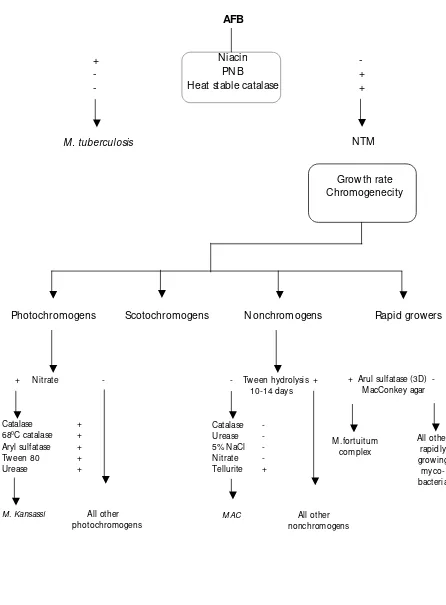
Identification
The identification of strains isolated as belonging to M .tuberculosis complex or Non-Tuberculous mycobacteria (NTM ) can be ascertained by performing a few simple tests i.e.
susceptibility to p-nitrobenzoic acid (PNB), niacin production test and catalase activity of at 680C /pH7 and nitrate reduction test, besides looking at their growth rate, temperature
Figure 3. Speciation of M ycobacteria
- Tween hydrolysis + 10-14 days
Catalase -
Urease -
5% NaCl -
Nitrate -
Tellurite +
M AC All other nonchromogens
All other rapidly growing
myco-bacteria
+ Nitrate -
Catalase +
680C catalase +
Aryl sulfatase +
Tween 80 +
Urease +
M . Kansassi All other photochromogens
W ork flow of speciation of M ycobacteria
+
-
-
M . tuberculosis
-
+
+
NTM
AFB
Niacin
PNB
Heat stable catalase
Growth rate
Chromogenecity
Photochromogens
Scotochromogens
Nonchromogens
Rapid growers
+ Arul sulfatase (3D) - M acConkey agar
Identification of mycobacteria
(6)Differentiation between Mycobacterium tuberculosis and Non-Tuberculous Mycobacterium (NTM).
Characteristic M . tuberculosis NTM
Growth rate Slow grower Slow/Rapid grower
Temperature 370C 250C - 450C
Colony morphology Dry, rough Dry
Colony on solid media Eugonic Dysgonic
Colour of colony Buff Yellow, orange or creamy
Emulsify Difficult Easy
Cord formation + -
Niacin test + -
Nitrate reduction test + + / -
at 680C Catalase test - +
Growth on p-nitrobenzoic acid (PNB) 500 µg/ml
- +
Identification of non-tuberculous mycobacteria
M ycobacteria have been classified as obligate pathogens, facultative or opportunistic pathogens, or free living saprophytes. O bligate pathogens are species that do not appear to multiply outside their hosts and include M .tuberculosis, M .bovis M .africanum, M .asiaticum, M .farcinogens, M .haemophilum, M .leprae, M .malmoense, M .microti, M .paratuberculosis, M .shimodei, M .simiae and M .szulgai.
The habitat of majority of the mycobacteria responsible for NTM disease is primarily environmental in existence and most of them exhibit inherent resistance to anti-TB drugs, making treatment difficult. The NTM strains may be broadly divided into those as the ones which are potentially pathogenic and those which are saprophytic in existence and usually are nonpathogenic, although they may be responsible for disease under exceptional situations. Hence, criteria for diagnosis of pulmonary disease due to NTM are rather more stringent. The American Thoracic Society has given the following criteria for diagnosis of disease due to NTM :
(1) Evidence such as infiltrate, visible on chest skiagram of disease, the cause of which has not been determined by careful clinical and laboratory studies; and
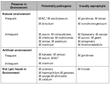
Table 1. M ost frequently identified non-tuberculous mycobacteria in medical laboratories and their presence in the environment(7)
Presence in
Environment Potentially pathogenic U sually saprophytic
Natural environment
Frequent
Infrequent
M AC,*M .scrofulaceum,
M .fortuitum
M .avium, M .intracellulare, M .chelonae, M .malmoense, M .simiae, M .asiaticum, M .marinium
M .gordonae, M .terrae
M .nonchromogenicum
M .flavescens, M .vaccae M .aurum, M .gastri, M .smegmatis, M .thermoresistibile
Artificial environment
Frequent
Infrequent
M .kansasii, M .xenopi, M .avium, M AC*
* The M .avium complex (M AC) includes several environmental species that resemble M .avium and M .intracellulare or that have intermediate characteristics common to these species.
Before the AIDS pandemic, non-tuberculous mycobacteria rarely caused serious illness, even in the immunocompromised. The prolonged immunosuppression of the cell mediated immune system caused by the HIV provided the opportunity for these relatively avirulent organisms to cause disease. M .avium complex (M AC) was recognized in the W estern countries in the early AIDS pandemic as a cause of serious disseminated infection and is now the most common cause of systemic bacterial infection in AIDS, affecting 15%-40% of patients. Besides M AC, many other nontuberculous mycobacteria such as
M .kansasii, M .celatum, M .chelonei, M .fortuitum, M .genavense, M .gordonae, M .haemophilum, M .malmoense, M .mariinum, M .scrofulaceum, M .simiae and M .xenopi
have been reported to cause disease.
M ycobacterium avium
complex
(M AC)
Prior to the AIDS epidemic, M AC infection or disseminated M AC infection was extremely rare. O n the other hand, M AC contributes significantly to mortality and morbidity nowadays. M AC infections are environmentally acquired diseases and the route of infections can be by inhalation, ingestion or direct contact with damaged skin.
Natural habitat
Clinical manifestations of M AC infection in AID S patients
Between 1981 and 1987, 2 269 cases of disseminated mycobacteriosis in patients with AIDS were reported to the Centre for Disease Control, Atlanta, USA. In 96% of cases, infection was caused by M . avium complex. These patients survived a shorter time (median 7.4 months) than did other AIDS patients (median 13.3 months; p < 0.0001.) It was concluded that disseminated mycobacteriosis was acquired by unavoidable environmental exposure.
Severe immunosuppression is the most significant risk factor for disseminated M AC. It rarely occurs in patients with CD4 counts above 100/mm3.
M AC infection is often under-diagnosed because of its non-specific symptoms and signs such as fever, night sweats, weight loss, nausea, vomiting, intractable crampy abdominal pain, diarrhoea and malaise. The onset of illness is usually insidious, and symptoms are often present for many weeks before the diagnosis is established. Accompanying clinical signs and laboratory abnormalities frequently included cachexia, hepatosplenomegaly, anaemia, neutropenia and elevation of alkaline phosphatase level.
O rgans most commonly affected by M AC are the liver, spleen, bowel and bone marrow. M AC usually originates from the gastrointestinal tract and eventually disseminates along the reticulo-endothelial system which predominately affects the lymph nodes, liver and spleen. Endobronchial lesions and focal pneumonia may occur with or without bacteraemia. M AC may occasionally produce skin lesions. Its clinical presentation on the skin may be in the form of papules, nodules, or ulcers at the antecubital fossa and perianal area. The untreated disseminated M AC has a short survival, with a mean of 5.6 ± 1.1 months (median 4 months).
Essential laboratory investigations and their interpretations
M icroscopy
A presumptive diagnosis of M AC infection can be made quickly by demonstration of acid fast bacilli (AFB) in smears from tissue e.g. skin, bone marrow, lymph node, liver biopsy or buffy coat of blood in patients with clinical pictures mentioned above, but definite diagnosis still requires culture confirmation.
Culture
The definite diagnosis of disseminated M AC infection is usually made by isolation of organisms from blood or bone marrow cultures. Cultures of M AC require 2 to 6 weeks to become positive. The BACTEC 9240 system is faster but still requires a minimum of 8-10 days to become positive and may require up to four weeks in samples with low colony counts.
H aemoculture for M AC
technique with the DuPont Isolator tube has successfully recovered M .chelonei, M .avium
complex and M .tuberculosis from blood. A 10 ml volume of blood is added to the Isolator tube; the tube is inverted several times to mix the contents, and then it is allowed to stand for one hour to lyse the blood cells. After lysis, the tube is centrifuged at 3000g or more to concentrate the material into 1.5 – 2.0 ml of sediment, or of which is spread over the surface of several tubes or plates of isolation media (e.g. 7H 10, 7H 11, L-J). By using appropriate dilutions of sediment (or diluting lysed specimens prior to centrifugation), it is also possible to quantify the extent of the bacteremia.
O ther rapid methods for culture
Today, there are few rapid methods for the culture and measurement of the susceptibility of mycobacteria. These include microcolony detection on solid media, the Septi-Check AFB method, mycobacterial growth indicator tube (M GIT) system, BACTEC radiometric method, and M B/Bact mycrobacteria detection system.
Laboratory methods based on D NA technology
Nucleic acid probes with non-isotopic detection systems are available for the M . tuberculosis complex, M . avium, M . intracelluare, M . avium complex, M . kansasii and M . gordonae and more will become sooner available. These can be used to identify organisms directly from clinical samples, cultures from conventional media and from radiometric vials. H owever, these tests are expensive. Since the use of these techniques requires higher technical expertise and training, their implementation is possible only in a reference laboratory.
Q uality assurance programme
Q uality control procedures should be performed on a regular and periodic basis in the mycobacteriology laboratory to assure reproducibility and reliability of laboratory results.
Sputum smear M icroscopy
This remains the basis for diagnosis of TB in most SEAR countries. The importance of correct readings of sputum smears at the local level, where the diagnosis is usually made, is critical. The IUATLD currently recommends that a systematic sample of sputum smear microscopy specimens be selected for review. The sample should include both positive and negative specimen smears, and the slides should be re-read by a second individual who did not perform the initial specimen slide reading; the second individual should not know the result of the first reading (blinded re-reading).16
Culture
Records must be kept on all “ homemade” culture media and should be checked for sterility and sensitivity. The latter must be carried out by using the standard suspension of
Likewise Q C must be ensured for procedures such as digestion and decontamination, media, reagents and biochemical tests employed for the isolation and identification of mycobacteria.
D rug susceptibility
Besides internal quality control (IQ C) procedures, each centre must participate in an external quality assessment scheme (EQ AS) adhering to the norms prescribed by W H O in the Global Drug Resistance Surveillance (DRS) programme.
Storage of cultures
From each patient, at least one culture must be expanded, tested for its purity and then stored at -700C till the end of the study.
Grade of TB laboratory diagnostic procedures
to be performed at different levels
Level I: Peripheral These are usually single room or parts of another laboratory at the primary health care or local hospital level. Tuberculosis investigation is restricted to collection and smear examination and despatch of specimens to a higher level laboratory for culture.
Level II: Intermediate These laboratories may be in larger hospitals, such as district hospitals and be part of pathology laboratory complex. The work includes examination of direct smear and, in some culturing specimens, but the cultures are usually sent to a higher grade laboratory for incubation and further tests.
Level III: Central These more specialized laboratories examine direct smears and culture specimens for mycobacteria. Some may do no more than this and send the cultures to other TB level III laboratories. O thers continue with the identification of the mycobacteria to species level and do antituberculosis drug susceptibility tests. The highest grade of TB level III laboratories usually do research and function as training centres and as reference laboratories.
References
(1) Smithwick, R.W . Laboratory manual for acid-fast microscopy, Second edition (July 1976). U.S. Department of Health, Education, and W elfare CDC Atlanta, Georgia.
(3) Petroff, S.A. (1915). A new and rapid method for the isolation and cultivation of tubercle bacilli directly from the sputum and faeces. J. Exp. M ed. 21:38-42.
(4) Allen, B.W . and Baker, F.J. (1968). M ycobacteria - Isolation, identification and sensitivity testing. Butterworths, London, Page 10.
(5) Canetti et al, (1963) M ycobacteria : Laboratory method for testing drug sensitivity and resistance. Bull W H O ;29:565-578.
(6) H awkins, J.E., R.C. Good, P.R. Gangadharan, H .M . Gruft, and K.D. Stottmeier (1983). Levels of service concept in mycobacteriology. ATS News. 9:19-25.
(7) Portaels. F. (1995) Epidemiology of M ycobacterial Diseases : Clinics in Dermatology, 13, 207-22.
(8) Gutierrez-Vazquez, J.M . (1960) Further studies on the spot test for the differentiation of tubercle bacilli of human origin from other mycobacteria. Amer.Rev.Resp.Dis., 81, 412.
(9) Venkataraman, P and Prabhakar R., (1977) Niacin production test in mycobacteria: Replacement of benzidine-cyanogen bromide reagent by o-tolidine - cyanogen bromide. Ind.J.Tuberc., 24, 153.
(10) Kubica, G.P and Pool, G.I. (1960) Studies on the catalase activity of acid-fast bacilli. I An attempt to subgroup these organisms on the basis of catalase activities at different temperatures and pH . Am.Rev.Resp. Dis., 81, 387-391.
(11) Laboratory services in Tuberculosis control Part I, II, and III W H O /TB/98.258.
(12) H orsburgh CR Jr., Selik RM . (1989) The epidemiology of disseminated nontuberculous mycobacterial infection in the acquired immunodeficiency syndrome (AIDS). AmRevRespir Dis. 139:47.
(13) Kirschner RA Jr., Parker BC, Falkinham III JO (1992) Epidemiology of infection by nontuberculous mycobacteria. M ycobacterium avium, M . intracellulare, and M . scrofulaceum in acid, brown- water swamps of the Southeastern United States and their association with environmental variables. Am Rev Respir Dis. 145:271-5.
(14) Rippon JW . (1988) M edical M ycology : The Pathogenic Fungi and The Pathogenic Actinomycetes. 3rd ed. W .B. Saunders Company, Philadelphia, pp. 53-68.
(15) Vithayasai P. (1996) Atlas of M ycobacterium avium complex in AIDS. H ua Nam Printing Co. Ltd., Thailand, pp. 53.
Appendix
Preparation of stain
Preparation of 1% carbol fuchsin
(1) W eigh 5 grams of basic fuchsin dye in a balance and transfer it to a 250 ml Erlenmeyer glass flask.
(2) Add 50 ml of methylated spirit and shake to dissolve the dye.
(3) Heat 25 grams of phenol until it melts and add to the above solution.
(4) Heat the flask containing basic fuchsin dye dissolved in spirit and phenol in a water bath at about 600C. D o not heat directly on a flame.
(5) Transfer the contents into a 500 ml measuring cylinder.
(6) Add distilled water to make up a final volume of 500 ml.
(7) Pour the solution through filter paper (W hatman No.1) and store filtered solution in a glass bottle. Label the bottle as 1% carbol fuchsin with the date of preparation.
Preparation of 25% sulphuric acid
(1) Pour 375 ml of distilled water into a 1 litre glass flask.
(2) M easure 125 ml of concentrated sulphuric acid and transfer it slowly into the flask containing water.
Always add acid to water. Never add water to acid.
(3) Store the sulphuric acid solution in a labelled glass bottle.
Preparation of 0.1% methylene blue solution
(1) W eigh 0.5 grams of methylene blue and transfer to a 1 litre glass flask.
(2) Add 500 ml of distilled water.
(3) Shake well to dissolve.
(4) Store in a glass bottle with the label showing name of the reagent and date of preparation.
Sodium hydroxide
Preparation of culture media
Lowenstein Jensen M edium-D rug free
Salt solution w ith malachite green (SSM G)
Potassium dihydrogen orthophosphate, 0.4% 14.4 g
M agnesium sulphate, anhydrous, 0.04% 1.44 g
M agnesium citrate, 0.1% 3.6 g
L-asparagine, 0.6% 21.6 g
Glycerol, 2% 72 ml
M alachite green, 2% solution 120 ml
Distilled water, to 3,600 ml
Distribute in 600 ml amounts, autoclave and store in refrigerator at 2-80C.
Complete medium
To 1 litre of egg fluid, add 600 ml of SSM G, mix well and distribute in approx. 6 ml amounts. Inspissate at 85-900C for 50 minutes. Re-inspissate after overnight storage at
room temperature.
Lowenstein-Jensen with sodium pyruvate
For this prepare the salt solutions with malachite green as above omitting glycerol. Proceed as above and to every 1 600 ml of complete medium, add 8 gm sodium pyruvate (0.5 %). Distribute and inspissate.
Susceptibility to PNB
This test is included along with the DS tests. PNB is incorporated in L-J medium at a concentration of 500 mg/l and inoculated with one loopful of a standard suspension.
M .tuberculosis complex is inhibited by PNB while all other mycobacteria are resistant .
Nitrate reduction tests
Classical method with liquid reagents
M edia and supplies
− Sodium nitrate (NaNO3)
− M onopotassium phosphate (KH2PO4)
− Disodium phosphate (Na2H PO4.12H2O )
− Hydrochloric acid (HCl)
− N-naphthylethylenediamine dihydrochloride
− Distilled water
− Zinc dust
− Screwcap test tubes , 16x125 mm
− W ater bath or constant-temperature block heater , 370C
Preparations
Substrate
To prepare 0.01 M sodium nitrate in 0.022 M phosphate buffer, pH 7.0 dissolve in order the following chemicals in 100 ml of distilled water : 0.085 g NaNO3, 0.117 g KH2PO4,
0.485 g Na2H PO4.12H2O . Sterilize by autoclaving.
Reagent
#1
Carefully add 50.0 ml of concentrated HCl to 50.0 ml of distilled water. N ever add water to acid.
Reagent
#2
Dissolve 0.2 g of sulphfanilamide in 100.0 ml of distilled water.
Reagent
#3
Dissolve 0.1 g of N-naphthylethylenediamine dihydrochloride in 100.0 ml of distilled water.
Store the substrate and reagents in the dark at 50C. Discard the reagants if the color
changes or a precipitate forms and prepare afresh.
Controls
Positive = M . tuberculosis (3+ to 5+ ).
Negative = M . bovis (BCG) or M .intracellulare.
Negative = reagents without organisms.
Procedure
(1) Add 0.2 ml of sterile distilled water to a 16 x 125 mm screwcap tube.
(2) Use a sterile spade or applicator stick to emulsify in the water two spadesful of growth from a four week old culture on Lowenstien -Jensen or egg-base medium.
(3) Add 2.0 ml of the NaNO3 substrate to the tube.
(4) Shake by hand and incubate upright for two hours in a 370C water bath.
(6) Add one drop reagent # 1.
(7) Add two drop reagent # 2.
(8) Add two drop reagent # 3.
(9) Examine immediately for a pink to red color.
Results and interpretation
Positive = M ay range from pale pink to deep red
Negative = No color.
Niacin production test
Autoclave (15 lb for 30 minutes) a control drug-free slope after the final reading of the sensitivity or identification test. Add 0.5 ml distilled water to the culture bottle if no condensation water is present before autoclaving.
W ork in a protection cabinet.
To a 75 x 12 mm test tube add 0.25 ml of the culture extract followed by 0.25 ml of 1.5% o- toluidine solution and an equal volume of 10% cyanogen bromide solution. Shake by hand to mix and read the result within one minute. Pink to red colour of the precipitate indicates positive, white precipitate indicates negative.
Strains of M .tuberculosis are almost always niacin-positive. All other types are negative.
Reagents
(1) Approximately 10% solution of cyanogen bromide in distilled water. (A saturated solution of cyanogen bromide is approximately 10%). Store in a brown bottle at 40C.
(2) 1.5% o-tolidine in ethyl alcohol, freshly prepared just before use.
Stability of catalase at pH 7.0/68
0C
Reagents
(1) M /15 phosphate buffer, pH 7.0: Sterilise in 20ml. amounts in universal containers.
(2) Hydrogen peroxide, 30% AR.
(3) Tween-80, 10% solution.
M ethod
(1) W ith a sterile 1ml pipette, add 0.5 ml of buffer to sterile screw-capped test tubes.
(3) Place the tubes with the emulsified suspension in a water bath maintained at 680C
for 20 minutes. The time and temperature are critical.
(4) Remove from the water bath and cool to room temperature.
(5) Add to each tube 0.5 ml of the peroxide-tween mixture inside a safety cabinet.
(6) O bserve for bubble formation on the surface of the liquid, without shaking. Hold tubes for 20 minutes before discarding.
Report as positive, if bubbles are observed and as negative if no bubbles are seen.
M . tuberculosis are negative for heat stable catalase.
Controls
4. Standard O perating Procedures for the
Laboratory Diagnosis of other Bacterial
Infections in HIV/AIDS Patients
Introduction
In H IV-infected patients, the bacterial infection is one of the causes of severe complications. AIDS patients can also be infected by the pathogenic bacteria that infect normal persons. M oreover, these patients may be infected by the opportunistic bacteria. Examples of the most common opportunistic bacteria in AIDS patients are Salmonella, and Rhodococcus equi.
Clinical association and diagnosis
Salmonellosis is a disease most commonly caused by ingestion of food, water or milk contaminated with Salmonella. Three types of infections are gastroenteritis, bacteraemia or septicemia and enteric fever. Routine culture using selective and differential culture media usually is sufficient for isolating Salmonella. The identification can be done by conventional biochemical tests.
Rhodococcus equi, an aerobic gram-positive coccobacilli, formerly known as
Corynebacterium equi, was considered as an extremely rare opportunistic bacterial pathogen in man. In recent years, there has been a dramatic increase in the number of reported isolations in HIV/AIDS patients. Patients with rhodococcus present with bronchopneumoniae, bacteraemia, skin infection, endophthalmitis, peritonitis, catheter-associated sepsis and prostatic abscess. Lethality is high, particularly in H IV-infected patients. Laboratory diagnosis is made by Gram stain, modified acid fast stain and culture from the specimen.
Safety considerations
Specimen collection • Universal precautions and aseptic technique to be followed.
Specimen transport and Storage
• Sterile leak proof and appropriate container in a sealed plastic bag.
Specimen processing • Universal precaution to be followed.
• At least biosafety level II with good laboratory practice.
Specimen collections
O ptimum time ofspecimen collection
• Ideally before initiation of antimicrobial therapy.
Correct specimen type • Depends upon the clinical criteria and the organism suspected e.g. haemoculture, sputum, stool or urine.
• Appropriate specimen from the site of infection in appropriate container and adequate amount.
Specimen transport and storage
Time between specimencollection and processing.
• Ideally the specimen should reach the laboratory within 30 minutes of collection.
• Urine which arrives at the laboratory more than two hours after collection is not suitable for culture and should be rejected.
• Specimens in the transport medium should be sent to the laboratory as fast as possible otherwise the pathogenic bacteria will be overgrown by normal flora.
• Specimens should be processed promptly after receipt.
Storage • Any specimen that cannot be sent to the laboratory
within 30 minutes or cannot be processed immediately should be stored in the refrigerator or freezer (except CSF, body fluids and hemoculture) or put in an appropriate transport medium where applicable.
Specimen processing
Proper documentation upon receipt of specimen• Record the correctness of the specimen on the requisition form.
• M aintenance of laboratory records with lab number. Initial processing • Depends upon the types of specimen collected and
organism suspected.
• Haemoculture (blood culture) should be incubated at 350C immediately.
• CSF and other body fluids should be centrifuged at 2500g for 15 minutes. The sediment is used for culture and staining.
M icroscopy • Perform staining as required.
• Gram stain is a common stain for bacteria
• Acid fast stain for M ycobacterium
• M odified acid fast stain for Rhodococcus and
Nocardia
Reporting procedure • Accuracy and timeliness of reporting procedure to be practised.
• Report results by telephone in critical care patient.
• W ritten report
• Staining: report one day
• Culture and susceptibility: report 3-5 days
Preservation of specimens • Specimen should be kept for confirmation wherever applicable.
Disposal of specimen • All specimens must be autoclaved or disinfected before being discarded.
• Applicator sticks and gauge pieces should be incinerated.
• All used glassware should be put into the disinfectant pan for at least one hour before washing.
Isolation and identification of
Rhodococcus equi
Isolation
The isolation of Rhodococcus can be performed by plating the specimen onto blood agar medium. Incubate the plates at 35oC in ambient air. O n primary culture, the colonies are
usually visible after 24-48 hours and attain a diameter of 1-2 mm. O lder colonies tend to become larger and mucoid with a characteristic salmon-colored pigment. Appearance of the pigment may be delayed.
Identification
Rhodococcus can be presumptively identified via the staining technique. Any specimen that shows gram-positive pleomorphic coccobacilli and stained red with modified acid fast stain may be suspected as Rhodococcus.
The characteristic colonies that are gram-positive pleomorphic coccobacilli or diphtheroid-like should be subcultured. The pure culture of the suspected organism can be identified by conventional biochemical tests.
The modified acid fast staining from the colonies usually gives negative results.
Antimicrobial susceptibility test
According to the NCCLS, there still have no standard zone interpretation using the Kirby-Bauer susceptibility test for this organism. In cases of clinical importance, the M IC technique may be performed where applicable.
Isolation and identification of
Salmonella
Isolation
The organisms belonging to genus Salmonella are gram-negative, non acid fast, non capsulated and non-sporing bacilli, which measure approximately 2-4 µm x 0.6 µm. Almost al species of Salmonella are motile. The organisms grow rapidly on ordinary media and optimum temperature of growth is 370C. The Salmonella spp. other than S. typhi and