A JOURNAL ON TAXONOMIC BOTANY,
PLANT SOCIOLOGY AND ECOLOGY
Vol. 13(3): 221 —315, April 11, 2012
Chief Editor
KARTINI KRAMADIBRATA
Editors
DEDYDARNAEDI (INDONESIA)
TUKTRIN PARTOMIHARDJO (INDONESIA) JOENI SETIJO RAHAJOE (INDONESIA) TEGUHTRIONO (INDONESIA)
MARLINAARDIYANI (INDONESIA) EIZI SUZUKI (JAPAN)
JUN WEN (UNITED STATE OF AMERICA)
Managing editor
HIMMAH RUSTIAMISecretary
ENDANG TRI UTAMI
Lay out
DEDEN SUMIRATHIDAYAT
Illustrators
SUBARIWAHYUDI SANTOSO ANNE KUSUMAWAIY
Reviewers
BRYAN SIMON (AUSTRALIA), EVE J. LUCAS (UNITED KINGDOM), J.F.VELDKAMP (NETHERLANDS), LAUR-ENCE SKOG (USA), PIETER BAAS (NETHERLANDS), RUTH KIEW (MALAYSIA), ROBERT J. SORENG (USA), HE-LENA DUISTERMAAT (NETHERLANDS), LYN A. CRAVEN (AUSTRALIA), RUGAYAH (INDONESIA), MARK HUGHES (UNITED KINGDOM), MARTIN CALLMANDER (USA), PETER C. VAN WELZEN (NETHERLANDS), WAYNE TAKEUCHI (USA), NOBUYUKI FUKUOKA (JAPAN).
Correspondence on editorial matters and subscriptions for Reinwardtia should be addressed to:
HERBARIUM BOGORIENSE, BOTANY DIVISION,
RESEARCH CENTER FOR BIOLOGY-LIPI,
CIBINONG 16911, INDONESIA
REINWARDTIA Vol 13, No 3, pp: 305 − 313
305
LEAF ANATOMY OF PANDANUS
SPECIES (PANDANACEAE) FROM JAVA
Received August 25, 2011; accepted December 27, 2011
SRI ENDARTI RAHAYU
Biology Department, National University, Jl. Sawo Manila 61, Pejaten – Pasar Minggu , Jakarta Selatan, Indonesia. Email: endarti [email protected]
KUSWATA KARTAWINATA
Herbarium Bogoriense, Botany Division, Research Center for Biology-LIPI, Cibinong Science Center, Jl. Raya Bogor−Jakarta Km. 46, Cibinong 16911, Bogor, Indonesia.
Botany Department, Field Museum , Chicago, Illionis, USA
TATIEK CHIKMAWATI
Biology Department, Bogor Agricultural Institute, Jl. Raya Dramaga, Bogor, Indonesia.
ALEX HARTANA
Biology Department, Bogor Agricultural Institute, Jl. Raya Dramaga, Bogor, Indonesia.
ABSTRACT
RAHAYU, S. E., KARTAWINATA, K., CHIKMAWATI, T., HARTANA, A. 2012. Leaf anatomy of Pandanus species (Pandanaceae) from Java. Reinwardtia 13(3): 305−313. –– The leaf epidermis and mesophyll of fifteen species of Pandanus from Java were investigated to assess the value of anatomical features in species identification and classification. Characters of diagnostic importance are epidermal cell shape, differentiation of the abaxial epidermis into costa and intercosta, adaxial anticlinal cell wall outline, occurrence of raphides in the mesophyll, distribution of cubical crystals, pallisade cell shape, papillae on epidermal cells, and the stomatal complex. Leaf epidermal anatomy was found to be useful in the identification at species level.
Keywords: anatomy, Pandanus, Pandanaceae, Java.
ABSTRAK
RAHAYU, S. E., KARTAWINATA, K., CHIKMAWATI, T., HARTANA, A. 2012. Anatomi daun Pandanus
(Pandanaceae) dari Jawa. Reinwardtia 13(3): 305−313. –– Studi tentang epidermis daun dari 15 jenis Pandanus di Jawa dilakukan untuk menentukan kegunaan dari karakter-karakter anatomi ini dan untuk menetapkan nilai manfatnya di dalam identifikasi jenis dan klasifikasi. Karakter-karakter diagnostik yang penting di dalam klasifikasi Pandanus di Jawa adalah bentuk sel epidermis, demarkasi atau diferensiasi epidermis abaksial menjadi kosta dan interkosta, dinding antiklinal sel adaksial, adanya raphid pada potongan melintang, penyebaran kristal berbentuk kubus, bentuk jaringan palisade, adanya papila pada sel epidermis, kompleksitas stomata. Hasil penelitian ini menunjukkan bahwa anatomi dari epidermis daun berguna secara taksonomi dalam identifikasi Pandanus pada tingkat jenis.
Kata kunci: anatomi, Pandanus, Pandanaceae, Jawa.
INTRODUCTION
Pandanus Parkinson is characterized by a number of very obvious features, forming a unique combination of characters. These distinctive characters are generally an erect stem (sometimes sprawling), basally supported by many long stilt and prop roots, branching more or less dichotomously, usually rather prickly because of short, blunt or pointed specialized adventitious roots; leaves in 3 regular, close-set spirals, on the rounded or slightly 3-angled trunks, leaves entire, long, narrow, deeply channelled along the midrib and pleated once on each side, the tip, margin, midrib (below) and the pleats (above, sometimes) set with stout or fine
prickles (Stone, 1965).
Pandanus, with three other genera, Freycinetia
Gaud., Sararanga Hemsl., and Martellidendron
Callm. & Chassot consititute the family
Pandanaceae. Pandanus contains more than 600 species which are distributed throughout the tropics of the Old World and this large genus is very diverse (Kam, 1971). The last revisions of the
(Kam, 1971).
Edeoga and Ikem (2001) showed that leaf epidermal features are as useful in systematic botany as for instance DNA sequences or chemical compounds. The taxonomic value of leaf anatomy features was considered in some detail by Stace (1965). Examining the shape of epidermal, guard and subsidiary cells of stomata may prove useful for the identification of selected plant species (Jakubska, 2007). Stone (1976) constantly reaffirmed that the variation in the epidermal tissue (including stomata) is of great value in systematic botany. Tomlinson (1965) proposed a classification of stomatal types based on progressive complexity, and this system was used by Kam (1971) to demonstrate that there is a correlation between stomatal characters and the sectional delimitation within Pandanus. The aims of the present study are to describe the epidermal variation in 15 Javanese species of Pandanus and to evaluate the usefulness of the characters for identification and classification purposes.
MATERIALS AND METHOD
The survey was based on fresh material collected from the field, from plants cultivated at the Bogor Botanical Garden and from herbarium material supplied by the Herbarium Bogoriense, Bogor.
Dried leaves were boiled in water for a few minutes to soften the leaf until they became unfolded and were ready for epidermal scrapping. Fresh leaves were fixed in 70% FAA. Leaf samples were prepared according to the modified method of Johansen (1940). The fresh or dried leaves were by brushing away unwanted tissue with the help of a pointed needle and forceps, after which the epidermis was stained with safranin. An excess of safranin was washed off and the epidermis was temporarely mounted in an aqueous glycerol solution.
Embedded leaf segments were used for sectioning with a rotary microtome to prepare 15-20 µm thick sections. The ribbons were placed on
clean slides smeared with a thin film of Haupt’s
albumen and allowed to dry, after which a drop of water was added prior to mounting. The slides were placed on a hot plate at 40oC for a few minutes to let the ribbons expand and they were stored overnight. The next day the slides were immersed for 2-5 minutes in a solution of xylene and absolute alcohol (1:1 ratio v/v). The slides were then
transferred to another solution of xylene and alcohol in the ratio 1:3 (v/v) for a few minutes, after which they were washed in a series of 95%, 90%, 70% and 50% alcohol. The slides were stained with a few drops of fast green and counterstained with safranin for two minutes, then dehydrated in a series of 50%, 70%, 80%, 90% xylene/alcohol solution and labyrinthicus, P. pseudolais and P. scabrifolius, the leaf is truly isolateral: adaxially the palisade tissue is much more developed than abaxially, while in some others, e.g. P. multifurcatus and P. tectorius
cv. sanderi, the leaf is somewhat dorsiventral which abaxially the palisade tissue is not so developed. This result agrees with earlier conclusion of Tomlinson (1965) and Kam (1971) that the structure of Pandanus leaf was dorsiventral.
Epidermis
Pandanus labyrinthicus Kurz has a thick cuticle (5 μm), while the remaining species have a thin cuticle (less than 2 μm). The adaxial epidermis is never differentiated into costal and intercostal regions. The shape of the adaxial and abaxial epidermis cells is similar in P. amaryllifolius Roxb.,
P. multifurcatus Fagerl., P. nitidus Kurz,
P. tectorius var. littoralis and P. tectorius cv.
sanderi, whereas the adaxial epidermal cells have a very different shape than the abaxial cells in
P. bantamensis Koord., P. dubius Spreng., P. kurzii
Merr., P. labyrinthicus Kurz, P. odoratissimus L.f.,
P. polycephalus Lam., P. pseudolais Warb., P. scabrifolius Martelli, P. spurius Miq. cv. putat and
P. utilis Bory.
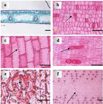
Six types of epidermal cells could be distinguishedadaxially. Each species always shows only a single type. The five types of the epidermal cells of the adaxial surfaces are: (1) rectangular in
P. dubius and P. kurzii (2) squarish in P. bantamensis (Fig. 1b), P.pseudolais, P. scabrifolius
and P. spurius cv. putat, (3) elongated in P.
amaryllifolius. (5) long cells elongated in P. nitidus,
P. multifurcatus, P. odoratissimus and P. tectorius
RAHAYU et al.: Leaf anatomy of Pandanus species (Pandanaceae) from Java
2012] 307
recorded in P. labyrinthicus, P. polycephalus, P. tectorius var. littoralis and P. utilis.
The anticlinal walls of the adaxial epidermal cells are straight or undulate. The type of anticlinal walls is constant at the species level. Pandanus kurzii shows adaxially undulate anticlinal walls (Fig. 1c), while the remaining species have straight anticlinal walls.
The distribution of cubical crystals in the fifteen species is variable. Cubical crystals are absent in P. amaryllifolius, P. dubius, P. kurzii, P. labyrinthicus,
P. tectorius var. littoralis, and P. bantamensis (Fig. 1d); present in both epidermides in P. multifurcatus
and P. nitidus; only abaxially present in P. odoratissimus, P. polycephalus, P. pseudolais, P. scabrifolius, P. spurius cv. putat, and P. tectorius
cv. sanderi; and finally, only adaxially present in P. utilis.
Stomata occur in the adaxial and abaxial epidermis, but the stomata are always more abundant in the abaxial than in the adaxial epidermis, and the polar subsidiary cells of the latter are slightly larger than those of the abaxial stomata. All species have the tetracytic type of stomata.
There are four subsidiary cells adjacent to the guard cells: two terminal (or polar) subsidiary cells, situated at the ends of the guard cell pairs and smaller in size than the other two, lateral subsidiary cells.
In most species studied the adaxial epidermis has stomata without papillae, but P. utilis has stomata with lobed papillae situated on the subsidiary and guard cells (Fig. 1e). Stomatal complex sunk with one ring of papillae over the guard cells and one ring of larger lobes extending from the neighbouring cells above the stomata complex.
For a classification of the stomatal complex in
Pandanus into 5 classes, see below.
The abaxial epidermis is variable throughout the species studied. It may or may not be differentiated into costal and intercostal regions. The demarcation into zones is very clear-cut in P. amaryllifolius (Fig. 1f), P. dubius, P. kurzii, P. multifurcatus, P. nitidus,
P. odoratissimus, P. polycephalus, P. spurius cv.
putat, P. tectorius var. littoralis, P. tectorius cv.
sanderi, and P. utilis. However, in P. labyrinthicus
the demarcation is not so clear, and in P. bantamensis, P. pseudolais and P. scabrifolius the
Figure 1. Light micrographs of leaves: (a) Transverse leaf section of P. multifurcatus Fagerl.; (b) Squarish abaxial epidermal cells of P. bantamensis Koord.; (c) Undulate adaxial anticlinal cell walls of P. kurzii Merr.; (d) Adaxial epidermis cells with cubical crystals of P. bantamensis Koord.; (e) Adaxial stomata with lobed papillae of P. utilis Bory; (f) Costal zone in the abaxial epidermis of P. amaryllifolius Roxb. Scale bar for a & f = 100 µm.; Scale bar for b, c and d = 50 µm; scale bar for e = 20 µml.
a
b
c
d
abaxial epidermis shows no differentiation into costal and intercostal regions.
The shape of the abaxial epidermal cells can only be divided into three classes, i.e. (1) long cells elongated in P. amaryllifolius, P. bantamensis, P. polygonal in P. polycephalus
In most of species, the abaxial epidermis is frequently associated with papillae. The distribution, size and shape of the papillae is highly variable throughout all species studied and can be used with caution for diagnostic purposes.
Papillae, when present may occur on all normal epidermal cells or only on certain cells. Lateral and terminal subsidiary cells of the stomatal apparatus often are papillate. Papillae which occur in lateral subsidiary cells are always arranged in a linear row of three to four as found in P. tectorius var.
The number of papillae per epidermal cell varies from one to several. One to three globose papillae are found on each epidermal cell of P. spurius cv.
putat (Fig. 2c). In P. labyrinthicus, P. tectorius var.
littoralis and P. utilis only one papilla is found on each epidermal cell, but the papillae are elaborately lobed at the distal end. P. kurzii and P. polycephalus
have dendritic papillae on the abaxial epidermal cells, while in P. scabrifolius and P. dubius papillae are absent on the abaxial epidermis.
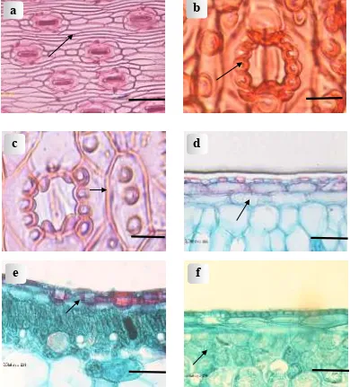
Hypodermis
One or more cell layers immediately beneath the epidermis are developed as a colourless hypodermis. A hypodermis is most conspicuous in species in which the hypodermis of the lamina is multiseriate, e.g. P. amaryllifolius, P. multifurcatus,
P. pseudolais and P. tectorius cv. sanderi consists of at least two layers of colourless cells, three layers of colourless cells are usually present in P. labyrinthicus (Fig. 2d).
The adaxial hypodermis is more uniform than the abaxial hypodermis, because it is not interrupted by many stomatal chambers. The adaxial hypodermal layers are usually somewhat thicker than the abaxial ones. The outermost hypodermal layers are sclerotic, while the inner cells are larger and isodiametric and remain thin-walled. Hypodermal cell rows do not coincide with the
epidermal rows. The number of epidermal cells above one of the outermost hypodermal cells in transverse direction can be used as a diagnostic feature for certain species. For example, in P. labyrinthicus the cells of the first hypoderrmal layer are long and correspond to 4 or 5 epidermal cells, 5 or 6 cells in P. amaryllifolius, and 7 or 8 epidermal cells in P. tectorius cv. sanderi.
Crystalliferous cells with rhombic crystals are found in some of the species studied, viz. P. amaryllifolius, P. bidur, P. labyrinthicus, P. multifurcatus, P. pseudolais, P. tectorius var.
littoralis and P. tectorius cv. sanderi. Crystal cells are generally found in the outermost hypodermis. The crystal cells are distributed uniformly, either solitary or in pairs. In P. amaryllifolius and P. pseudolais the large rhombic crystals occurs in the outermost layer of the hypodermis (Fig. 2e). while the inner cells are larger and isodiametric and thin-walled. Hypodermal cell rows do not coincide with the epidermal rows. The number of epidermal cells above one of the outermost hypodermal cells in transverse direction can be used as a diagnostic feature for certain species. For example, in P. labyrinthicus the cells of the first hypoderrmal layer are long and correspond to 4 or 5 epidermal cells, 5 or 6 cells in P. amaryllifolius, and 7 or 8 epidermal cells in P. tectorius cv. sanderi.
Mesophyll
The leaves of all species are isolateral (i.e. in TS the adaxial side is similar to the abaxial side) to weakly bilateral (adaxial part contains more chlorenchyma and the cells are more palisade like- than the abaxial mesophyll). The mesophyll comprises parallel veins separated by large colourless cells, which desintegrate in mature leaves, resulting in the formation of air cavities. In mature leaves there is a wide air cavity between each adjacent pair of veins. The adaxial chlorenchyma may be a two-layered palisade as in
P. pseudolais or even four-layered palisade in P. amaryllifolius; abaxially the palisade is always 1 (or 2) layered. The shape of the palisade cells is variable. Most species show columnar and compactly arranged palisade cells (P. multifurcatus,
P. pseudolais and P. tectorius cv. sanderi); the only exception is P. amaryllifolius with isodiametric palisade cells (Fig. 2f).
RAHAYU et al.: Leaf anatomy of Pandanus species (Pandanaceae) from Java
2012] 309
3a), or in the palisade and spongy tissue as in P. labyrinthicus. The fibre cells are characterized by concentric wall layering with narrow, circular to oval lumina and in some of the cells also have cone-shaped silica bodies that project into the lumina.
Raphides
Calcium oxalate usually occurs in the form of raphide clusters in distinct raphide sacs. Raphids are bundles of narrow, elongated needle-shape crystals usually of similar orientation, with pointed ends at maturity. They are usually found in crystals idioblast in parenchymatous tissues (Tomlinson, 1961; Prychid & Rudall, 1999).
Raphids are known to occur in at least 49 monocotyledons and 27 dicotyledon family
worldwide (Dahlgreen & Clifford, 1982). These includes bananas (Musaceae), cordyline (Laxmannia) and Pandanus (Pandanaceae) (Osuji & Ndukuwu, 2005).
Idioblastic raphide sacs are present in the palisade e.g. in P. amaryllifolius (Fig. 3b), P. multifurcatus and P. pseudolais; in the spongy tissue, e.g. in P. tectorius cv. sanderi or in the palisade and spongy tissue, e.g. in P. labyrinthicus.
The individual raphides are as pencil-shaped, i.e.
flat at one end and pointed at the other.
Stomata
Tomlinson (1965) recorded a considerable range in stomatal structures found in 30 Pandanus
species, and he provided a general description of the
Figure 2. Light micrographs of leaves: (a) Abaxial epidermal cells of P. dubius Spreng.; (b) Abaxial papillae in lateral subsidiary cells of P. tectorius var. littoralis; (c) Abaxial papillae on epidermal cells of P. spurius Miq. cv.
putat; (d) Hypodermis of P. labyrinthicus Kurz; (e) Crystal cells in outermost hypodermis of P. pseudolais
Warb.; (f) Isodiametric palisade cells of P. amaryllifolius Roxb. Scale bar for a, d, e. & f = 50 µm.; scale bar for b & c = 20 µm
a
b
c
d
a
stomatal apparatus. The variation in the stomatal structure depends on the number of papillae that develop on the subsidiary and neighbouring cells. Tomlinson (1965) and Kam (1971) considered stomata without associated papillae to represent the unspecialized condition, and they grouped such stomata into Class 1. A series of stomatal types, which become increasingly more papillate, can be recognized, whereby the most complex class of stomata has guard cells that are completely obscured by overarching papillae. Tomlinson (1965) and Kam (1971) recognized five arbitrary classes, which are applied in this study.
Class 1. Unspecialized stomata. Each guard cell is more or less symmetric in transverse view. The lateral subsidiary cell are thin-walled, and conspicously different from normal epidermal cells. Terminal subsidiary cells are short, but otherwise less distinct from normal epidermal cells. The subsidiary and neighbouring cells lack papillae.
b
c
d
e
f
g
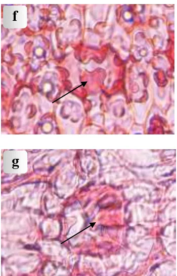
Figure 3. Light micrographs of leaves: (a) Sclerenchyma in spongy tissue of P. tectorius Parkinson cv.
sanderi, (b)Raphide sacs in internal palisade of
P. amaryllifolius Roxb.; (c)Unspecialized abaxial stomata of P. tectorius Parkinson cv.
sanderi; (d) Abaxial papillose lateral subsidiary cells of P. odoratissimus L.f; (e) Abaxial papillose terminal and lateral subsidiary cells of P. scabrifolius Martelli; (f) Abaxial papillose neighbouring and subsidiary cells of
P. kurzii Merr; (g) Abaxial, overarching, lobed or dendritic papillae of P. utilis Bory. Scale bar for a & b = 100 µm; scale bar for c, d, e, f & g
RAHAYU et al.: Leaf anatomy of Pandanus species (Pandanaceae) from Java similar to that of class 1 except for the addition of a row of four to six papillae on the outer surface of each lateral subsidiary cell. There is no other stomatal outgrowth, except for a tendency for the terminal subsidiary cells to overarch the lateral subsidiary and guard cells to a greater extent than in class 1. Class 2 stomata are observed together with intermediate stomata, in which papillae occur on one of the two lateral subsidiary cells belonging to a single stoma. At least one full row of papillae is stomata for the terminal subsidiary cells to protrude over the lateral subsidiary cells is much more pronounced in class 3. Each of the terminal subsidiary cells has prominent papillae, which overarch the stomatal pore. Frequently, the papillae from opposite poles meet and overlap, their ends only mutually overlap or the papillae may even fork to produce short interdigitating branches. Such forking papillae are usually closely adpressed to the stomatal pore, in between the opposite rows of papillae on the lateral subsidiary cells. A species in this category is P. scabrifolius (Fig. 3e).
Class 4. Papillose neighbouring and subsidiary cells. One step further than class 3 is the development of papillae, which protrude from neighbouring epidermal cells. The external stomatal cavity is larger than in class 3, because the complete stomatal apparatus is sunken into the epidermis. Class 4 stomata are very diverse, because the size and frequency of the papillae varies considerably. In the less papillate types the papillae are not large, so that the outer chamber is shallow, but in more papillate types the papillae are very tall and form a distinct wall surrounding a very deep outer chamber. Tall papillae further show a marked tendency to overarch and hide the outer chamber. The papillae themselves are also diverse. They may be the result of protrusions of the whole outer wall of the epidermal cell, or they may only be formed by a part of the outer wall. Finally, some papillae surrounding the stomata show a tendency to become lobed or shortly branched. Usually this is first noticeable in the terminal subsidiary cells, as in P. utilis. This is a transition to class 5. In P. labyrinthicus the papillae are very low but often distinctly lobed. The following species included in class 4 are P. amaryllifolius, P. kurzii (Fig. 3f), P. partly or wholly covered by branched papillae of the terminal subsidiary and neighbouring cells. In the least dendritic members the papillae are short and lobed. Increased branching can be seen in a number of species studied, whereby the papillae become
Stomatal complex sunk with one ring of papillae over the guard cells and one ring of larger lobes investigate the possibility to identify species using anatomical characters. The information gathered in the present study can only give a rough indication of the anatomical characters, which may prove to be of value taxonomically.
Several anatomical characters that can be used for this purpose (see also key below and results above) are the shape of the epidermal cells, costal-intercostal zones in the abaxial epidermis, the outline of the anticlinal walls of the epidermal cells, the distribution of rhomboidal crystals in the epidermis and hypodermis and the stomatal structure, especially the presence of papillae.
Kam & Stone (1970) and Kam (1971) reported that stomata structure, epidermal zonation, silica-body presence and arrangement, and epidermal papillosity may have value for delimiting sections. By contrast, in this study six species of Javanese
Pandanus, all belonging to Section Rykia, did not have the same features. Zonation is very clear cut in
P. multifurcatus and P. nitidus, but indistinct in P. labyrinthicus, and zonation is absent in P. bantamensis, P. pseudolais and P. scabrifolius. Thus, for the Javanese species, zonation can only be used for the identification of species, not sections.
shapes for the adaxial epidermal cells, and three shapes for the abaxial cells. Kam (1971) also stated that the transverse length of first layer of hypodermis corresponds to the space accupied by 10 to 12 rows of epidermal cells, but our materials only showed an equivalence of 4-8 cells.
In general, the stomatal structure of the fifteen present study (P. utilis from Java) elaborate stomata in P. utilis should be classified as class 5 (papillae overarching and lobed or dendritic). So the shape of elaborate stomata in P, utilis could be different if the sample source is different.
In Javanese Pandanus anatomical characters appear to provide reliable characters for the identification of species as was already pointed out by Stone (1978). The key underneath can only tentative because their constancy of the features within species was tested on very few specimen only.
KEY TO THE SPECIES OF JAVANESE PANDANUS
1. Costal zonation distinct in abaxial epidermis
2. Cuticle thick (5µm ) ... P. labyrinthicus 1. Costal zonation absent in abaxial epidermis
13. Papillae on stomata absent ... P. pseudolais curators of the following herbaria: BO, L and Kew. Prof. Dr.Mien A Rifai for providing valuable suggestion. We wish to thank Dr. Sunaryo, Eka Fatmawati Tihurua, Widoyanti and Ujang Hapid of Herbarium Bogoriense for helping during laboratory and photographic work. This work was funded by The Directorate General of Higher Education of Indonesia through fundamental research grant number 109/SP2H/PP/DP2M/III/2008. Monocotyledons. A comparatice study. Academic Press. London.
EDEOGA, H. O. & IKEM, C. I. 2001. Comparative morphology of leaf epidermis in three species of
Boerhevia L. J. Econ.Tax. Bot. 19: 197-205.
JAKUBSKA, A. 2007. The analysis of morphological differentiation of the epidermis of selected species of the genus Epipactis ZINN, 1757 (Orchidaceae: Neottieae). Wroclaw 15(2): 41-45.
JOHANSEN, D. A. 1940. Plant microtechnique. McGraw-Hill, Book Company, Inc. New York. KAM, Y. K. & STONE, B. C. 1970. Morphological
studies in Pandanaceae IV. Stomata structure in some Mascarene and Madagascar Pandanus and its meaning for infrageneric taxonomy. Adansonia 10 (2): 219-246.
KAM, Y. K. 1971. Comparative systematic foliar anatomy of Malayan Pandanus. Bot. J. Linn. Soc. 64: 315-353.
MBAGWU, F. N., NWACHUKWU, C. U. & OKORO, O. O. 2007. Comparative leaf epidermal studies on
Solanum mascrocarpon and Solanum nigrum. Nature and Science 5(3): 1-4.
OSUJI, J. O. & NDUKUWU, B. C. 2005. Probable functions and remobilisation of calcium oxalates in
RAHAYU et al.: Leaf anatomy of Pandanus species (Pandanaceae) from Java
2012] 313
PRYCHID, C. J. & RUDALL, P. A. 1999. Calcium oxalate crystals in monocotyledons: a review of their structure and systematics. Annals of Botany 84: 725-739.
STACE, C. A. 1965. Cuticular studies as an aid to plant taxonomy. Bull. Brit. Mus.(Nat.Hist) Bot. 4: 1 - 78. STONE, B. C. 1965. Pandanus Stickman in the
Malayan Peninsula, Singapore and lower Thailand.
Malay. Nat. J . 19(4): 203-213.
STONE, B. C. 1972. A review of Javanese
Pandanaceae with notes on plants cultivated in Hor-tus Bogoriensis. Reinwardtia 8: 309-318.
STONE B. C. 1976. The morphology and systematics of Pandanus today (Pandanaceae). Gardens’ Bulletin Singapore 29: 137-142.
STONE, B. C. 1978. Studies in Malesian Pandanaceae XVII on the taxonomy of “Pandan Wangi” A Pandanus cultivar with scented leaves. Economic Botany 32: 285-293.
TOMLINSON, P. B. 1961. Anatomy of the Monocotyledons II Palmae. Clarendon Press. Oxford. TOMLINSON, P. B. 1965. A study of stomatal
ERRATUM
REINWARDTIA Vol. 13, Part 2, 2010
1. Please change the existing word in p. 213, LINE 7 on ABSTRAK (written in Bahasa Indonesia version) with the following:
Keberadaan dua jenis terakhir melampaui distribusi yang sebelumnya hanya diketahui di barat garis
Wallace.
2. Please change the existing epithet name in p, 214, COLUMN 1, LINE 40 on Key to the species of Marantaceae in Sulawesi number 5.a. after Phrynium:
INSTRUCTION TO AUTHORS
Reinwardtia is a scientific journal on plant taxonomy, plant ecology, and ethnobotany. Manuscript intended for a publication should be written in English represent an article which has not been published in any other journal or proceedings. Every manuscript will be sent to two blind reviewers.
Two printed copies (on A4 paper) of the manuscript of not more than 200 pages together with an electronic copy prepared on Word Processor computer program using Time New Romance letter type and saved in Rich Text File must be submitted.
For the style of presentation, authors should follow the latest issue of Reinwardtia very closely. Title of the article should be followed by authors name and mailing address in one-paragraphed English abstract of not more than 250 words. Keywords should be given below each abstract. On a separated paper, author(s) should send the preferred running title of the article submitted.
Taxonomic identification key should be prepared using the aligned couplet type.
Strict adherence to the International Code of Botanical Nomenclature is observed, so that taxonomic and nomenclatural novelties should be clearly shown. Latin description for new taxon proposed should be provided and the herbaria where the type specimens area deposited should be presented in the long form that is name of taxon, authors name, year of publication, abbreviated journal or book title, volume, number and page.
Map, line drawing illustration, or photograph preferably should be prepared in landscape presentation to occupy two columns. Illustration must be submitted as original art accompanying, but separated from the manuscript. On electronic copy, the illustration should be saved in jpg or gif format at least 350 pixels. Legends or illustration must be submitted separately at the end of the manuscript.
Vol. 13. No. 3. 2012 CONTENTS Page
W.J.J.O. DE WILDE & BRIGITTA E.E. DUYFJES. Trichosanthes (Cucurbitaceae) in Malesia: additions and corrections, including a new species and a new variety 221
DEDEN GIRMANSYAH. Two new species of Begonia (Begoniaceae) from Bukit Tiga-puluh National Park, Riau, Sumatra 229
PUDJI WIDODO. New nomenclature in Syzygium (Myrtaceae) from Indonesia and its vicinities 235
ALEX SUMADIJAYA & JAN FRITS VELDKAMP. Non-Bambusoid Grasses (Gramineae) from Raja Ampat Archipelago, Papua Barat Province, Indonesia 241
ARY PRIHARDYANTO KEIM. New variety, records & discoveries of some species of Pandanus
(Pandanaceae) in Sumatra and Kalimantan, Indonesia 255
HARRY WIRIADINATA. A new species of Begonia (Begoniaceae) from Sagea Lagoon, Weda Bay, Halmahera Island, North Moluccas, Indonesia 263
ARY PRIHARDYANTO KEIM. The Pandan flora of Foja-Mamberamo Game Reserve and Baliem Valley, Papua-Indonesia 271
JAN FRITS VELDKAMP. Koordersiochloa Merr. (Gramineae), the correct name for Streblochaete Hochst. exPilg. 299
SRI ENDARTI RAHAYU, KUSWATA KARTAWINATA, TATIEK CHIKMAWATI & ALEX HARTANA. Leaf anatomy of Pandanus species (Pandanaceae) from Java 305
Reinwardtia is a LIPI acredited Journal (258/AU 1/P2MBI/05/2010)
Herbarium Bogoriense Botany Division