www.elsevier.com / locate / bres
Research report
The effects of phosphodiesterase inhibition on cyclic GMP and cyclic
AMP accumulation in the hippocampus of the rat
*
W.C.G. van Staveren , M. Markerink-van Ittersum, H.W.M. Steinbusch, J. de Vente
Maastricht University, Department of Psychiatry and Neuropsychology, Universiteitssingel 50, POB 616, 6200 MD Maastricht, The Netherlands Accepted 3 October 2000
Abstract
The effects of selective and non-selective 39,59-cyclic nucleotide phosphodiesterase (PDE) inhibitors on cGMP and cAMP accumulation were studied in rat hippocampal slices incubated in vitro. The following PDE inhibitors were used: vinpocetine and calmidazolium (PDE1 selective), erythro-9-(2-hydroxy-3-nonyl)adenine (EHNA, PDE2 selective), SK&F 95654 (PDE3 selective), rolipram (PDE4 selective), SK&F 96231 (PDE5 selective), the mixed type inhibitors zaprinast and dipyridamole, and the non-selective inhibitors 3-isobutyl-1-metylxanthine (IBMX) and caffeine. cGMP levels were increased in the presence of different concentrations of IBMX, EHNA, dipyridamole, vinpocetine and rolipram. cGMP immunocytochemistry showed that incubation with different inhibitors in the presence and / or absence of sodium nitroprusside resulted in pronounced differences in the extent and regional localization of the cGMP response and indicate that PDE activity in the hippocampus is high and diverse in nature. The results suggest an interaction between cGMP and cAMP signalling pathways in astrocytes of the rat hippocampus. 2001 Elsevier Science B.V. All rights reserved.
Theme: Neurotransmitters, modulators, transporters, and receptors
Topic: Second messengers and phosphorylation
Keywords: Phosphodiesterase; cGMP; cAMP; Hippocampus; Nitric oxide; Inhibitor
1. Introduction cGMP synthesis. cGMP and the other second messenger cAMP are inactivated by hydrolytic cleavage of their Nitric oxide synthase (NOS) has a widespread dis- 39-phosphoester bonds to form 59-GMP and 59-AMP by tribution in the central nervous system (CNS) [4,46]. In the the superfamily of enzymes known as the cyclic nucleotide hippocampus, two constitutive isoforms of NOS have been phosphodiesterases (PDEs). At present, the PDEs have described, i.e. the neuronal NOS (nNOS) and the endo- been classified into 11 different families, i.e. PDE1 to thelial NOS (eNOS), also referred as NOS-I and NOS-III PDE11, based on their substrate and inhibitor profiles respectively [13]. In this brain area, both isoforms are together with their structural characteristics [1,6,14,43]. activated through calcium / calmodulin dependent path- Within families multiple splice variants of those isozymes ways, which are triggered by the N-methyl-D-aspartate exist which makes the number of PDE isozymes more than
(NMDA)-type glutamate receptors [21]. NO diffuses from 30 [1,23]. A number of these enzymes were shown to be the site of synthesis to the target structures which are localized regionally [2,19,24,25,27,31,36–39,47] and may neurons and astrocytes close to the site of the NO be expressed to different degrees even within one cell type production [10,20,21,29,35]. The soluble isoform of [1,25].
guanylyl cyclase (sGC) presents an important target mole- From previous studies it is known that the PDE activity cule for NO [5,28]. NO activates sGC by binding to the in the hippocampus is high (e.g. [12]). We also found heme group of this enzyme which leads to an increased evidence for the presence of cGMP-hydrolyzing PDE activity in the hippocampus which is not or only partly inhibited by IBMX [12]. In addition, it was shown in brain
*Corresponding author. Tel.: 131-43-388-1168; fax: 1
31-43-367-slices that zaprinast, an inhibitor of cGMP-specific PDE
1096.
E-mail address: [email protected] (W.C.G. van Staveren). activity (PDE5 and PDE9), increased NO-mediated cGMP
276 W.C.G. van Staveren et al. / Brain Research 888 (2001) 275 –286
accumulation especially in the CA2 / CA3 region and the (DMSO), therefore a final concentration of 1% DMSO was stratum lacunosum moleculare of the hippocampus. In present in all experiments. In an earlier study it was found contrast, IBMX, a non-selective PDE inhibitor, increased that 1% DMSO had no effect on cGMP levels in hip-cGMP levels in varicose fibers and astrocytes throughout pocampal slices (unpublished results).
the hippocampus [11]. Thus, the choice of the PDE inhibitor is a very important factor when cyclic nucleotide
2.2.1. Radioimmunoassay levels are studied in complex tissues. In this respect it is
cGMP and cAMP levels were determined in individual striking that in the canine proximal colon only a
combina-hippocampal slices, incubated as described above, using a tion of zaprinast and IBMX was effective in inhibiting
radioimmunoassay as published previously [9]. Briefly, the NO-mediated cGMP in smooth muscle cells [40].
incubations were terminated by placing the slices into a In order to study the possibilities of locally increasing
solution of 5% trichloroacetic acid. Subsequently, the cyclic nucleotide levels using selective inhibitors of PDE
samples were sonicated and centrifuged. The supernatant activity [11], we measured the effects on cGMP and cAMP
was used for the determination of the cGMP and cAMP accumulation in hippocampal slices, after the incubation
content, measured by a radioimmunoassay according to with a number of PDE inhibitors with different selectivity
Steiner and coworkers [44]. The pellet was used for the profiles. In addition, we visualized the effect of these
analysis of the protein content according to Lowry and inhibitors on the accumulation of NO-mediated cGMP
coworkers [30]. synthesis using cGMP immunocytochemistry. A
compari-Each condition was measured in two different slices per son was made between the determination of cGMP levels
animal and for each PDE inhibitor three different rats were by the use of a radioimmunoassay and by the evaluation of
tested. In each slice, both cGMP and cAMP levels were cGMP immunofluorescence intensity using an image
anal-measured in triplicate and the median was taken for each ysis system.
sample. In the radioimmunoassay, a level of 0.6 fmol cGMP and 5 fmol cAMP could be detected. cGMP and cAMP levels were corrected for the protein content of each
2. Materials and methods
slice.
2.1. Animals
2.3. Immunocytochemistry Experiments were performed on hippocampal slices
obtained from adult male Lewis rats (200–240 g). The After the incubation, slices were fixed with ice-cold effect of zaprinast on two different rat strains was studied fixative solution of 4% freshly prepared depolymerised in female Lewis and Wistar rats (obtained from Charles paraformaldehyde in 0.1 M phosphate buffer (pH 7.4) for River). The animals were housed under standard conditions 30 min at 48C. The slices were then fixed for another 90 at the local animal facility. All experiments were approved min with 4% paraformaldehyde containing 10% sucrose. by the committee on animal welfare according to Dutch After washing overnight at 48C in 0.1 M phosphate buffer governmental rules. (pH 7.4) containing 10% sucrose, the slices were frozen in CO . Cryostat sections (102 mm) were cut, thawed onto 2.2. Tissue preparation chrome–alumn / gelatin coated slides and processed for
immunocytochemistry.
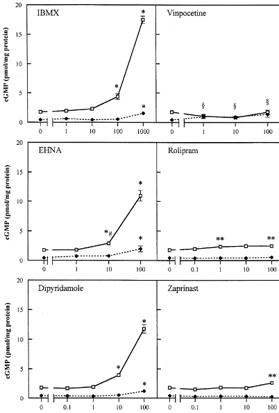
without any bleeding of the other fluorescent marker in the presence of 0.1 mM SNP (mean6S.E.M.; sig-through the filter [10]. nificantly different from control (0 M) without SNP, Glial fibrillary acidic protein (GFAP) was stained with Student t-test, P,0.01). As shown in Fig. 1, in the absence mouse anti-GFAP serum (Innogenetics), diluted 1:10 in of SNP the cGMP content in hippocampal slices was TBS-T and visualised with donkey anti-mouse Cy3 (Jack- increased by IBMX (1 mM), EHNA (100 mM), son), diluted 1:800 in TBS-T. dipyridamole (100 mM) and vinpocetine (1 mM and higher) compared to its controls. No changes in cGMP 2.4. Image analysis levels were found when slices were incubated with
roli-pram or zaprinast.
For the semi-quantitative measurement of cGMP, sec- Incubation of slices with 0.1 mM SNP in the presence of tions were stained for cGMP as described above and the different concentrations of IBMX, EHNA or dipyridamole, primary antibody was visualised by the incubation of resulted in a concentration dependent increase of cGMP sections for 1 h at room temperature with the Alexa 488 levels. Rolipram increased cGMP levels significantly at 1 donkey anti-sheep IgG (H1L) conjugate (Molecular mM and 100mM and the cGMP content was only raised in Probes), diluted 1:100 in TBS-T. Each condition was tested the presence of the highest dose of zaprinast (100mM). On in hippocampal slices obtained from three different ani- a molar basis EHNA and dipyridamole appeared to be the mals and from each slice, three different sections of the most potent inhibitors. In the presence of SNP we did not hippocampal area were studied per animal. All sections find a significant increase in cGMP accumulation by were stained and analysed at the same time under standard vinpocetine (Fig. 1) or calmidazolium (not shown). conditions. Pictures of the stratum lacunosum moleculare
and the CA1 area were made at a magnification of 203 3.2. cAMP levels in hippocampal slices after incubation using a Sony Power HAD 3CCD Color Video Camera. All with different PDE inhibitors
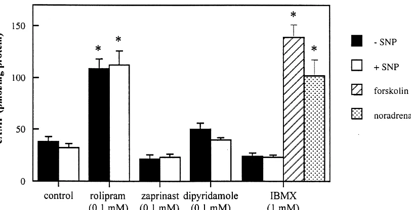
pictures were analysed with the computer program
analy-SIS Vers. 3.0. For each image, a color separation of the As shown in Fig. 2, in the absence of PDE inhibitors the green image was done and the mean grayvalue of each basal cAMP content of hippocampal slices was area was estimated as a measure for the cGMP content of 37.9464.94 pmol / mg protein (determined in six animals). the hippocampal area. All measurements were corrected This is in the range of reported hippocampal levels [17]. for control sections which were incubated without the Rolipram strongly increased cAMP levels while no effect primary antibody. on the cAMP content was found in the presence of zaprinast, dipyridamole or IBMX. There was no effect of 2.5. Statistical analysis SNP on cAMP levels regardless of the PDE inhibitors being present. Incubation of hippocampal slices with 10 To determine whether different concentrations of the mM forskolin (an activator of adenylyl cyclase) or 10mM PDE inhibitor tested, differed from its control, a Student noradrenalin, both in the presence of IBMX, resulted in Newman–Keuls test and a Student t-test were used. large increases of cAMP levels.
2.6. Chemicals 3.3. cGMP immunostaining after incubation with
different PDE inhibitors
IBMX was from Janssen Chimica; zaprinast,
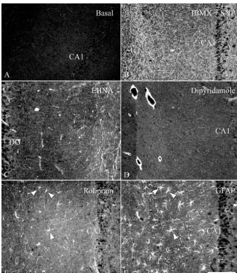
dipyridamole and EHNA from Sigma; rolipram and cal- When hippocampal slices were incubated in vitro with-midazolium from RBI; vinpocetine from Tocris; SK&F out PDE inhibitors, cGMP immunostaining was nearly 96231 and SK&F 95654 were kindly donated by absent (Fig. 3A). No effect on cGMP immunocyto-SmithKline Beecham. L-NAME and SNP were obtained chemistry was observed when slices were treated with
from Fluka. vinpocetine, calmidazolium, SK&F 95654, SK&F 96231 or zaprinast (data not shown). In the presence of 1 mM IBMX isolated fibers were observed distributed at random
3. Results throughout the hippocampal slice, with a cluster of thin, punctate fibers in the stratum lacunosum moleculare (data
G
3.1. cGMP levels in hippocampal slices after incubation not shown). The NOS inhibitor N -nitro-L-arginine (L
278 W.C.G. van Staveren et al. / Brain Research 888 (2001) 275 –286
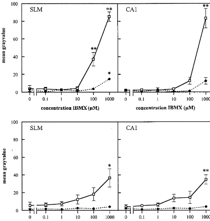
Fig. 1. Effect of PDE inhibitors on cyclic GMP levels in hippocampal slices of the rat, in the absence (♦- - -♦) or presence (h——h) of 0.1 mM SNP, measured by a radioimmunoassay. Each value is the mean (6S.E.M.) of three animals. Each concentration was performed in duplicate per animal and assayed in triplicate in a radioimmunoassay. Values which were different from control (0 M) (Student Newman–Keuls test; P,0.01) were marked with an asterisk (*). Values which were different from control tested with a Student t-test were marked as **(P,0.05) and *[(P,0.01). §, different from control
Fig. 2. Effect of PDE inhibitors on cAMP levels in hippocampal slices of the rat, in the presence or absence of 0.1 mM SNP, measured by a radioimmunoassay. Forskolin and noradrenalin were both used in a concentration of 10mM. Each value is the mean (1S.E.M.) of three animals. Each concentration was performed in duplicate per animal and assayed in triplicate in a radioimmunoassay. Values which were different from control (Student
t-test; P,0.01) were marked with an asterisk.
rolipram induced cGMP immunostaining (data not shown). (Fig. 5). In contrast, in the radioimmunoassay the effect of Dipyridamole (0.1 mM) strongly increased cGMP in the 1 mM of these inhibitors on the cGMP content was not smooth muscle layer of what appeared to be the larger significantly different from controls (Fig. 1).
blood vessels of the hippocampus (Fig. 3D). Incubation of slices with rolipram and SNP resulted in cGMP immunostaining in astrocyte-like cells and a few 3.4. cGMP accumulation after stimulation of sGC by isolated varicose fibers scattered throughout the
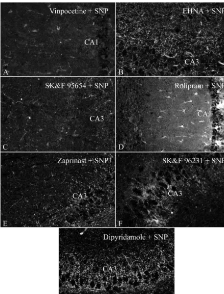
hippocam-SNP in the presence of PDE inhibitors pus (Fig. 4D). Using double immunostaining of cGMP in combination with GFAP, these cells could indeed be Incubation of hippocampal slices with 0.1 mM SNP in identified as astrocytes (see also Fig. 3E and 3F). When the absence of PDE inhibitors, showed a few intensely slices were incubated with rolipram combined with IBMX stained fibers which, taking into account all experiments and SNP, cGMP staining was observed similar as in the (.10), might be observed in any region of the hippocam- case of IBMX and SNP, although the cGMP immuno-pus (not shown). cGMP immunocytochemistry after in staining in the astrocytes was more pronounced (Fig. 6A). vitro incubation of the slices in the presence of IBMX and Strikingly, only a subpopulation of astrocytes showed SNP has been described previously (e.g. [11]). This cGMP immunoreactivity (Fig. 6B).
combination of drugs resulted in cGMP accumulation in a multitude of varicose fibers throughout the hippocampus
(Fig. 3B). Similar staining patterns were observed when 3.5. Image analysis of cGMP immunostaining after EHNA or dipyridamole were used (Fig. 4B and 4G). In treatment with IBMX or zaprinast
contrast, the combination of SNP with the inhibitors
molecu-280 W.C.G. van Staveren et al. / Brain Research 888 (2001) 275 –286
Fig. 3. Localization of cGMP immunoreactivity in hippocampal slices incubated in vitro: (A) in the presence of 1 mM IBMX; (B) combination of 1 mM IBMX and 0.1 mM SNP; (C) 0.1 mM EHNA; (D) 0.1 mM dipyridamole; (E) 0.1 mM rolipram. The immunostaining in (F) with GFAP shows the same field as depicted in (E) indicating that rolipram increases cGMP in astrocytes (arrowheads). DG, dentate gyrus. Bar represents 100mm for all pictures.
Fig. 4. Localization of cGMP immunoreactivity in hippocampal slices incubated in the presence of 0.1 mM SNP and: (A) 0.1 mM vinpocetine; (B) 0.1 mM EHNA; (C) 0.1 mM SKF 95654; (D) 0.1 mM rolipram; (E) 0.1 mM zaprinast; (F) 0.1 mM SKF 96231; (G) 0.1 mM dipyridamole. Bar represents 100
282 W.C.G. van Staveren et al. / Brain Research 888 (2001) 275 –286
Fig. 5. Localization of cGMP immunoreactivity in the hippocampal slice incubated in the presence of 0.1 mM SNP and 1–10mM of EHNA or dipyridamole. Bar (c) represents 100mm for all pictures.
Fig. 7. The effect of IBMX and zaprinast on the fluorescence intensity of the cGMP immunostaining in the stratum lacunosum moleculare (SLM) and the CA1 area of hippocampal slices of the rat, in the absence (♦- - -♦) or presence (h——h) of 0.1 mM SNP, measured by image analysis. Each value is the mean (6S.E.M.) of three animals. For each concentration three different sections were analysed per animal. Values which were different from control (0 M) (Student Newman–Keuls test) were marked as *(P,0.05) and **(P,0.01).
284 W.C.G. van Staveren et al. / Brain Research 888 (2001) 275 –286
At present, several PDE types have been localized in the metabolism. These factors are not involved when the rat hippocampus. It has been shown with in situ hybridisa- purified enzyme is used. Furthermore, it is not known tion or immunocytochemistry that PDE1, PDE2, PDE3 and whether more than one PDE type is present in one cell. PDE4 are present in this brain area of the rat Therefore, it is possible that if a certain PDE is inhibited [19,31,37,38,45]. In an attempt to gain more insight in the this could have an effect on another PDE type in this cell localization of PDEs with different selectivity profiles, as well, which in turn can influence the cyclic nucleotide several PDE type inhibitors were used in this study. The levels also.
PDE inhibitors studied include the highly selective in- Using immunocytochemistry for cGMP, it was found hibitors vinpocetine, calmidazolium (both PDE1 in- that the largest part of the NO-mediated cGMP immuno-hibitors), EHNA (PDE2) [34] and rolipram (PDE4). In staining was present in varicose fibers. As shown previous-addition, we used zaprinast and dipyridamole, which were ly [11,12], inhibition of the PDE activity using non-until recently, considered to be highly selective inhibitors specific PDE inhibitors as methylxanthines like IBMX of PDE5 [7,32]. Recent evidence showed that zaprinast (caffeine and theophylline not shown) in combination with inhibited PDE9 also [16] and dipyridamole has affinity for an NO donor resulted in cGMP-accumulation in varicose PDE7 [22], PDE8 [15,41], PDE10 [18,42] and the recently fibers throughout the hippocampus. As a somewhat vary-cloned PDE11 [14]. In this study IBMX was used as a ing response between experiments, cGMP immunostaining non-specific PDE inhibitor. was also found in astrocytes and a few interneurons under Selective inhibitors have a high affinity for a particular these conditions. In some experiments a very low level of PDE. In our study, the effects in the radioimmunoassay are cGMP immunoreactivity was possibly observed in pyrami-found at a relative high concentration of most inhibitors dal cells in unstimulated slices. However, in the stimulated which could point to unspecific inhibition. However, if all slice the cGMP staining in these cells could not normally the effects found would be due to unspecific reactions then be observed, probably due to the intensity of the cGMP it was expected that all the staining patterns were the same. immunofluorescence in the surrounding fibers (Fig. 3B), Boulton and coworkers [3] reported that 2 mM zaprinast although a redistribution of cGMP through an as yet increased the cGMP content in the hippocampus. As unknown mechanism might also be involved. If cGMP shown in Fig. 1 we did not measure an effect of zaprinast immunoreactivity is to be present in cell somata in the at such low concentrations. To investigate if strain or sex pyramidal cell layer, as has been suggested [3], the differences might cause this difference we repeated the concentration must be very low.
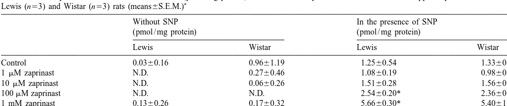
experiments using female Wistar rats, as used by Boulton The cGMP immunoreactive fibers in the hippocampus and coworkers, and compared them with female Lewis have not yet been characterized in terms of the major rats. As shown in Table 1, we were not able to reproduce neurotransmitters and it is known neither from which the effects that were found with zaprinast. projections area(s) these fibers originate. cGMP immuno-In literature, it has been shown that the potency of a staining has been observed only in an occasional NOS PDE inhibitor might be higher on purified enzymes than immunoreactive hippocampal interneuron. The hippocam-found in cells or tissues. Coste and Grondin [7] reported pal cGMP-positive fibers in our study do not correspond in differences in the potency of the PDE5 inhibitor 1,3- any aspect to the localization and direction of pyramidal dimethyl-6-(2-propoxy - 5 - methanesulfonylamidophenyl) - dendritic or axonal fibers as visualized using eNOS pyrazolo[3,4d]-pyrimidin-4-(5H)-one (DMPPO) when an antibodies or the modified NADPH–diaphorase stain [13]. enzymatic PDE5 assay was compared to a cell model. A It must be noted that EHNA, dipyridamole and IBMX lower potency for inhibitors in a cell model or slices might exert different actions in adenosine pharmacology. EHNA be explained by cell permeability or the intracellular is a potent inhibitor of adenosine deaminase whereas
Table 1
The effects of zaprinast and IBMX on cGMP levels (pmol / mg protein) in the absence or presence of 0.1 mM SNP in hippocampal slices from female a
Lewis (n53) and Wistar (n53) rats (means6S.E.M.)
Without SNP In the presence of SNP
(pmol / mg protein) (pmol / mg protein)
Lewis Wistar Lewis Wistar
dipyridamole is an inhibitor of adenosine uptake, and pocampus was unaffected by the presence of L-NAME,
suggesting that NO was not involved. IBMX is an adenosine receptor antagonist. If adenosine
Our results permit the following conclusions. First, our was involved in the cGMP response, we would expect
results indicate that several PDE isoforms are present in EHNA and dipyridamole to have similar effects, because
the rat hippocampus and function as part in the NO–cGMP both compounds increase the adenosine concentration
signal transduction pathway. Nevertheless, the non-selec-outside the cell (see e.g. [8]). Indeed, the cGMP
immuno-tive component in the pharmacological profile of the staining is rather similar when EHNA or dipyridamole is
inhibitors used in the present study, makes it unlikely that used in the presence of SNP. IBMX is an adenosine
the approach of cGMP immunocytochemistry can be used antagonist and, following the above reasoning, in the
for further characterization of the localization of PDE presence of IBMX it might be expected that the cGMP
isoforms. Secondly, the results obtained with rolipram staining would be less than the immunoreactivity obtained
strongly suggest that PDE4 is present in a subpopulation of in the presence of EHNA or dipyridamole. However, in the
astrocytes in the hippocampus. presence of IBMX, cGMP immunoreactivity was found in
the hippocampus and was strongly increased in the pres-ence of SNP. Furthermore, we did not observe any effect
References
with a non-selective adenosine receptor agonist 59 -N-ethylcarboxamidoadenosine (NECA) in a concentration of
[1] J.A. Beavo, Cyclic nucleotide phosphodiesterases: functional
impli-0.1 mM, in the presence or absence of different PDE
cations of multiple isoforms, Physiol. Rev. 75 (1995) 725–748.
inhibitors on cGMP immunostaining in the hippocampus [2] M.L. Billingsley, J.W. Polli, C.D. Balaban, R.L. Kincaid, Develop-slice. Therefore, we conclude tentatively that adenosine in mental expression of calmodulin-dependent cyclic nucleotide
phos-not involved in the cGMP-response in the hippocampus phodiesterase in rat brain, Dev. Brain Res. 53 (1990) 253–263. [3] C.L. Boulton, A.J. Irving, E. Southam, B. Potier, J. Garthwaite, G.L.
slice.
Collingridge, The nitric oxide-cyclic GMP pathway and synaptic
Mayer and coworkers (1992) [33] reported that
especial-depression in rat hippocampal slices, Eur. J. Neurosci. 6 (1994)
ly the calcium dependent PDE1 activity is responsible for 1528–1535.
cGMP breakdown in the rat brain. Although we did [4] D.S. Bredt, P.M. Hwang, S.H. Snyder, Localization of nitric oxide
observe a small increase in cGMP levels in the presence of synthase indicating a neural role for nitric oxide, Nature 347 (1990) 768–770.
vinpocetine alone, no effect was measured in the presence
[5] D.S. Bredt, S.H. Snyder, Nitric oxide mediates glutamate-linked
of SNP and vinpocetine. In addition, we did not find any
enhancement of cGMP levels in the cerebellum, Proc. Natl. Acad.
effect of calmidazolium on cGMP levels in the hippocam- Sci. USA 86 (1989) 9030–9033.
pus, the frontal brain, or the cerebellum of the rat. Under [6] M. Conti, S.L.C. Jin, The molecular biology of cyclic nucleotide
the conditions used, our results indicate that the inhibition phosphodiesterases, Prog. Nucl. Acid Res. Mol. Biol. 63 (1999) 1–38.
of PDE1 does not have a major effect on cGMP
break-[7] H. Coste, P. Grondin, Characterization of a novel potent and specific
down in the hippocampus. In addition, no effect was found
inhibitor of type V phosphodiesterase, Biochem. Pharmacol. 50
of the inhibition of PDE3 by SK&F 95654 in our study (1995) 1577–1585.
also. [8] R.A. Cunha, A.M. Sebastiao, J.A. Ribeiro, Inhibition by ATP of
The results obtained with rolipram were unexpected. It hippocampal synaptic transmission requires localized extracellular catabolism by ecto-nucleotidases into adenosine and channeling to
was found that rolipram influenced both cGMP and cAMP
adenosine A1 receptors, J. Neurosci. 18 (1998) 1987–1995.
levels. Rolipram alone increased the cAMP concentration
[9] J. De Vente, J.G.J.M. Bol, L. Hudson, J. Schipper, H.W.M.
Stein-in the hippocampus slice till a very high level, whereas a busch, Atrial natriuretic factor-responding and cyclic guanosine small raise in the cGMP content was detected also. When monophosphate (cGMP)-producing cells in the rat hippocampus: a
the effect of rolipram was studied by immunocyto- combined micropharmacological and immunocytochemical ap-proach, Brain Res. 446 (1988) 387–395.
chemistry, cGMP positive astrocytes were found. These
[10] J. De Vente, D.A. Hopkins, M. Markerink-Van Ittersum, P.C. Emson,
data suggest an interaction between cGMP and cAMP
H.H.H.W. Schmidt, H.W.M. Steinbusch, Distribution of nitric oxide
signalling pathways in the hippocampus, resulting in a synthase and nitric oxide-receptive, cyclic GMP-producing struc-massive increase in cAMP and a more restricted increase tures in the rat brain, Neuroscience 87 (1998) 207–241.
in cGMP in a subpopulation of astrocytes. This effect of [11] J. De Vente, D.A. Hopkins, M. Markerink-van Ittersum, H.W.M. Steinbusch, Effects of the 39,59-phosphodiesterase inhibitors
iso-rolipram on cGMP levels does not appear to be a general
butylmethylxanthine and zaprinast on NO-mediated cGMP
accumu-effect in all brain tissue as we found little, if any, cGMP
lation in the hippocampus slice preparation: an immunocytochemical
immunostaining in frontal cortical areas after incubation of study, J. Chem. Neuroanat. 10 (1996) 241–248.
slices in the presence of rolipram and SNP (not shown). [12] J. De Vente, H.W.M. Steinbusch, On the stimulation of soluble and
Nevertheless, this observation is not completely without particulate guanylate cyclase in the rat brain and the involvement of nitric oxide as studied by cGMP immunocytochemistry, Acta
precedent, because recently it was reported that rolipram
Histochem. 92 (1992) 13–38.
increased cGMP levels in endothelial cells through an
[13] J.L. Dinerman, T.M. Dawson, M.J. Schell, A. Snowman, S.H.
NO-dependent mechanism [26]. A difference between our Snyder, Endothelial nitric oxide synthase localized to hippocampal results and the observations of Kessler and Lugnier [26] is pyramidal cells: implications for synaptic plasticity, Proc. Natl.
286 W.C.G. van Staveren et al. / Brain Research 888 (2001) 275 –286
[14] L. Fawcett, R. Baxendale, P. Stacey, C. McGrouther, I. Harrow, S. cyclic nucleotide phosphodiesterase is localized predominantly at Soderling, J. Hetman, J.A. Beavo, S.C. Phillips, Molecular cloning postsynaptic sites in the rat brain, Neuroscience 44 (1991) 491–500. and characterization of a distinct human phosphodiesterase gene [32] C. Lugnier, P. Schoeffter, A. Le Bec, E. Strouthou, J.C. Stoclet, family: PDE11A, Proc. Natl. Acad. Sci. USA 97 (2000) 3702–3707. Selective inhibition of cyclic nucleotide phosphodiesterases of [15] D.A. Fisher, J.F. Smith, J.S. Pillar, S.H. St Denis, J.B. Cheng, human, bovine and rat aorta, Biochem. Pharmacol. 35 (1986)
Isolation and characterization of PDE8A, a novel human cAMP- 1743–1751.
specific phosphodiesterase, Biochem. Biophys. Res. Commun. 246 [33] B. Mayer, P. Klatt, E. Bohme, K. Schmidt, Regulation of neuronal 21
(1998) 570–577. nitric oxide and cyclic GMP formation by Ca , J. Neurochem. 59 [16] D.A. Fisher, J.F. Smith, J.S. Pillar, S.H. St Denis, J.B. Cheng, (1992) 2024–2029.
Isolation and characterization of PDE9A, a novel human cGMP- [34] A.M. Michie, M. Lobban, T. Muller, M.M. Harnett, M.D. Houslay, specific phosphodiesterase, J. Biol. Chem. 273 (1998) 15559– Rapid regulation of PDE-2 and PDE-4 cyclic AMP phosphodies-155564. terase activity following ligation of the T cell antigen receptor on [17] J.C. Fowler, J.M. O’Donnell, Antagonism of the responses to thymocytes: analysis using the selective inhibitors erythro-9-(2-isoproterenol in the rat hippocampal slice with subtype-selective hydroxy-3-nonyl)-adenine (EHNA) and rolipram, Cell. Signal. 8 antagonists, Eur. J. Pharmacol. 153 (1988) 105–110. (1996) 97–110.
[18] K. Fujishige, J. Kotera, H. Michibata, K. Yuasa, S. Takebayashi, K. [35] T.J. O’Dell, R.D. Hawkins, E.R. Kandel, O. Arancio, Tests of the Okumura, K. Omori, Cloning and characterization of a novel human roles of two diffusible substances in long-term potentiation: evi-phosphodiesterase that hydrolyses both cAMP and cGMP dence for nitric oxide as a possible early retrograde messenger, Proc. (PDE10A), J. Biol. Chem. 274 (1999) 18438–18445. Natl. Acad. Sci. USA 88 (1991) 11285–11289.
[19] T. Furuyama, Y. Iwahashi, Y. Tano, H. Takagi, S. Inagaki, Localiza- [36] J.W. Polli, R.L. Kincaid, Expression of a calmodulin-dependent tion of 63-kDa calmodulin-stimulated phosphodiesterase mRNA in phosphodiesterase isoform (PDE1B1) correlates with brain regions the rat brain by in situ hybridization histochemistry, Mol. Brain Res. having extensive dopaminergic innervation, J. Neurosci. 14 (1994)
26 (1994) 331–336. 1251–1261.
[20] J. Garthwaite, Glutamate, nitric oxide and cell–cell signalling in the [37] R.R. Reinhardt, C.A. Bondy, Differential cellular pattern of gene nervous system, Trends Neurosci. 14 (1991) 60–67. expression for two distinct cGMP-inhibited cyclic nucleotide phos-[21] J. Garthwaite, C.L. Boulton, Nitric oxide signaling in the central phodiesterases in developing and mature rat brain, Neuroscience 72
nervous system, Annu. Rev. Physiol. 57 (1995) 683–706. (1996) 567–578.
[22] J.M. Hetman, S.H. Soderling, N.A. Glavas, J.A. Beavo, Cloning and [38] D.R. Repaske, J.G. Corbin, M. Conti, M.F. Goy, A cyclic GMP-characterization of PDE7B, a cAMP-specific phosphodiesterase, stimulated cyclic nucleotide phosphodiesterase gene is highly ex-Proc. Natl. Acad. Sci. USA 97 (2000) 472–476. pressed in the limbic system of the rat brain, Neuroscience 53 [23] M.D. Houslay, The N-terminal alternately spliced regions of PDE4A (1993) 673–686.
cAMP-specific phosphodiesterases determine intracellular targeting [39] H. Sakagami, Y. Sawamura, H. Kondo, Synchronous patchy pattern and regulation of catalytic activity, Biochem. Soc. Trans. 24 (1996) of gene expression for adenylyl cyclase and phosphodiesterase but 980–986. discrete expression for G-protein in developing rat striatum, Mol. [24] Y. Iwahashi, T. Furuyama, Y. Tano, I. Ishimoto, Y. Shimomura, S. Brain Res. 33 (1995) 185–191.
Inagaki, Differential distribution of mRNA encoding cAMP-specific [40] C.W. Shuttleworth, C. Xue, S.M. Ward, J. De Vente, K.M. Sanders, phosphodiesterase isoforms in the rat brain, Mol. Brain Res. 38 Immunohistochemical localization of 39,59-cyclic guanosine mono-(1996) 14–24. phosphate in the canine proximal colon: responses to nitric oxide [25] D.M. Juilfs, H.J. Fulle, A.Z. Zhao, M.D. Houslay, D.L. Garbers, J.A. and electrical stimulation of enteric inhibitory neurons,
Neuro-Beavo, A subset of olfactory neurons that selectively express cGMP- science 56 (1993) 513–522.
stimulated phosphodiesterase (PDE2) and guanylyl cyclase-D define [41] S.H. Soderling, S.J. Bayuga, J.A. Beavo, Cloning and characteriza-a unique olfcharacteriza-actory signcharacteriza-al trcharacteriza-ansduction pcharacteriza-athwcharacteriza-ay, Proc. Ncharacteriza-atl. Accharacteriza-ad. tion of a cAMP-specific cyclic nucleotide phosphodiesterase, Proc. Sci. USA 94 (1997) 3388–3395. Natl. Acad. Sci. USA 95 (1998) 8991–8996.
[26] T. Kessler, C. Lugnier, Rolipram increases cyclic GMP content in [42] S.H. Soderling, S.J. Bayuga, J.A. Beavo, Isolation and
characteriza-L-arginine-treated cultured bovine aortic endothelial cells, Eur. J. tion of a dual-substrate phosphodiesterase gene family: PDE10A, Pharmacol. 290 (1995) 163–167. Proc. Natl. Acad. Sci. USA 96 (1999) 7071–7076.
[27] R.L. Kincaid, C.D. Balaban, M.L. Billingsley, Differential localiza- [43] S.H. Soderling, J.A. Beavo, Regulation of cAMP and cGMP tion of calmodulin-dependent enzymes in rat brain: evidence for signaling: new phosphodiesterases and new functions, Curr. Opin. selective expression of cyclic nucleotide phosphodiesterase in Cell Biol. 12 (2000) 174–179.
specific neurons, Proc. Natl. Acad.Sci. USA 84 (1987) 1118–1122. [44] A.L. Steiner, C.W. Parker, D.M. Kipnis, Radioimmunoassay for [28] R.G. Knowles, M. Palacios, R.M. Palmer, S. Moncada, Formation of cyclic nucleotides. I. Preparation of antibodies and iodinated cyclic
nitric oxide from L-arginine in the central nervous system: a nucleotides, J. Biol. Chem. 247 (1972) 1106–1113.
transduction mechanism for stimulation of the soluble guanylate [45] S. Suda, M. Nibuya, T. Ishiguro, H. Suda, Transcriptional and cyclase, Proc. Natl. Acad. Sci. USA 86 (1989) 5159–5162. translational regulation of phosphodiesterase type IV isozymes in rat [29] V. Lev-Ram, T. Jiang, J. Wood, D.S. Lawrence, R.Y. Tsien, brain by electroconvulsive seizure and antidepressant drug
treat-Synergies and coincidence requirements between NO, cGMP, and ment, J. Neurochem. 71 (1998) 1554–1563. 21
Ca in the induction of cerebellar long-term depression, Neuron 18 [46] S.R. Vincent, H. Kimura, Histochemical mapping of nitric oxide (1997) 1025–1038. synthase in the rat brain, Neuroscience 46 (1992) 755–784. [30] O.H. Lowry, N.J. Rosebrough, A.L. Farr, F.J. Randall, Protein [47] C. Yan, J.K. Bentley, W.K. Sonnenburg, J.A. Beavo, Differential
measurements with the Folin reagent, J. Biol. Chem. 193 (1951) expression of the 61 kDa and 63 kDa calmodulin-dependent
265–275. phosphodiesterases in the mouse brain, J. Neurosci. 14 (1994)