Upper Gastrointestinal Bleeding in Hasan Sadikin General
Hospital ; Analysis of 406 patients during periode 1996 – 1997.
A Djumhana,J Widjojo,S Hadi,SA Abdurachman,JRS Wibisono
Subunit Gastroenterology Departement of Internal Medicine Hasan Sadikin General Hospital-Medical Faculty University of Padjadjaran Bandung
Indonesia
Abstract
Background:Indonesia is a country with high prevalence of chronic liver
diseases (CLD).It may have a specific picture in morbidity and mortality of Upper Gastrointestinal (UGI) bleeding.The aim of this study are to get clinical and endoscopic picture of patients who had history of haematemesis and or melena.
Material and Methods:Retrospective study was done to medical record of all patients admitted to Hasan Sadikin General Hospital Bandung Indonesia withUGI bleeding from January 1996 to December 1997
Result and Discusion:Between January 1996 to December 1997,406 patients with history of hematemesis and or melena had performed endoscopic procedure,288 of them were male and 118 were female.Among 406 bleeding patients,51.7% was found CLD Male to female ratio were 3 in CLD and 2 in non CLD.Patients’ age ranged from 14 to 84 years.The mean age was 55.7513.15 years.The number of patients whose age > 55 years was 61.2%.Total mortality was 17.2% ;68.6%of them were > 55 years, mortality rate was same in both sex. Mortality was25.2% in CLD and 8.7% in non CLD(p<0.001).Mortality was related with age, systolic blood pressure, Hb concentration and red colour in nasogastric tube on admission. Sources of bleeding were esophageal varices (EV) 29.2%, gastroduodenal erosions 24.1%,ulcers 17%, portal-hypertensive gastropathy 8.4%,malignacies 6.2%,gastric varices 4.9%,esophagitis 2.2%,others 1%,no bleeding lesion in UGI 3.4% and difficulty identifying the source of bleeding 3%.
Endoscopic hemostasis were performed in 74 patients with active bleeding( 59 EV and 19 ulcers).Early hemostasis were achieved equally between sclerotheraphy and ligation(p>0.05).Rebleeding occurred in 10/70(14.9%)of patients .The causes of death in CLD were bleeding 35.8%,hepatic encephalopathy 35.8%. The causes of death in non CLD were pneumonia 41.2%,cardiovascular 36.3% and bleeding 17.6%.
Conclusion:UGI bleeding in male was 2.4 times more than in female. Mortality increased with age.Mortality in CLD was 3 fold greater than in non CLD. The major causes of bleeding were rupture of EV and erosions. The causes of death in CLD were bleeding and encephalopathy but in non CLD was not the actual bleeding.
Introduction
present in the emergency room and there fore may delay or fail to make the best initial decision. There are several factors that contribute to outcome of the treatment(2,3). Countries with high prevalence of chronic liver disease (CLD) caused by HBV and HCV infection may have different clinical and endoscopic picture of UGIB compare to western country with low prevalence of CLD. Several conditions in patient with CLD such as coagulation defect, low albumin serum, impairment of respiratory and renal functions may influence the outcome of standard treatment (4,5). The aim of our study were to get clinical and endoscopic pictures of patients who had history of hematemesis and or melena. We then would like to analyse clinical outcome of patients with UGIB.
Material and Methods
A retrospective study from medical record of all patients admitted to Hasan Sadikin General Hospital with history of hematemesis and or melena from January 1996 to December 1997.We used univariate methode for statistcal analysis.
Result
Between January 1996 to December 1997, we had performed endoscopic procedure to 406 patients with history of hematemesis and or melena.Two hundred and eighty eight patients were male and 118 were female(Table.1)
Table 1.General characteristic of patients with UGI bleeding.
Age range : 14-85
Mean : 55.7513.15 >55 years : 248
55 years : 158
Gender Male : 288
Female : 118
Source of bleeding
29.8% EV
24.1% Erosions 17 % Ulcers
8.4% Portal hypertensive Gastropathy(PHG) 6.2% Upper GI malignancies
4.9% GV
2.2% Esophagitis 0.7% Mallory Weiss 0.2% AV Malformation
3.4% No Bleeding lesion (NBL) 3 % Difficulty identify the source of bleeding (DITS)
causes of UGI bleeding. Portal hypertension related to bleeding was 43.1% of all patients.
The overall mortality was not related to gender, however it was related to systolic blood pressure <100 mm Hg(p=0.026), red aspirate in nasogastric lavage(p=0.05), ,hemoglobin concentration <10 g% (p=0.015)and age > 55 years(p=0.03).There was no significant difference of mortality in patients with CLD between age >55 years and 55 years(p=0.26)(Table.2.).Total mortality was 17.2%;sixty eight point nine percent of them were > 55 years.Mortality was 25.2% in CLD and 8.7% in non CLD(p<0.001).
Table.2. Mortality
Gender Male 50 p=0.92
Female 20 Age
Age >55 51
55 19 p=0.03
Syst BP <100 26.25%
>100 15.12% p=0.026
Hb concent <10 g% 26.83%
>10 g% 14.2% p=0.015
NGT Red colour 25%
Non red colour 17.95% p=0.05
CLD
Age >55 36
55 17 p=0.26 Non CLD
Age >55 15
55 2 p=0.04
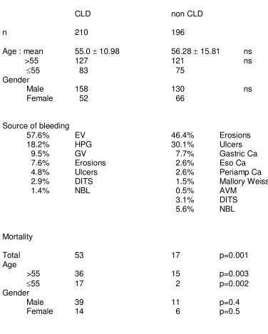
Table 3.UGI bleeding in CLD and non CLD
CLD non CLD
n 210 196
Age : mean 55.0 10.98 56.28 15.81 ns
>55 127 121 ns
55 83 75
Gender
Male 158 130 ns
Female 52 66
Source of bleeding
57.6% EV 46.4% Erosions
18.2% HPG 30.1% Ulcers
9.5% GV 7.7% Gastric Ca
7.6% Erosions 2.6% Eso Ca 4.8% Ulcers 2.6% Periamp Ca 2.9% DITS 1.5% Mallory Weiss
1.4% NBL 0.5% AVM
3.1% DITS 5.6% NBL
Mortality
Total 53 17 p=0.001
Age
>55 36 15 p=0.003
55 17 2 p=0.002
Gender
Male 39 11 p=0.4
Female 14 6 p=0.5
Table 4.The result of endoscopic hemostasis in patients with active bleeding.
Early hemostasis Rebleeding EV
Sclerotherapy 27/31 4/27
p=0.88 p=0.9
Ligation 25/28 3/25
Ulcers
Injection 18/19 3/18
Causes of death in CLD were mainly bleeding and encephalopathy. In non CLD patients the causes of death were respiratory and cardiovascular diseases (Table.5)
Table 5. The causes of death
CLD Non-CLD
Bleeding 19 3 p=0.0018
Encephalopathy 19 - -
Respiratory disease 7 7 p=0.89
Cardiovascular 6 6 p=0.86
Renal disease 2 1 p=0.9
Discussion
to age .This finding was not different with others studies(5,13). However in CLD patients, the mortality was not related to age, we assumed that the mortality in CLD patient may had relation with severity of liver diseases or with portal hypertension.The result of endoscopic treatment in EV with active bleeding was equal between scleroteraphy and ligation. Our results were similar with other study(14).The cause of death in non CLD patients was not the actual bleeding,but most of them died because of co-morbid illness. Conversely in CLD patients,the majority of patients died because of CLD related illnesses such as encephalopathy and coagulopathy.
References
1.Silverstein FE,Gilbert DA,Tedesco FJ et al.The national ASGE survey on Upper gastrointestinal bleeding I.Study design and base line data.
Gastrointest.Endosc.1981;27:73-79
2.Chang C,Chan C,Lin U.Endoscopy for upper gastrointestinal bleeding at emmergency unit.Chin.Med.J.1992;49:217-222
3.Bordley DR,Mashlin AI,Dolan JG,et al.Early clinical sign identfy low risk patients with acute upper gasteointestinal hemorrhage.JAMA.1985;253:3283 -3285.
4.Silverstein FE,Gibert DA,Tedesco FJ,et al.The national ASGE survey on upper gastrointestinal bleeding :II.Clinical prognostic factors.Gastrointest. Endosc.1981;27:90-93.
5.Graham DY,Smith JL.The course of patients after variceal hemorrhage. Gastroenterology.1981;80:800-809.
6Yavorsky RT,Wong Roy KH,maydonovitch C,et al.Analysis of 3,294 cases of Upper Gastrointestinal bleeding in Military Medical Facilities.Am.J. Gastroent.1995;90;568-573.
7.Hawkey CJ.Epidemiology and aetiology of upper gastrointestinal bleeding, in gastrointestinal bleeding.Krasner N (ed.).BMJ.Publishing group. London. 1996:3-13.
8.Hernomo K:Pengelolaan perdarahan masif varises esofagus pada sirosis hati.Thesis Unair.Surabaya.1983.
9.Sulaiman A,Julitasari.Virus Hepatitis A sampai E di Indonesia.Yayasan IDI. Jakarta.1995.
10.Mc CarthyD.Nonsteroidal antiinflammatory Drug-related gastrointestinal toxicity.Am.J.Med.1998;105:10S-16S.
11.Sippone P,Sappala K,Arisyne M, et al.Gastritis and gastroduodenal ulcer, a case control study of risk toxic duodenal or gastric ulcer in patients with artritis.Gut.1989;30:922-929.
12.Soll A. Pathogenesis of NSAID drug related upper gastrointestinal toxicity. Am.J.Med.1998;105:10S-16S.
13.Corley DA,Stefan AM,Wolf M,et al.Early indicators of prognosis in Upper Gastrointestinal hemorrhage.Am.J.Gastroenterol.1998;93:336-340.