P-ISSN.2503-0817, E-ISSN.2503-0825 is gingival disease, one of the gingival diseases that mostly disturbs tooth aesthetic and functional is the gingival enlargement, which can occur due to inflamma- tion, without-inflammation, a combina-tion of both, a systemic influence, the influence of drugs and neoplastic. It causes changes in the form of gingival clinically looked bigger than normal.1
Gingival esthetic problems commonly complained from patients include gingival enlarge-ment, gingival contour is abnormal, missing papillae and opened root surface. Gingival enlargement is a common clinical sign of the disease gingiva. There are many types of gingival enlargement, which are based on the etiological factors and pathological processes. Gingival enlargement is a condition in which there was an additional measure of gingival. In these circumstances, gingival tissue excessively bulging among the teeth and or on the neck area. The increase of this size can occur by way of hypertrophy, hyperplasia, or a combination of both.2
Gingival disease accompanied by an increase in the size of the gingiva (edema), called gingival enlargement (or gingival overgrowth). Gingival enlargement is divided into two kinds, namely
gingival enlargement caused by inflammatory and non-inflammatory. Histopathologic examination of the gingival enlargement due to inflammation found signs of increased exudate and proliferative from chronic inflammatory; fluid and inflamma-tory cells, with enlarged vascular and formation of new capillaries, as well as degenerative changes. Gingival enlargement because of inflammation can be treated with scaling and root planning treat-ment. Histopathologic features of non-inflamma-tory gingival enlargement showed connective tissue and epithelial hyperplasia, elongation of rete pegs, increase of fibroblasts and collagen fibers. Problems often complained by patients with gingival enlarge-ment is the aesthetic factor although the fact is that the health of the teeth supporting tissue has disorders. Gingival enlargement in the interden-tal papillae, thickened, rounded gingival contour and discomfort became major issues that must be treated in order to perform optimal appearance and function. Gingival enlargement that experienced fibrosis would not disappear with only plaque control, but required surgery that is gingivectomy and gingivoplasty.3,4
Gingival enlargement is also an indication of gingivectomies. gingival enlargement can be classified based on the etiological factors and pathological changes, they are: Inflammatory Abstract
Objective: Gingival enlargement in the interdental papillae, thickened, rounded gingival contour and discomfort became major issues that must be treated in order to be optimal appearance and function. Gingival enlargement that experienced fibrosis would not disappear with only plaque control, but required surgery that is gingivectomy and gingivoplasty.
Methods:The 24-year-old woman came to the periodonsia clinic with complaints maxillary anterior gingiva swelled at teeth 11-21 with plaque index 52%. The depth of the tooth pocket 11: labial (mesial: 4, medial: 1, distal: 3). Palatal (mesial: 3, medial: 2, distal 1). The depth of the tooth pocket 21 is labial (mesial: 4, medial: 1, distal: 1),
palatal (mesial: 3, medial: 1, distal 1). Gingivectomy treatment and gingivoplasty were performed with the aim of eliminating pockets and restore physiologic gingival contour which can help prevent the recurrence of the disease periodontal.
Results: In inflammatory gingival enlargement, clinical evalution pasca gingivectomy surgery performed optimum healing, without any inflammation signs.
Conclusion:In performing surgical gingivectomy and gingivoplasty, which must be considered is to minimize the disposal of gingival tissue to maintain the aesthetic, adequate access to the bone defect.
Keywords:Gingival enlargement, Gingivectomy, Gingivoplasty
Cite this Article:Tjiptoningsih UG. 2016. Enlargement gingival treatment on teeth 11 and 21. Journal of Dentomaxillofacial Science 1(3):
196-200. DOI: 10.15562/jdmfs.v1i3.317
Departement of Periodontic, Faculty of Dentistry, University of Prof. Dr. Moestopo (Beragama), Jakarta, Indonesia
Enlargement gingival treatment on teeth 11 and 21:
a case report
enlargement such as chronic and acute. Drug-induced enlargement (drugs). Associated with systemic diseases/special conditions: pregnancy, pubertal, vitamin-C deficiency, cell gingivitis plasma, non-specific (pyogenic granuloma), systemic diseases: Leukemia, Granulomatous diseases. Neoplastic enlargement (gingival tumors): benign Tumors, malignant Tumors, false enlargement, based on the location and distribu-tion: localized: one/a group of teeth, generalized, marginal; only the gingival margin affected, papil-lary: only interdental papilla, diffuse: margin, attached gingiva and papilla, discrete: peduncu-lated, sessile, like tumors.3,4
Gingivectomy is an excision or removal of gingiva tissue, with the aim of eliminating the pocket wall. Gingivectomy improve the visibility and accessibility to lift the calculus overall, facili-tate the smoothing the root surface, create a good environment for gingival healing process and the restoration of gingival physiologic contour.1
Gingivectomy indications are:1,4-6
eliminate suprabonipockets, regardless of the depth of the pocket, if the wall pockets fibrous and hard, elim-inate gingival enlargement, elimelim-inate supraboni periodontal abscess.
Gingivectomy contraindications are:1,4-6 bone surgery is required or the examination of bone
shape and morphology, position of the base pocket is more apical than the mucogingival junction, esthetic considerations, especially on the anterior maxilla.
Gingivoplasty was performed to eliminate periodontal pockets and included resharping as part of the technique. Gingivoplasti is resharping gingiva to create physiologic gingival contour, with the sole aim to recontur gingival without pockets. Gingivoplasti can be performed with a periodontal knife, scalpe, diamond stone, or electrodes. These techniques include procedures that represent what is performed in festooning dentures: sharpen the gingival margin, creating a line of scalloped marginal, deplete attached gingiva and create vertical interdental grooves, as well as form a papilla in order to provide floodgate road (sluiceway) for food way. The main object of gingivoplasti is to restore the physiologic gingival contour which can help to prevent the recurrence of the perodontal disease. Gingivoplasty is a way to improve the gingival esthetic. Gingivoplasty is usually indicated to an abnormal gingival contour. Tissues are chewy and fibrotic and can be easily excised and formed.4-6
Case Report
Main complaintA 24-year-old female patient came to the periodon-tic clinic with complaints of swelled maxilla front teeth gum so that patients feel disturbed in appearance. To the patient performed scaling by private dentists but the gums still enlarged. Patients wanted to be treated.
Intraoral and Extraoral Examination
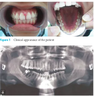
In the extra-oral examination found no abnor-mality and the patient had no systemic disorders. On intra-oral examination found circumstances of edema gingival in the midline of the anterior maxilla figure 1 and on panoramic view showed the teeth 12, 16, 26, 27, 28, with soft consistency, shiny rounded interdental condition, stippling (-), pitting test (+) and teeth premature contact 26 and 36 as well as 27 and 37 as well as horizontal resorption alveolar bone in teeth 25, 26, 27, 34, 35, 36, 37, 45, 46 with a value of plaque score 52% figure 2.
Based on poket depth examination showed diag-nosis local chronic periodontitis on teeth 25, 26, 27, 34, 35, 36, 45, 46 accompanied by inflammatory of gingival enlargement on 11-2. Table 1 It propbably caused by bacterial plaque, premature contact of teeth 26 and 36 as well as 27 and 37 and horizontal resorptionon alveolar bone on teeth 25, 26, 27, 34, 35, 36, 37, 45, 46.
Figure 1 Clinical appearance of the patient
Operation Procedures
Firstly preparation of the patient, operator, oper-ator assistant, tools and materials and informed consent. Secondly plaque scores and prophylaxis. Thirdly extra-oral and intra-oral aseptic action by using 10% betadine solution. Fourthly closure of the patient’s face using a perforated sterile towel except the area of operation. Finally local anesthe-sia by local infiltration technique 11, 21 and block anesthesia on nasopalatine nerve figure 3.
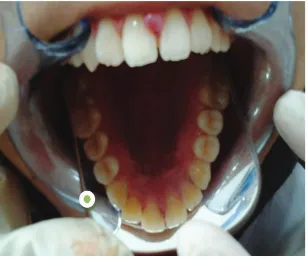
Injection in N. Nasopalatinus through cana-lis insisivum in the second rugive to anathesize mucosapoda regio anterior palatum durum (premaxila), by anathetic liquid as many as 0.1-0.2 cc figure 4, firstly gum surface which has pocket searched by probe and marking with pocket marker. Marking in someareas which are an outline for insisition figure 5. Secondly kirkland knife is used to cut the facial surface and lingual as well as distal. Orban periodontal knife isapplied for inter-dental interception. If needed Bard-Parker knive #1 and #2 as well as scissors are applied for complement instrument figure 6 and 7. Thirdly insision begin from apical of point marking, directed coronally to the point between based pocket and bone peak. Insision is conducted as close as the bone without Table 1. Pocket depth examination
Tooth 18 17 16 15 14 13 12 11 21 22 23 24 25 26 27
Facial X 113 123 322 323 312 211 413 411 212 212 312 315 415 323
Palatal X 113 122 211 111 111 111 321 311 111 112 112 111 111 112
Mobility - - -
-Tooth 48 47 46 45 44 43 42 41 31 32 33 34 35 36 37
Facial X 245 435 521 111 211 111 111 211 121 111 111 335 514 212
Lingual X 111 211 121 111 111 211 211 111 111 111 111 111 212 212
Mobility - - -
-Figure 3 Anesthesia in the teeth 11 and 21 labial part
Figure 4 Anesthesia in the teeth 11 and 21 in palatal part
causing bone exposed and the angle about 45° to the teeth surface. Fourthly take off the exsision pocket surface, clean that area and pay attention to the root teeth surface. In coronal, we found the claculus residu, root caries or bone resorption. Granulated tissue can be found in smooth tissue. Fifthly tissue’s smoothing using diamond bar with 2 cm in diam-eter figure 8. The next, take the granulated tissue, necritic cement and remained calculus by Gracey curete until the bone surface is clean and smooth, irigated with NaCl 0.9% saline solution and H2O2 3% figure 9, finally if the bloody occurs, solved by
pressing the tampon that was full of adrenaline solution (added with aquades) in operated area.
Operated area irrigated with NaCl phisiologist 0.9% and H2O2 3%. Clean and dry the operated area with sterile tampon figure 10. Operated area clearence. Cover the operated area with periodontal pack figure 11. Instruction for post operation and prescription. Clearence of the operation room. Figure 6 Insision with kirkland knife
Figure 7 Insision with orban knife
Figure 8 Gingival smoothing in 11 and 21
Figure 9 Irrigation with NaCl 0.9% and H2O2 3%
Figure 10 Clinical appearance after gingivectomy
Control I, II and III
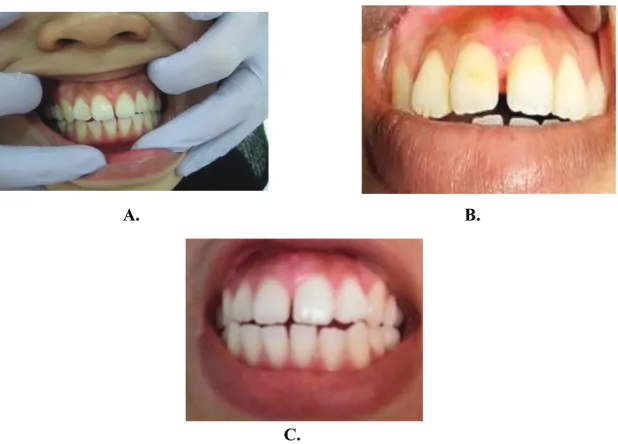
In inflammatory gingival enlargement, clinical evalution pasca gingivectomy surgery performed optimum healing, without any inflammation signs figure 12 A, B and C. Factors such as mantain kera-tine tissue in gingiva, loss gingival tissue minimumly to keep the elasticity, enough way in conducting repairement of osseous damage, and minimalize uncomfortability and bloody after operation must be considered to reduce minimumly over gingival tissue.
Discussion
Inflammatory gingival enlargement mainly clas-sified into 2, those are chronic and acute, chronic inflammatory gingival enlargement commonly marked with false pocket. In the initial cure enlargement in this case, we should eliminate and control plaque, to prevent continous periodontal damage. Plaque accumulation related to inflamma-tory gingival enlargement and remove local factor ethiology and preservation of gingival enlargement as well as improve the gingival teeth.
In corrective phase, before operation was conducted, evaluation of plaque control must be
performed and decline occured, this result perfoms that home care, for teeth and mouth cleanliness which conducted to the patient show a significant influence, which can be a reference for the gingi-vectomy and gingivoplasty application, in hoping that there will be no complication afterward and its prognosa is optimum. Sealing cause to the reduc-tion of inflammareduc-tion in gingival enlargement at control week 1 evaluation pasca initial phase ther-apy (initial phase).3,4
The result of the clinical evaluation performs that the healing occurs in a week pasca operation, there is tissue generation and gingival contour advance in the operated area. The healing of gingi-val tissue conduct for 3-4 weeks in 1 week control and in 1 month, the patient’s gingival performs a significant healing.
Conclusion
Gingivectomy has some applications to reduce minimumly over gingival tissue but many factors inhibited must be considered. This cure is support-ing the patient because it reduces uncomfortability of gingival enlargement and better ethical appear-ance can be reached.
Re
ferences
1. Affandi Y. Pembesaran gingiva pada pasien leukemia. USU 2011.
2. Axelsson P. Diagnosis and risk prediction of periodontal disease. Quintessence Publishing Co Inc; 2002. p. 317-324. 3. Carranza FA, Takei HH. Gingival surgical techniques. In
Newman M, Takei HH, Klokkevold PR, Carranza FA, edi-tors. Carranza’s clinicall periodontology. 11th ed. St Louis: W.B. Saunders Co; 2010.
4. Newman MG, Takei HH, Klokkevold PR, et al. Carranza’s clinical periodontology. 10th ed. St. Louis: Elsevier; 2006. 5. Rateitschak KHEM, Wolf HF, Hassell TM. Color atlas of
periodontology. New York: Thieme; 1985.
6. Lindhe J, Karring T, Lang NP. Clinical periodontology and implant dentistry. 4th ed. Iowa: Blackwell Publishing Ltd; 2003.
This work is licensed under a Creative Commons Attribution Figure 12 Clinical evalution pasca gingivectomy; A. 1 week control,
performed, this shows that home care for teeth and mouth cleanliness conducted to the patient show a
significant influence, which can be a reference for
the gingivectomy and gingivoplasty application, hoping that there will be no complication afterward and its prognosis is optimum. Sealing cause to the reduction of inflammation in gingival enlargement at control week 1 evaluation pasca initial phase therapy (initial phase).3,4
The result of the clinical evaluation shows that the healing occurs in a week pasca operation, there is tissue generation and gingival contour advances in the operated area. The healing process of gingival tissue continued for 3–4 weeks with 1 week control in 1 month, the patient’s gingiva showed a signifi-cant healing.
CONCLUSION
In inflammatory gingival enlargement, clinical eval-uation post gingivectomy surgery was performed for optimum healing without any inflammation signs. Gingivectomy has some applications to reduce minimally over gingival tissue, but other inhibitory factors must be considered. Periodontal surgery must be considered to (1) maintain keratin tissue in gingiva, (2) Minimal loss gingival tissue to keep the elasticity, (3) enough way in conducting repairment of osseous damage, and (4) minimalize some discomfort and blood loss after operation.
This cure is encouraging the patient because it reduces discomfort of gingival enlargement and better ethical appearance can be reached.
REFERENCES
1. Affandi,Y., 2011, Pembesaran gingiva pada pasien Leukemia. USU.
2. Axelsson P. 2002. Diagnosis and risk prediction of periodontal disease. Quintessence Publishing Co Inc; P.317–24.
3. Carranza, F.A., and Takei, H.H., 2010 Gingival Surgical
Techniques, dalam M. Newman, H.H. Takei, P.R. Klokkevold dan F.A. Carranza (eds): Carranza’s clinical
periodontology, 11th ed., W.B. Saunders Co., St Louis.
4. Newman MG, Takei HH, Klokkevold PR, Carranza FA.
Carranza’s clinical periodontology. 10th ed. St. Louis:
Elsevier; 2006.
5. Rateitschak KH & EM, Wolf HF, Hassell TM. Color atlas of
periodontology. New York: Thieme; 1985.
6. Lindhe J, Karring T, Lang NP. Clinical periodontology and
implant dentistry. 4th ed. Iowa: Blackwell Publishing Ltd;
2003.